

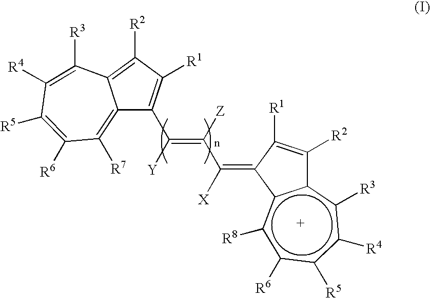
Azulene dimer-quenched, near-infrared fluorescent probes
a fluorescent probe and dimer technology, applied in the field of in vivo optical imaging, can solve the problems of inability to achieve high-efficiency quenchers for these nir fluorochromes, inability to develop nir probes, and inability to meet the needs of in vitro and in vivo applications, etc., to achieve efficient self-quenching, reduce background fluorescence, and facilitate simultaneous multiple probe use
- Summary
- Abstract
- Description
- Claims
- Application Information
AI Technical Summary
Benefits of technology
Problems solved by technology
Method used
Image

Examples
example 1
Preparation of Tropolone tosylate
[0124] Triethylamine (114.30 μl, 0.82 mmol) was added dropwise to a round, dried flask containing a colorless solution of tropolone 1 (100 mg, 0.82 mmol) and tosyl chloride (171.90 mg, 0.90 mmol) in CH2Cl2 (5 mL) at room temperature and stirred for 3 hours. The reaction was poured onto ice-cold water (20 mL), and the crude product was extracted with CH2Cl2, dried over MgSO4, filtered, and concentrated by rotavapor. The crude product was recrystallized from 8:2 CH2Cl2 / hexanes to afford 190 mg (84%) of a hygroscopic brown solid: Rf=0.25 (9.5:0.2 CH2Cl2 / MeOH); 1H NMR (400 MHz, CDCl3) δ 2.45 (s, 3H), 6.95-7.23 (m, 4H), 7.36 (d, J=8.0 Hz, 2H), 7.45 (d, J=8.5 Hz, 1H), 7.92 (d, J=6.7 Hz, 2H); 13C NMR (400 MHz, CDCl3) δ 21.8, 128.6, 129.6, 130.0, 131.0, 134.6, 136.3, 141.2, 145.0; LRMS (FAB+) calcd (M+H)+ (C14H12O4S) 277.31, found 277.13
example 2
2H-3-methoxycarbonylcyclohepta[b]furan-2-one (2)
[0125] A solution of NaOMe (67 mg, 1.24 mmol) in anhydrous MeOH (5 ml) was canulated to a dried flask containing a clear solution of tropolone tosylate (170 mg, 0.62 mmol) and dimethyl malonate (141.70 μl, 1.24 mmol) in MeOH (20 mL) at 0° C. The reaction turned yellow at the end of the addition. The reaction mixture was allowed to warm slowly to room temperature with stirring for 14 hours. The precipitate was collected by vacuum filtration and air-dried. The crude product was recrystallized from 8:2 CH2Cl2 / MeOH to provide 108 mg of yellow solid. The filtrate was concentrated to a yellow solid by rotavapor. Chromatography with 9.5:0.2 CH2Cl2 / MeOH afforded 18.6 mg. The total yield of yellow solid was 126.6 mg (100%): Rf=0.48 (9.5:0.2 CH2Cl2 / MeOH); 1H NMR (400 MHz, CDCl3) δ 3.95 (s, 3H), 7.34 (ddd, J=3.3, 3.8, 3.3 Hz, 1H), 7.50 (t, J=4.1 Hz, 2H), 7.64 (m, 1H), 8.86 (d, J=11.3 Hz, 1H); 13C NMR (200 MHz, CDCl3) δ 51.6, 96.3, 119.2, 130.6, ...
example 3
1-(methoxycarbonyl)-2-methylazulene (3)
[0126] A yellow suspension of lactone 2 (660 mg, 3.23 mmol) and 2,2-dimethoxy propane (2 mL, 16.20 mmol) in anhydrous toluene (3 mL) in an ACE pressure sealed tube was heated slowly from room temperature to 200° C. in a period of 2 hours. The temperature was kept constant for a period of 24 hours. The brownish-red solution was introduced directly onto a silica gel flash column using 1:1 CH2Cl2 / hexanes to afford the brownish-red viscous liquid 646.5 mg (100%): Rf=0.63 (CH2Cl2); 1H NMR (400 MHz, CDCl3) δ 2.83 (s, 3H), 3.98 (s, 3H), 7.13 (s, 1H), 7.39 (t, J=11.1 Hz, 1H), 7.50 (t, J=11.1 Hz, 1M), 7.69 (t, J=11.1 Hz, 1H), 8.28 (d, J=10.6 Hz, 1H), 9.48 (d, J=10.6 Hz, 1H), 13C NMR (200 MHz, CDCl3) δ 18.1, 50.8, 86.2, 115.1, 120.2, 126.8, 127.7, 135.8, 137.2, 142.1, 143.2, 154.1, 166.6; LRMS (EI) calcd M+ (Cl3H12O2) 200.2366, found 200.0833; LRMS (MALDI-TOF) found 201.22; UV-vis (MeCN) λmax=524 nm.
PUM
| Property | Measurement | Unit |
|---|---|---|
| Magnetic field | aaaaa | aaaaa |
| Nanoscale particle size | aaaaa | aaaaa |
| Nanoscale particle size | aaaaa | aaaaa |
Abstract
Description
Claims
Application Information