Ex vivo verification of biopsy tissue samples

a biopsy tissue and ex vivo technology, applied in the field of biopsy tissue verification, can solve the problems of not easily fitting into standard biopsy procedures, remains a degree of uncertainty as to whether the excised tissue does in fact include part or all, and clinical application of these agents has not yet been realized. , to achieve the effect of improving confidence and improving biopsy accuracy and speed

Active Publication Date: 2006-08-03
PAWLUCZYK OLGA +1
View PDF18 Cites 24 Cited by
  • Summary
  • Abstract
  • Description
  • Claims
  • Application Information

AI Technical Summary

Benefits of technology

[0040] Based on the concentration of the contrast agent within the obtained sample, information relating to the likelihood that the particular sample was within the region of interest on the guiding image can be provided from non-destructive, or minimally destructive testing of the sample to observe the presence of the contrasting agent in relative concentrations indicative of abnormal tissue. This will in turn increase confidence that the biopsy sample obtained is actually that from the region of concern. Due to the potential for non-destructive activity in the proposed verification, an improvement in the biopsy accuracy and speed is possible without greatly influencing the established procedures now in place.

Problems solved by technology

One limitation of these methods is that optimally an excised sample should be checked whether it contains the lesion structure, before the patient can be released.
This is a time consuming procedure, requires transfer of the biopsy sample from the procedure room, and does not easily fit into standard biopsy procedures.
In most cases this can not be accommodated, and therefore there remains a degree of uncertainty as to whether the excised tissue does in fact include part or all of the tissue identified by the original imaging technique.
The clinical application of these agents has not yet been realized.
This is always a difficult procedure, and it is prone to error.
difficulties with confirmation that the sample actually being taken from the appropriate location determined with an imaging modality
However, there is little done to validate whether the actual excised tissue matches the region viewed with the imaging method guiding the biopsy.
Furthermore, no attempt has been made in this patent to measure or detect the specific signature of an exogenous contrast agent.
Many of these techniques perform well ex vivo, however once applied in vivo, in a much more challenging clinical environment, the techniques breakdown and lose their clinical utility.
As there may be uncertainty to whether the correct volume of tissue has been sampled, the results from the pathologist may also reflect this uncertainty.
Therefore, the decision whether to obtain more tissue samples, or remove a larger section of tissue can not be guided by a close examination of the tissue during a typical procedure.
Performing this verification after the patient has been removed from the apparatus is not as beneficial as it cannot guide clinical management decisions at that time; however, it may impact the degree of confidence to which the radiologist may accept a pathological diagnosis that does not match the imaging presentation.
This practice can often be helpful; however, many lesions are not well visualized using X-ray and do not present an X-ray evident presentation [M. Kriege, C. T. M. Brekelmans, C. Boetes, J. Klijn, et al.
This is becoming more problematic as modalities other than X-ray are increasingly being used to detect lesions and guide interventions (i.e., MRI, US [ultrasound], CT [computed tomography]).
There is no simple way of verifying whether the biopsy sample obtained matches with the area of interest on an image used to guide the biopsy.
Although these methods are very accurate and sensitive to the measurement of trace amounts of chemical compounds and could be used to determine the presence of contrast agent within the sample, they are not appropriate in a clinical setting where further histopathological analysis is required for normal patient management.
The spectral overlap of signal obtained from the contrasting agent present for a specific modality and the endogenous signal of the biological sample, including its water signal, creates significant difficulties in the detection of the alien molecules of the contrasting agent; however, it is possible to overcome these problems by using high performance spectroscopy methods such as absorption spectroscopy, MR spectroscopy or other spectroscopic methods such as scattered light spectroscopy, Raman spectroscopy and other similar methods.
The difficulty in the spectroscopic determination of the presence of a compound arises when multiple compounds are present within a sample or when some compounds such as water have predominant contribution to the measured spectrum.
However, the majority of substances used as contrast agents for imaging modalities have structures substantially different from those contained organic matter; therefore, they produce signals different from those produced by components of organic substances.
However, an area of the field of medicine that remains unexplored is the use of advanced spectrographic techniques to validate whether tissue removed from a patient in a clinical setting contains tissue of clinical interest that was seen with a given imaging modality when a contrasting agent was used.

Method used


Image

  • Ex vivo verification of biopsy tissue samples
  • Ex vivo verification of biopsy tissue samples
  • Ex vivo verification of biopsy tissue samples

Examples


Embodiment Construction

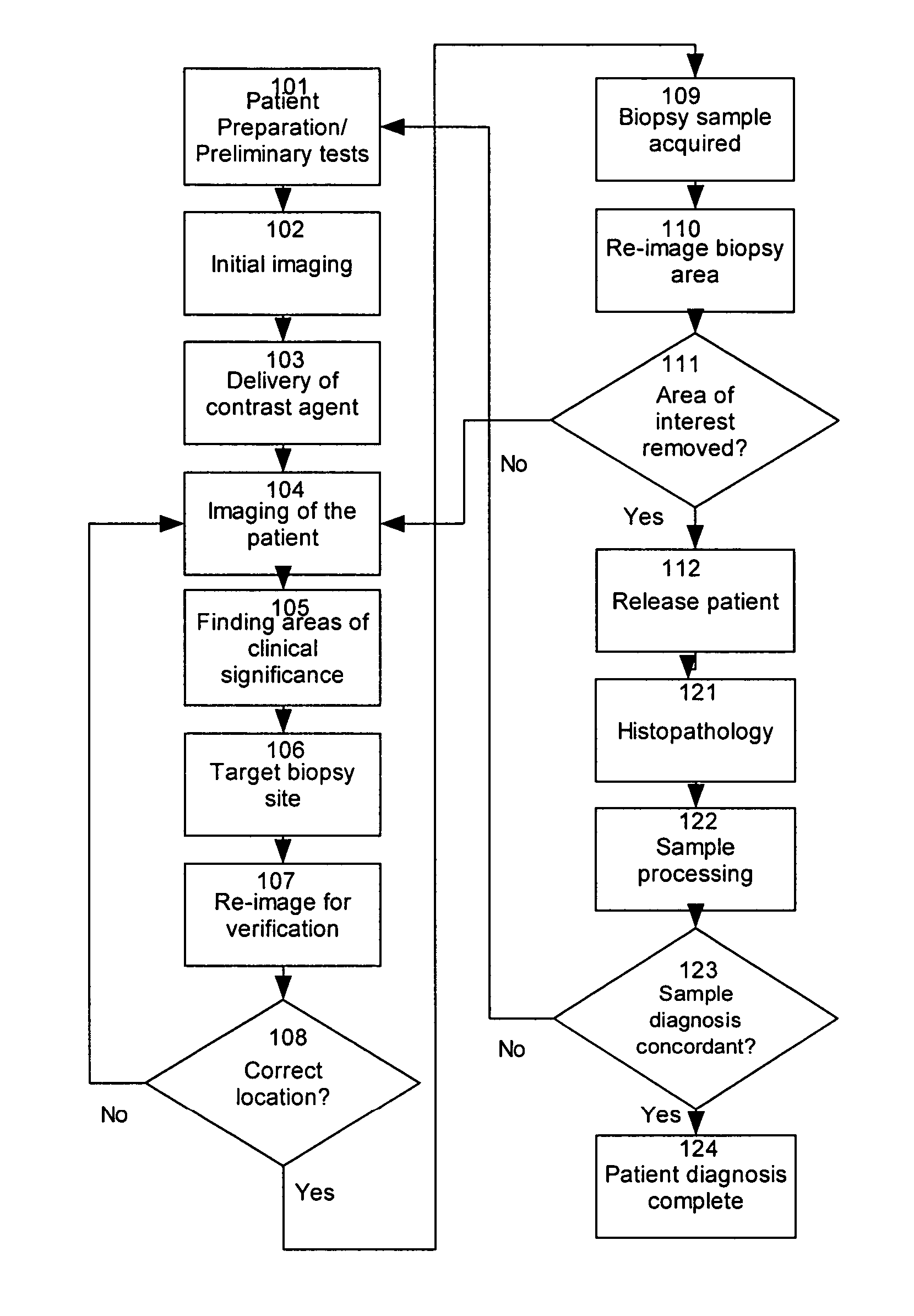
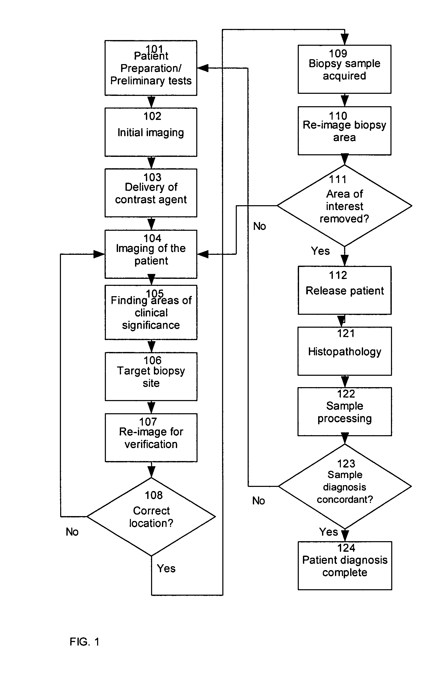
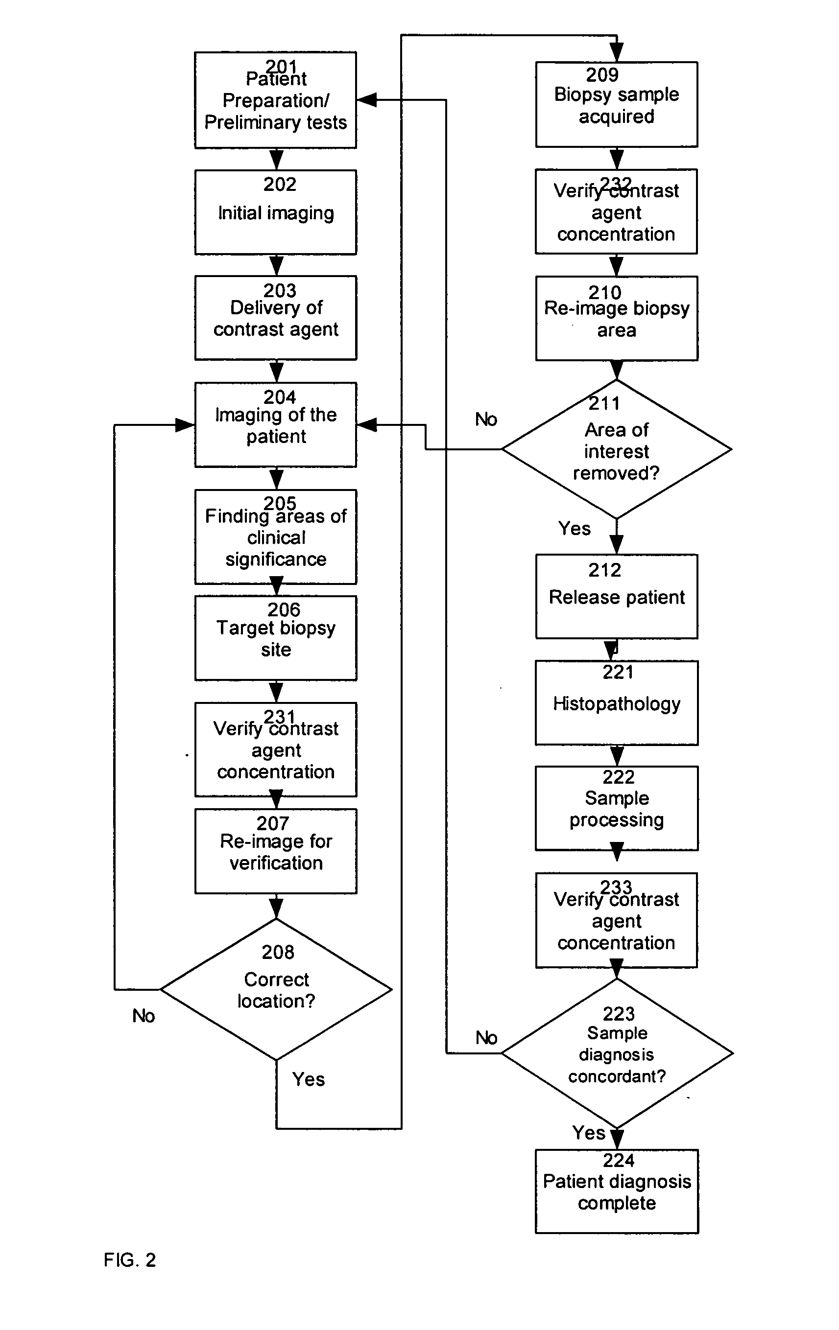
[0048] A method indicates a likely presence of abnormal tissue within an ex vivo sample of tissue by providing observable agent to a region of interest of a patient; removing from the patient a tissue sample from the region of interest to provide a removed tissue sample; non-destructively or minimally destructively observing the removed tissue samples under conditions that allow observation or detection of the non-destructively observable agent; and evaluating results of the observation or detection of the observable agent within the removed tissue sample. Observation may be by destructive, partially non-destructive or non-destructive techniques, as described herein. The method may evaluate results by assessing a likelihood of the presence of abnormal tissue within a sample or evaluating results to determine whether the removed tissue sample has been taken from the region of interest. After removing the sample, and preferably before or after evaluating results, histopathic examinati...


PUM


Abstract

An instrument for verification of presence of image enhancing, contrasting agent in a biopsy sample which was obtained by imaging the lesion area with an imaging modality which is sensitive to the contrasting agent. In one embodiment of the invention, an optical spectrometer is used to analyze biopsy samples for the presence of gadolinium enhanced Magnetic Resonance Imaging contrasting agent which typically pools in breast tumours.

Description

BACKGROUND OF THE INVENTION [0001] 1. Field of the Invention [0002] The present invention relates to biopsy tissue verification and more specifically to devices and methods which inspect excised tissue for the presence of endogenous or exogenous contrasting agents used to better image clinically interesting regions of the body, preferably by non-destructive observation or testing of excised tissue. [0003] 2. Background of the Art [0004] The field of detection and diagnosis of cancerous tumors, pre-malignant, malignant and other lesions and disorders is very broad and has been the subject of much research. Typically, an imaging modality including, but not limited to X-ray imaging, tomography, MRI, ultrasound, PET, nuclear imaging, palpation and visual inspection is first used to locate an area of clinical significance within the patient. Many of these techniques use the inherent biophysical contrast unique to the pathology to visualize, or diagnose the pathology. These endogenous tec...

Claims


Application Information

Patent Timeline
Patent Type & Authority Applications(United States)
IPC IPC(8): A61B5/05
CPCG01N21/64G01N21/65G01N24/08G01N29/0672G01N29/449G01N29/46G01N33/58G01N2291/02466G01N2291/02475
Inventor PAWLUCZYK, OLGAPIRON, CAMERON
Owner PAWLUCZYK OLGA
Why Eureka
Try Eureka