Coring device for preserving living tissue

a coring device and living tissue technology, applied in the field of cartilaginous and bony tissue removal, can solve the problems of limited availability and possible damage of donor sites, devices used in such a manner are not consistent with preserving cell viability in affected tissue areas, and cannot deal with large and deep osteochondral defects

Active Publication Date: 2006-08-03
DSM IP ASSETS BV
View PDF33 Cites 26 Cited by
  • Summary
  • Abstract
  • Description
  • Claims
  • Application Information

AI Technical Summary

Benefits of technology

[0022] It is another object of the invention to create a strong tool, ca

Problems solved by technology

Devices used in such a manner are not consistent with preserving cell viability in the affected tissue areas, as will be discussed below.
Limitations of the technique include the inability to deal with large and deep osteochondral defects, limited availability and possible damage to donor sites, non-filled spaces between the circular grafts and incomplete integration of the donor and recipient cartilage.
The process for creating a plug or core typically results in an unfavorable level of cell necrosis.
For these reasons, articular cartilage is a difficult tissue to repair.
It has long been recognized in orthopedic operations that excess heat derived from drilling results may result in thermal injury and/or necrosis of nearby tissues.
Elevated temperatures, even for relatively brief periods are capable of causing cell death.
These types of devices using high speed drilling generate excessive heat due to friction between the blade and the material.
Furthermore, research has shown that powered trephine devices, when utilized for cartilage grafting, frequently cause gross damage, such as shredding the soft tissue adjacent the bone, creating ragged graft edges, and possibly separating the cartilage from the subchondral bone.
Damage to this intricately oriented cartilage tissue, whether structural or mechanical, alters the tissue's ability to transmit loads.
The creation of these ragged edges, architecturally affects the tissues ability to transmit the forces generated, for example during gait, and therefore increases the localized forces the surrounding cells are exposed to, leading to cell necrosis.
Further study of these ragged edges demonstrate de

Method used


Image

  • Coring device for preserving living tissue
  • Coring device for preserving living tissue
  • Coring device for preserving living tissue

Examples


Embodiment Construction

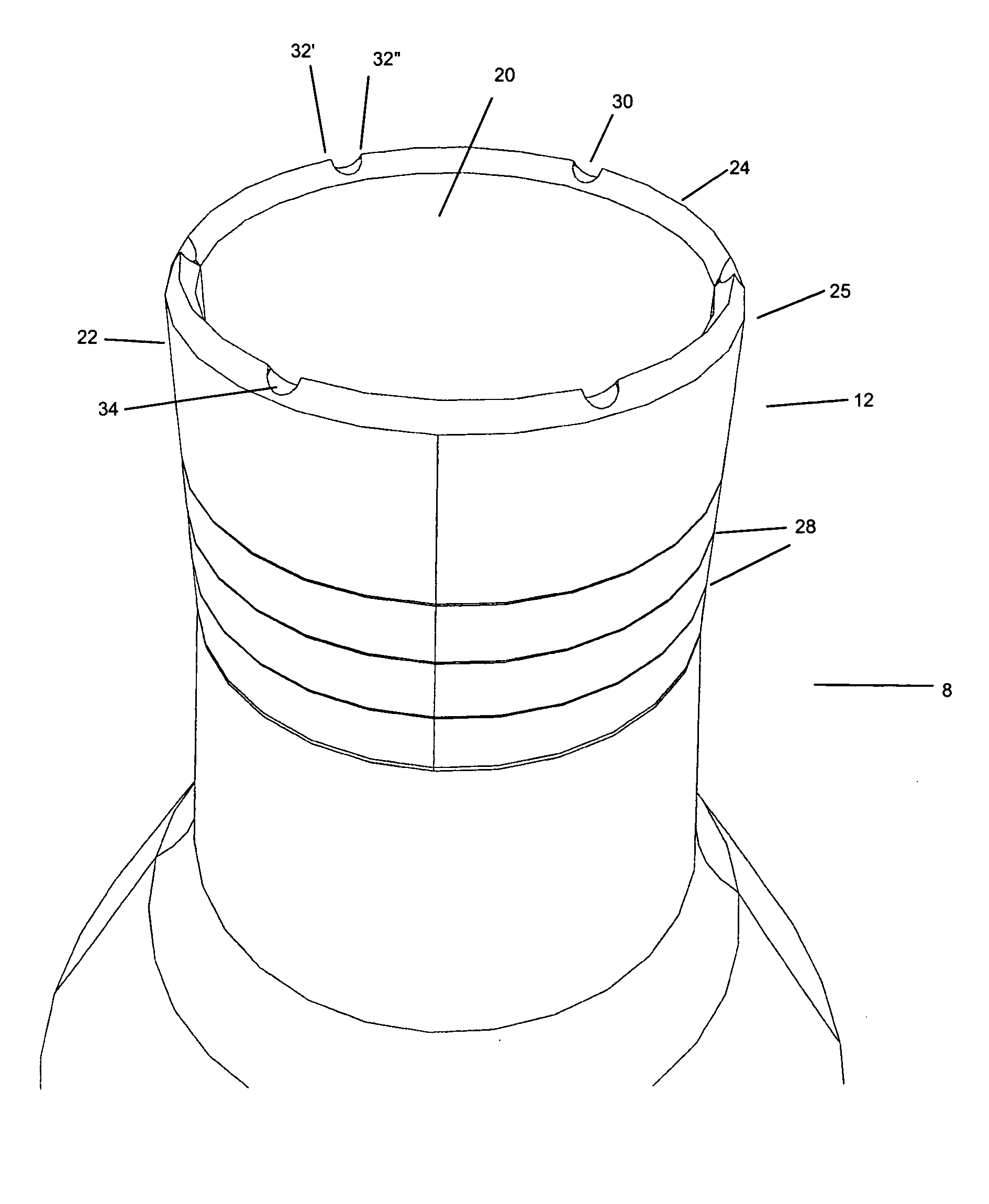
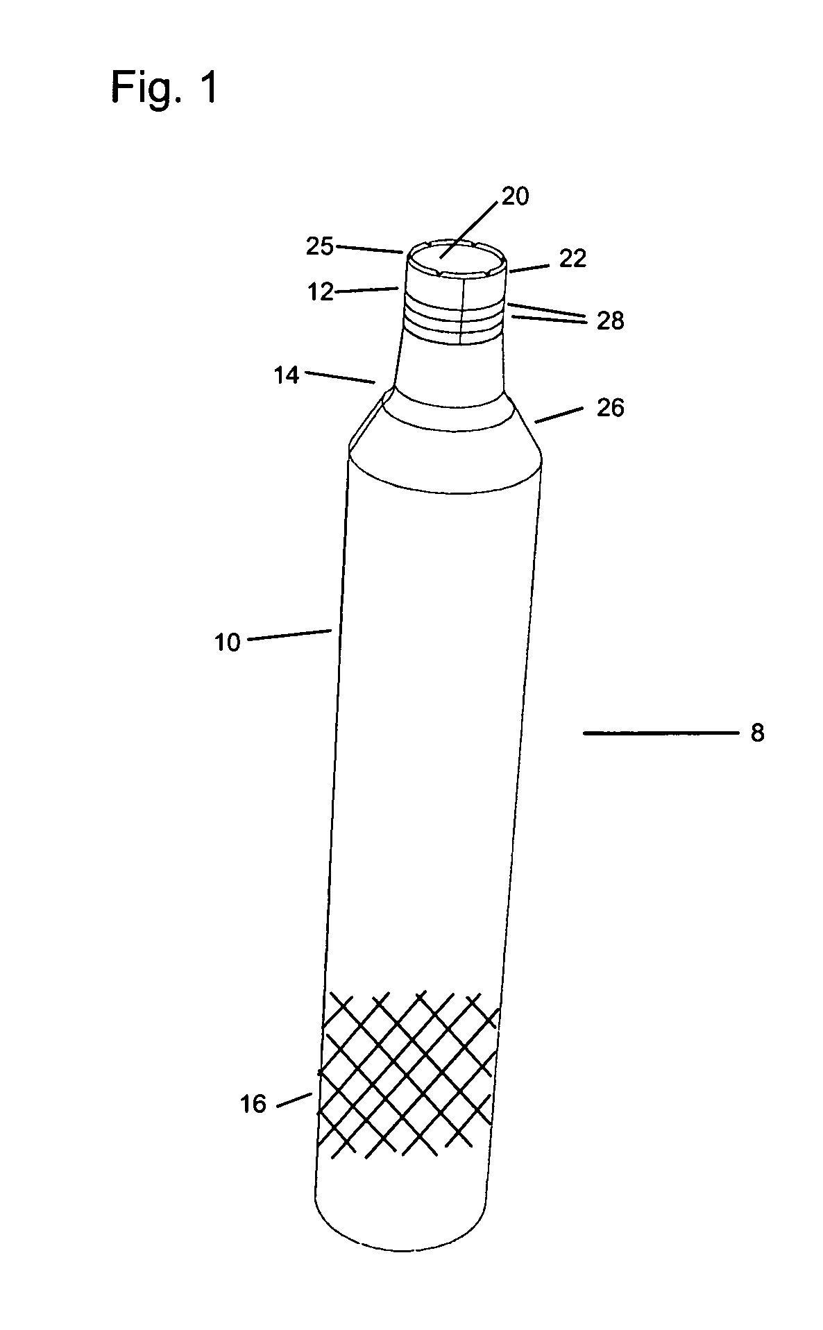
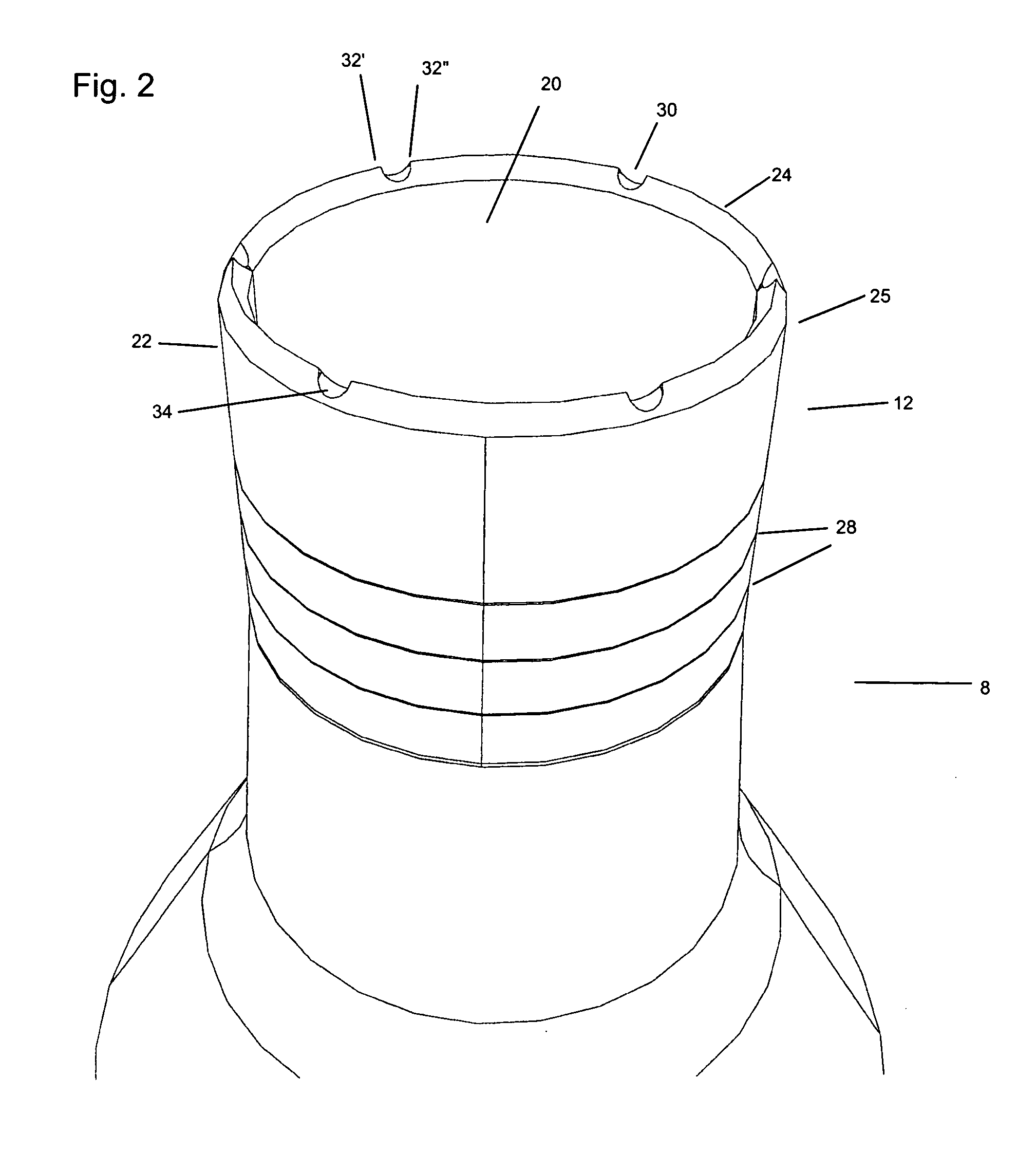
[0029] The present invention provides devices and methods for coring tissue of exact dimensions, wherein the viability of the cells in the tissue is substantially preserved. It has surprisingly been found that to facilitate effective integration of an implanted osteochondral core plug, not only must the osteochondral cell viability of the core plug material be preserved, but further the osteochondral cell viability of the surrounding tissue ought to also be preserved. In this manner, a plug of living material is able to be integrated into a surrounding area of living material. The present invention utilizes low speed, low impact techniques to neatly slice through soft tissue, such as cartilage, dividing the soft tissue with a cutting element having a sharpened level cutting edge surface, and preserving the viability of soft tissue cells. The present invention is also capable of cutting into to rigid tissue, such as bone, as the cutting element also features a plurality of serrations...


PUM


Abstract

Improved coring devices suitable for articular cartilage and bone, wherein the cutting device is capable of slicing through a tough protective tangential zone, delicately separating the shock absorbing columns of cells in the radial zone of the cartilage, and finally cutting into the hard underlying bone in a manner that preserves the viability of osteochondral cells. The coring device features an annulus having a flat annular cutting edge interrupted by at least one serration having neutral cutting angles. A method for concurrently removing cartilaginous and bony tissue using an improved coring device that preserves the viability of osteochondral cells.

Description

BACKGROUND OF THE INVENTION [0001] This application relates generally to medical instruments and methods of use for surgery and tissue excision, specifically relating to the removal of cartilaginous and bony tissue. A preferred embodiment more particularly concerns a device useful for preventing tissue damage and cell necrosis during a coring procedure in cartilaginous and bony tissue. [0002] It is well known to utilize a hollow, annular drill bit, such as a trephine cutter, to cut into hard tissue, especially bone, as has been well documented in the prior art. For example, U.S. Pat. Nos. 3,609,056; 4,142,517; 4,782,833; 5,324,300; 5,423,823; 5,697,935; and 6,488,033, all describe annular cutting devices with saw teeth. Frequently, these devices are utilized in combination with either high speed drilling, axially impacting with a mallet, or a combination of both. Devices used in such a manner are not consistent with preserving cell viability in the affected tissue areas, as will be ...

Claims


Application Information

Patent Timeline
IPC IPC(8): A61B17/32
CPCA61B17/1635A61B17/32002A61B17/32053A61B2017/00464A61B2017/00858
Inventor BRADICA, GINORINGEISEN, TIMOTHY A.
Owner DSM IP ASSETS BV
Why Eureka
Try Eureka