Screening antibodies using an optical fiber array device capable of simultaneously performing multiple functional assays

a technology of optical fiber array and antibodies, which is applied in the field of monoclonal antibodies, can solve the problems of false negatives, antibody binding by only a few antibodies may fail to initiate a recognizable change in the cell, and achieve the effects of increasing the number of assays, rapid, and high throughpu

Inactive Publication Date: 2006-09-21
ANGELIDES KIMON
View PDF27 Cites 1 Cited by
  • Summary
  • Abstract
  • Description
  • Claims
  • Application Information

AI Technical Summary

Benefits of technology

[0020] The preferred assays include functional assays, which determine the effect that an antibody has on the function of a cell. The assays can be used to determine any of a number of cell function, including but not limited to: (i) cytotoxic activity toward cancerous cells; (ii) intra-cellular signaling, including G protein activation, phosphatidyl inositol signaling, or ion channel effects; (iii) Ca2+ regulation in live cells; (iv) effects on the JAK-STAT pathway (related to apoptosis); and (v) effects on tyrosine kinase activity which is indicative of growth factor signaling. Simultaneously with a determination of function, assays can be included to determine antibody binding (a conventional enzyme-linked immunoadsorbant assay, “ELISA”), or to determine specificity, i.e., that it binds only to the target cells and not to other cell or tissues.
[0021] If desired, one could also perform screenings of different types of cells, or different subpopulations of cells, using the device. One method to screen different cell types is by performing a sequential screening, first with one cell type coated on the beads, which are then assayed for antibody reactivity, light excited and the outcomes recorded; and then with another cell type on the beads, which are again assayed, excited and recorded. In the alternative, a microbead(s) can be coated wit...

Problems solved by technology

If one was using only one cell per fiber well, false negatives are more likely because of failure of the target antigen on the cell surface to come into contact with a targeting antibody; or, even if there is contact, anti...

Method used


Image

  • Screening antibodies using an optical fiber array device capable of simultaneously performing multiple functional assays
  • Screening antibodies using an optical fiber array device capable of simultaneously performing multiple functional assays
  • Screening antibodies using an optical fiber array device capable of simultaneously performing multiple functional assays

Examples


example i

Screening with One Cell Type

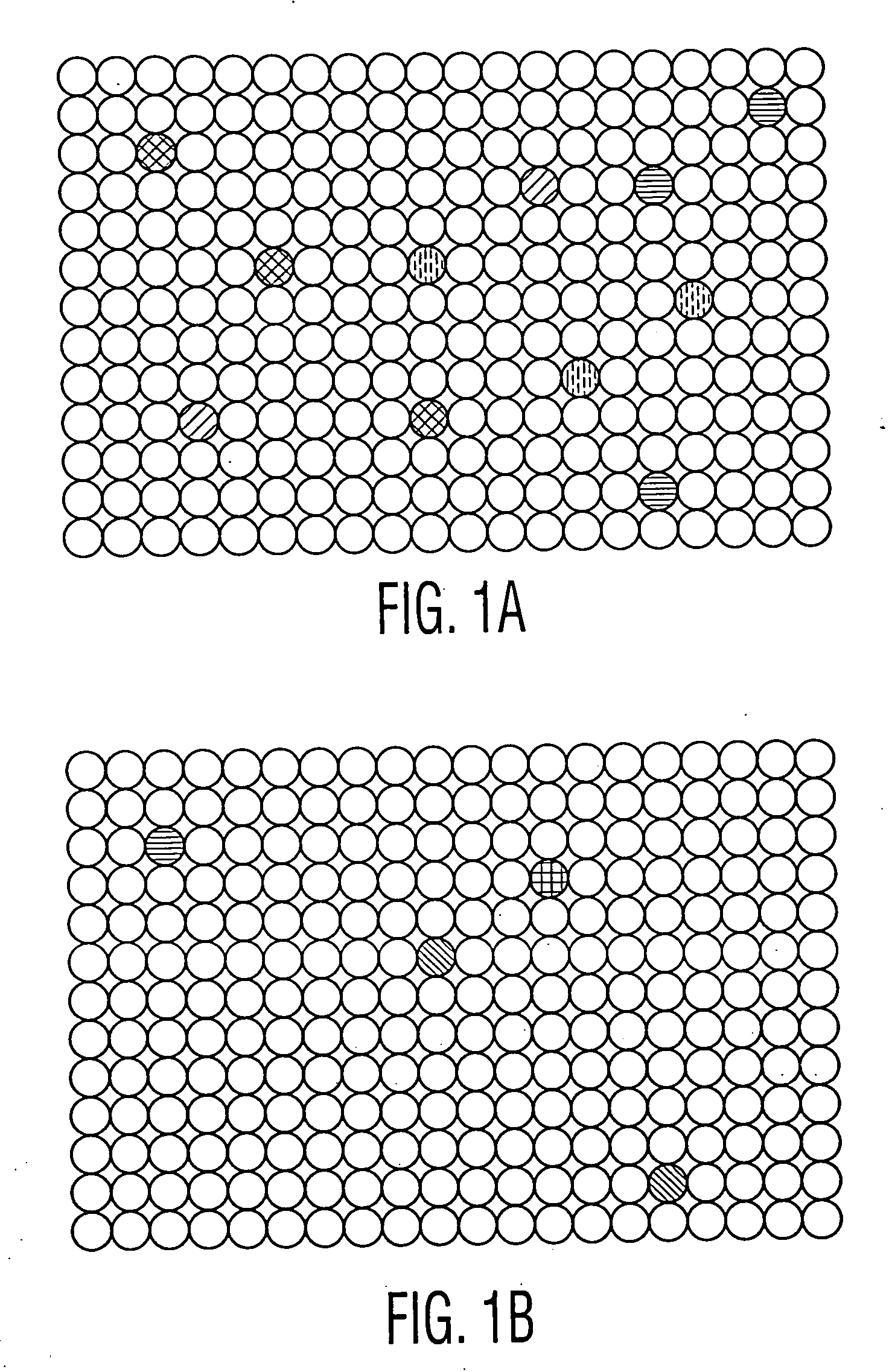
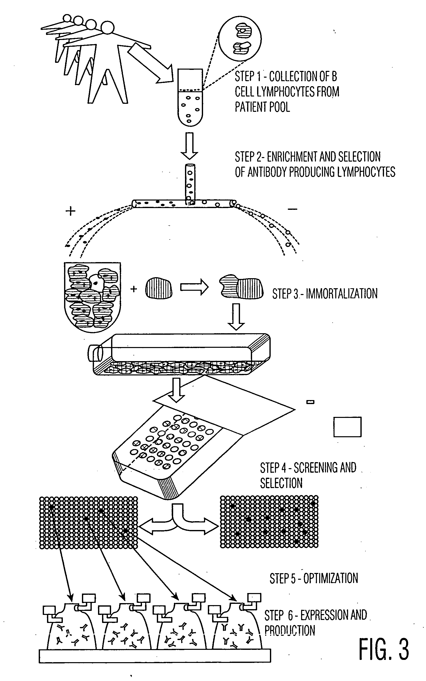
[0058] After generating a series of hybridoma cell lines, they are plated into 96 well microtiter plates, with each well likely to contain only one hybridoma cell (following a limiting dilution of hybridoma cells). In the alternative, it is possible to perform only a partial limit dilution, or even no dilution, so that there is likely to be more than one hybridoma cells in each microtiter plate well. The methods described herein can still isolate the wells with reactive antibodies, and the hybridomas from such wells can then be isolated and further limit diluted and screened to find the candidates of interest.

[0059] An embodiment of the optical fiber device having 96 arrays, each of which aligns with one well in the microtiter plate, is employed for screening. The hybridoma cells were generated either by: (i) immunizing mice with tumor cells and then performing a conventional fusion, following collection of lymphocytes from their spleens, or (ii) fusing...

example ii

Screening with Two Cell Types

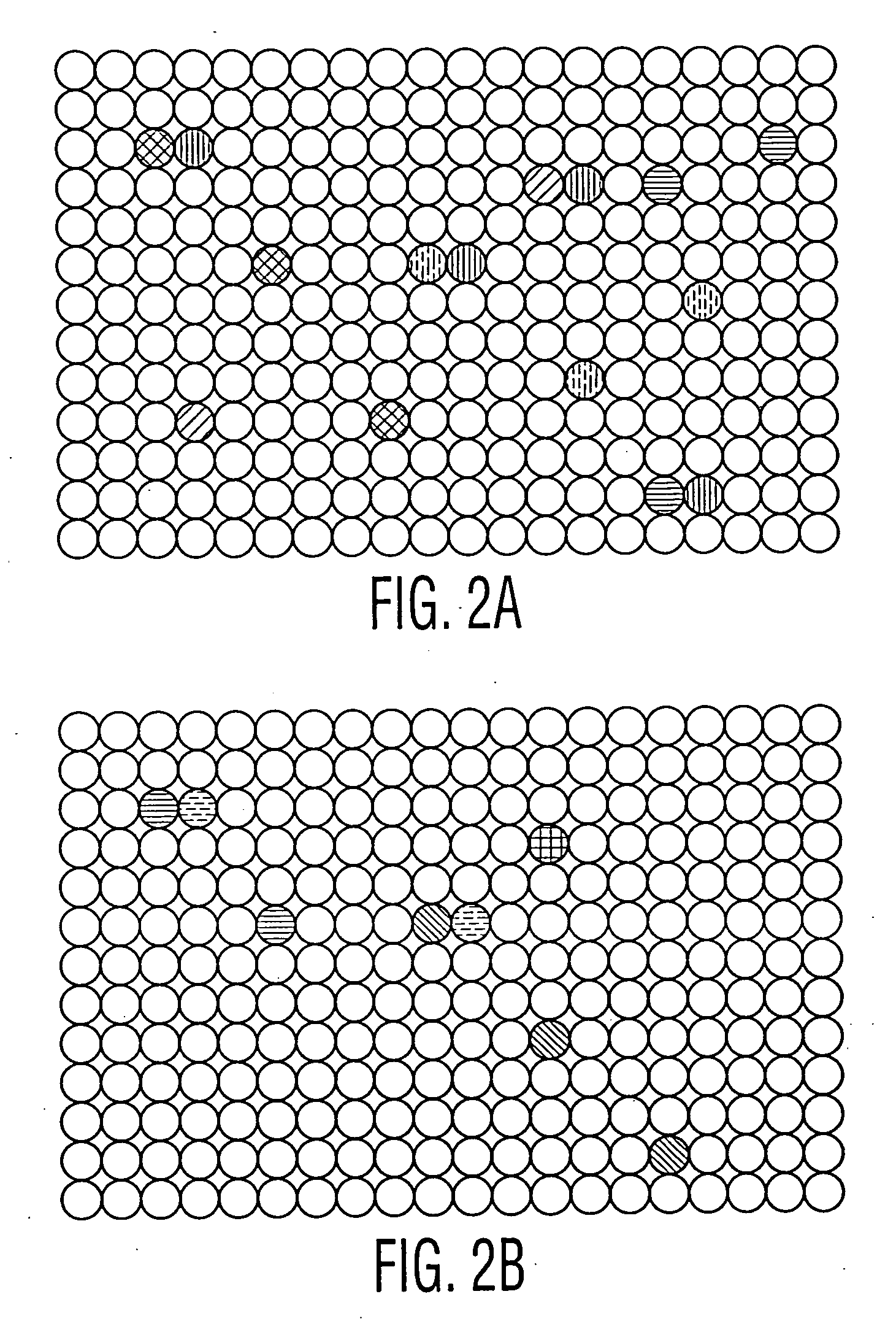
[0063] Hybridomas are generated and plated onto series of 96 well microtiter plates as described in Example I. In this case, however, two cell lines are adsorbed onto two different sets of color coded microbeads: (i) the same tumor cell line which was used to immunize the mice (or which the patients were exposed to) (ii) a non-tumor cell line, which is preferably the non-mutated counterpart of the tumor cell line. The two different sets of microbeads are separately encoded with a color or fluorescence marker. As described in Example I, the cells on each set of microbeads are then treated so as to report the results from one of several different assays, which results can be monitored by a fluorescent microscope following laser excitation. One or more of the assays is for cytotoxicity. The microbeads are then adsorbed into the wells in the fiber wells in the device.

[0064] The ends of the fibers are now placed into the microtiter plate wells, and allowed ...

example iii

Procedures Following Isolation of Desired Antibody-Producing Cells

[0066] Using the techniques in the examples and otherwise described herein, simultaneous assaying and recording of a number of properties of the antibodies within the 96 microtiter plate wells is provided. The cells in the wells which are determined to contain antibodies best suited for therapy are then extracted from the well, grown, subcloned and humanized or affinity matured, or otherwise optimized (if desired). Fully human hybridomas derived from human lymphocytes may not need any further manipulation, except perhaps subcloning to find high expressing, stable cell lines suitable for production. The same techniques described in Examples 1 and 2 may then be used on the subcloned, humanized or optimized cell lines to screen for suitable antibody-producing cell lines, produced following such steps. These subsequent screenings can also be performed in a high throughput manner, with a number of functional assays, and o...


PUM

PropertyMeasurementUnit
Fluorescenceaaaaaaaaaa

Abstract

Disclosed is a method of screening binding molecules, including Monoclonal Antibodies, using an optical fiber array device. Target cells are coated onto a population of microbeads, wherein each microbead is coated with several cells of the same cellular type and has an assay and an assay reporter associated with it. Each of the cell-coated microbeads is positioned in a particular well formed in one end of a particular fiber in an array of optical fibers. The microbeads are contacted with binding molecules, and the results of an assay associated with a microbead are reported to the distal end of the array of fibers.

Description

PRIORITY CLAIM [0001] Priority is hereby claimed to U.S. Provisional Application Ser. Nos. 60 / 406,510; 60 / 406,456; 60 / 406,457 (all of which were filed on Aug. 28, 2002), to Ser. No. 60 / 408,215, filed Sep. 4, 2002, and to Ser. Nos. 60 / 408,947; 60 / 408,948, both filed on Sep. 6, 2002.BACKGROUND OF THE INVENTION [0002] There are currently a number of different monoclonal antibodies approved for use in therapy by the United States Food & Drug Administration and other regulatory agencies. Monoclonal antibodies are deemed well-suited for therapy as they are specific for particular targets and do not bind to other cells or tissues, and because they can have any of a number of desired therapeutic effects, including cell-killing for tumor or infectious disease therapy. [0003] Monoclonal antibodies are derived from a single clone of B-lymphocytes. These B cells are immortalized to provide a cell. line able to indefinitely produce antibodies which are all specific to a particular target antigen...

Claims


Application Information

Patent Timeline
IPC IPC(8): G01N33/567C12M1/34G01N33/53C07K16/00C07K16/30C12P21/08G01N33/543
CPCC07K16/00C07K16/30C07K2317/21G01N33/54313G01N33/54373G01N2500/10
Inventor ANGELIDES, KIMON
Owner ANGELIDES KIMON
Why Eureka
Try Eureka