
High-density scintillators for imaging system and method of making same

a scintillator and high-density technology, applied in the field of imaging systems, can solve the problems of difficult fabrication, high cost of scintillator materials, and inability to meet the requirements of large-area detector assemblies,

Inactive Publication Date: 2006-10-05
GENERAL ELECTRIC CO
View PDF9 Cites 33 Cited by
  • Summary
  • Abstract
  • Description
  • Claims
  • Application Information

AI Technical Summary

Problems solved by technology

Although lutetium silicate (LSO) offers good light yield and decay times, these scintillator materials are costly and difficult to fabricate.
As a result, they are not economically viable for large area detector assemblies for a total body scanner using these scintillators.

Method used


Image

  • High-density scintillators for imaging system and method of making same
  • High-density scintillators for imaging system and method of making same
  • High-density scintillators for imaging system and method of making same

Examples


Embodiment Construction

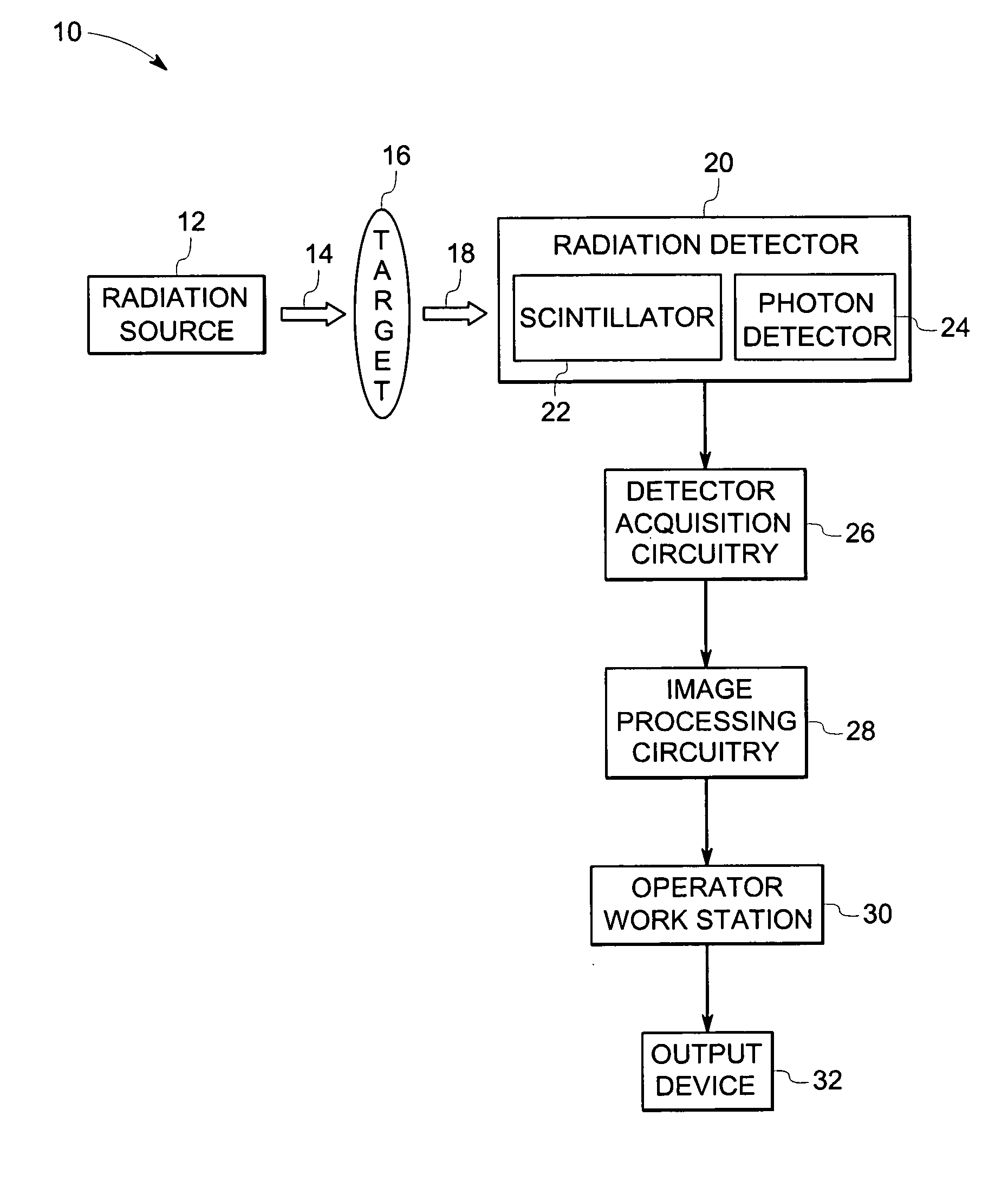
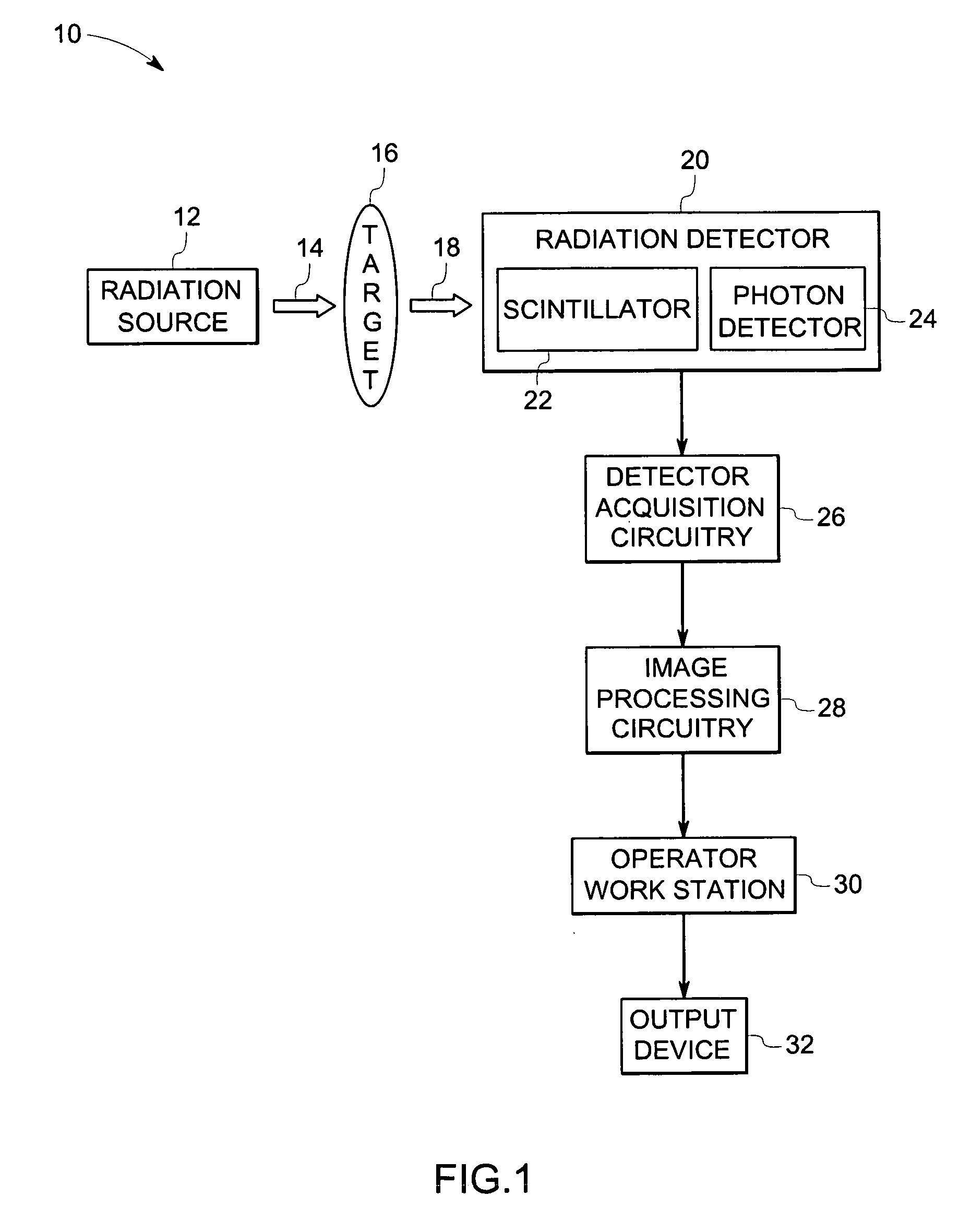
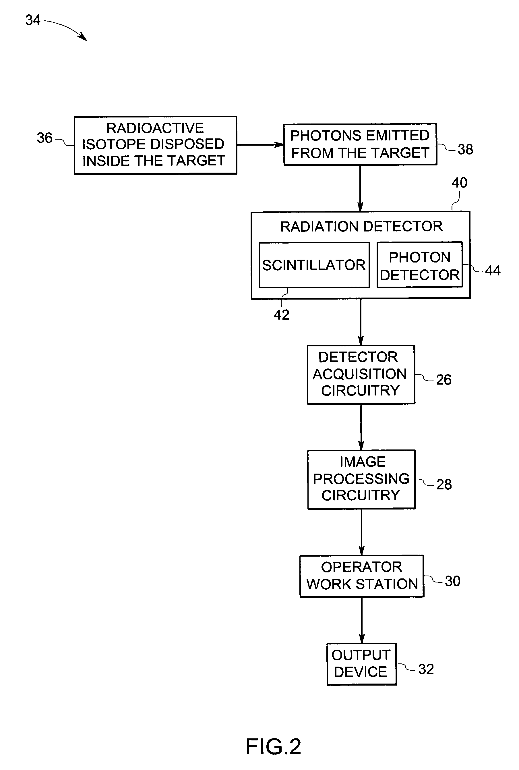
[0017]FIG. 1 illustrates an exemplary radiation-based imaging system, such as positron emission tomography, in accordance with certain embodiments of the present technique. In the illustrated embodiment, the imaging system 10 includes a radiation source 12 positioned such that a major portion of the radiation 14 emitted from the radiation source 12 passes through the target 16, such as an animal, or a human, or a baggage item, or any target having internal features or contents. In certain embodiments, the radiation 14 may include electromagnetic radiation such as, x-ray radiation, or beta radiation, or gamma radiation. A portion of the radiation 14, generally termed as attenuated radiation 18, passes through the target 16. More specifically, the internal features of the target 16 at least partially reduce the intensity of the radiation 14. For example, one internal feature of the target 16 may pass less or more radiation than another internal feature.

[0018] Subsequently, the attenu...


PUM

PropertyMeasurementUnit
pressureaaaaaaaaaa
temperatureaaaaaaaaaa
energyaaaaaaaaaa

Abstract

A scintillator composition comprising a garnet represented by (M1-x-yNxAy)3(Al5-a-bCaDb)O12, where M comprises yttrium, or terbium, or gadolinium, or holmium, or erbium, or thulium, or ytterbium, or lutetium, or combinations thereof, where N comprises additives including a lanthanide, or an alkali metal, or an alkaline earth metal, or combinations thereof, where A comprises a suitable activator ion including cerium, or europium, or praseodymium, or terbium, or ytterbium, or combinations thereof, where C or D comprises lithium, or magnesium, or gallium, or an element from group IIIa, or IVa, or Va, or IIId transition metal, or IVd transition metal, or combinations thereof, where x ranges from about 0 to about 0.90, y ranges from about 0.0005 to about 0.30, and a sum of a and b ranges from about 0 to 2.0.

Description

BACKGROUND [0001] The invention relates generally to imaging systems. In particular, the invention relates to a scintillator for use in an imaging system and a method of making the same. [0002] Positron emission tomography (PET) is a medical imaging technique in which a radioactive substance is administered to a patient and then traced within the patient's body by means of an instrument that detects the decay of the radioactive isotope. In PET, a chemical tracer compound having a desired biological activity or affinity for a particular organ is labeled with a radioactive isotope that decays by emitting a positron. The emitted positron loses most of its kinetic energy after traveling only a few millimeters in a living tissue. It is then highly susceptible to interaction with an electron, an event that annihilates both particles. The mass of the two particles is converted into 1.02 million electron volts (1.02 MeV) of energy, divided equally between two 511 keV photons (gamma rays). T...

Claims


Application Information

Patent Timeline
Patent Type & Authority Applications(United States)
IPC IPC(8): G01T1/20
CPCC09K11/7774C04B2235/77C04B35/44C04B35/62655C04B35/645C04B35/6455C04B2235/3203C04B2235/3206C04B2235/3208C04B2235/3224C04B2235/3225C04B2235/3229C04B2235/3239C04B2235/3244C04B2235/3284C04B2235/3286C04B2235/3287C04B2235/34C04B2235/3418C04B2235/442C04B2235/443C04B2235/449C04B2235/764G21K4/00
Inventor VENKATARAMANI, VENKAT SUBRAMANIAMDUCLOS, STEVEN JUDE
Owner GENERAL ELECTRIC CO
Learn More