Catheter-based delivery of Skeletal Myoblasts to the Myocardium of Damaged Hearts

a technology of skeletal muscle and myoblasts, which is applied in the direction of skeletal/connective tissue cells, drug compositions, biocide, etc., can solve the problems of organ or tissue replacement remains the only other possible option, irreversible cardiac cell loss, and substantial disability and productivity loss, so as to avoid tissue rejection problems and minimize invasive

Inactive Publication Date: 2006-11-23
MYTOGEN
View PDF18 Cites 34 Cited by
  • Summary
  • Abstract
  • Description
  • Claims
  • Application Information

AI Technical Summary

Benefits of technology

[0008] The present invention provides systems and methods that allow for the treatment of damaged / defective heart tissue, especially in individual suffering from disorders characterized by insufficient cardiac function, such as congestive heart failure or myocardial infarction. The inventive methods of treatment are simple, minimally invasive, and do not require general anesthesia or major surgical procedures. More specifically, in these methods, a cell composition is delivered to a patient's myocardium at or near the site of tissue damage using a catheter inserted into the patient's venous system. The cell transformation may be performed after identifying a region of the patient's myocardium in need of treatment. The cell compositions used in the transplantations of the invention comprise cells that are preferably isolated from the future recipient, thus avoiding tissue rejection problems. In certain embodiments, the cell composition comprises skeletal myoblasts. In other embodiments, the cell composition comprises different types of cells selected from the group consisting of skeletal myoblasts, cardiomyocytes, fibroblasts, and stem cells.

Problems solved by technology

One of the factors that renders ischemic heart disease so devastating is the inability (or weak capacity) of cardiac muscle cells to divide and repopulate damaged areas of the heart, making any cardiac cell loss irreversible.
When they do not lead to death, cardiac diseases may result in substantial disability and loss of productivity.
However, when these treatments fail, organ or tissue replacement remains the only other possible option.
Cardiac transplantation is so common that the primary limitation on patient outcome is not the surgical technique, but the scarcity of suitable donor organs.
The disadvantages of using autografts are their limited durability (E. Braunwald, in: “Heart Disease”, 4th Ed., E. Braunwald (Ed.
In addition, reconstructive surgery often involves using the body's tissues for purposes not originally intended, which can result in long-term complications.
However, since these mechanical valve substitutes are nonviable, they have no potential to grow, to repair or to remodel; therefore their durability is limited, especially in growing children (J. E. Mayer Jr., Semin. Thorac. Cardiovasc. Surg., 1995, 7: 130-132).
A major problem with transplantation of adult cardiac myocytes is that they do not proliferate in culture (P. D. Yoon et al., Tex.
While these methods of transplantation of skeletal myoblasts to the injured myocardium have produced promising results, they require open-heart surgery, i.e., a highly invasive procedure.

Method used


Image

  • Catheter-based delivery of Skeletal Myoblasts to the Myocardium of Damaged Hearts
  • Catheter-based delivery of Skeletal Myoblasts to the Myocardium of Damaged Hearts
  • Catheter-based delivery of Skeletal Myoblasts to the Myocardium of Damaged Hearts

Examples


example 1

Correlation of Autologous Skeletal Survival with Changes in Left Remodeling in Dilated Ischemic Heart Failure

Goals of the Study

[0114] Autologous skeletal myoblast (ASM) transplantation, or cardiomyoplasty, has been shown in multiple experimental studies to improve cardiac function after myocardial infarction (R. C. J. Chiu et al., Ann. Thorac. Surg., 1995, 60: 12-18; R. K. Li et al., Ann. Thorac. Surg., 1996, 62: 654-661; C. E. Murry et al., J. Clin. Invest., 1996, 08: 2512-2523; M. Scorsin et al., J. Thorac. Cardiovasc. Surg., 2000, 119: 1169-1175; K. Tambara et al., Circulation, 2003, 108 (suppl. II): 259-263; D. A Taylor et al., Nature Med., 1998, 4: 929-933; M. Jain et al., Circulation, 2000, 103: 1920-1927). Though the majority of studies have been performed in small animal models of myocardial injury, there is evidence of similar improvement in larger animal models (S. Ghostine et al., Circulation, 2002, 106(suppl. I): 131-136) and in the first patient trials (P. Menashé et...

example 2

Correlation of Autologous Skeletal Survival with Changes in Left Remodeling in Dilated Ischemic Heart Failure: Contribution of the Remote vs the Transplanted Myocardium

[0153] Introduction. Autologous skeletal myoblast (ASM) injection after myocardial infarction has been shown to improve left ventricular (LV) performance. However, the mechanism(s) behind such improvement remain(s) unclear.

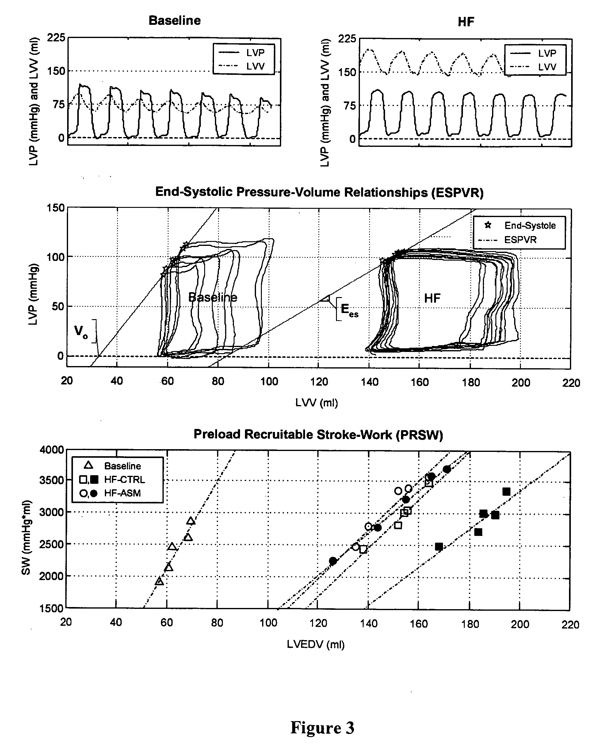
[0154] Methods. Ischemic heart failure (iHF) was induced in sheep (N=12) by selective microembolizations (circumflex artery). After iHF (LVEF: 33±2.2%; LVESV: 143±18 mL), animals were instrumented with sonomicrometers to assess global and segmental LV function. The infarcted myocardium (INF) was injected with either 5×10⁸ cells (ASM; N=6) or cell media (CM; N=6). Pressure volume analyses, hemodynamics and LV segment function (both INF and remote / anterior myocardium [RMT]), were evaluated weekly in unsedated animals for 10 weeks. Comparisons were made by 2-way ANOVA.

[0155] Results. ASM-derived myo...

example 3

Safety and Feasibility of Percutaneous Autologous Skeletal Myoblast Transplantation in the Coil-Infarcted Swine Myocardium

[0157] All experiments were conducted according to guidelines published in the “Guide for the Care and Use of Laboratory Animals” (DHHS publication number NIH 85-23, revised 1985) and Subchapter A of the Federal Animal Welfare Act written by the United States Department of Agriculture and in the spirit of FDA Good Lab Practices. The study protocol was approved by the Harrington Animal Care and Use Committee at Arizona Heart Hospital, Phoenix, Ariz., prior to the start of the study. A summary of the study design is shown in Table 3.

Materials and Methods

[0158] Animal Preparation. Ten (10) female Yorkshire swine between the ages of 3 and 6 months and weighing 91±25 lbs, underwent induced myocardial infarction. Three (3) died during or shortly after induction of the myocardial infarction. One (1) animal was used to evaluate short term retention and biodistributio...


PUM


Abstract

The present invention provides improved systems and methods for the minimally invasive treatment of heart tissue deficiency, damage and/or loss, especially in patients suffering from disorders characterized by insufficient cardiac function, such as congestive heart failure or myocardial infarction. In certain embodiments, a cell composition comprising autologous skeletal myoblasts and, optionally, fibroblasts, cardiomyocytes and/or stem cells, is delivered to a subject's myocardium at or near the site of tissue deficiency, damage or loss, using an intravascular catheter with a deployable needle. Preferably, the cell transplantation is performed after identifying a region of the subject's myocardium in need of treatment. The inventive procedure, which can be repeated several times over time, results in improved structural and/or functional properties of the region treated, as well as in improved overall cardiac function. In particular, the inventive therapeutic methods may be performed on patients that have previously undergone CABG or LVAD implantation.

Description

RELATED APPLICATIONS [0001] The present invention claims priority to Provisional Application No. 60 / 658,887 filed on Mar. 4, 2005 and entitled “Catheter-Based Delivery of Skeletal Myoblasts to the Myocardium of Damaged Hearts. The Provisional Application is incorporated herein by reference in its entirety.BACKGROUND OF THE INVENTION [0002] Cardiac diseases are responsible for a preponderance of health problems in the majority of industrialized countries as well as in many developing countries. In the United States, heart disease is the first leading cause of mortality, accounting for nearly 40% of all deaths (Heart and Stroke Statistical Update, American Heart Association 2002). About 85% to 90% of cardiac-related deaths are associated with ischemic heart disease, valvular disease, congenital heart disease, hypertensive heart disease and / or pulmonary hypertensive heart disease. In particular, ischemic heart disease, in its various forms, accounts for about 60-75% of all deaths cause...

Claims


Application Information

Patent Timeline
Patent Type & Authority Applications(United States)
IPC IPC(8): A61K35/12A61K35/33A61K35/34A61K35/545
CPCC12N5/0658A61K35/33A61K35/34A61K35/545A61K2300/00A61P9/00
Inventor JACOBY, DOUGLAS B.DINSMORE, JONATHAN H.
Owner MYTOGEN
Why Eureka
Try Eureka