Near infrared transrectal probes for prostate cancer detection and prognosis

a transrectal probe and prostate cancer technology, applied in the field of cancer detection, can solve the problems of low sensitivity and specificity for detecting prostate cancer, inability to detect early stages of prostate cancer, so as to achieve routine, early and more accurate detection, and enhance the knowledge of the prostate art.

Inactive Publication Date: 2006-11-23
BOARD OF RGT THE UNIV OF TEXAS SYST
View PDF16 Cites 29 Cited by
  • Summary
  • Abstract
  • Description
  • Claims
  • Application Information

AI Technical Summary

Benefits of technology

[0016] The present invention combines, for example, NIR tomographic imaging techniques with uniquely designed transrectal NIR probes. The present invention obtains optical and physiological signatures of prostate cancer in human, which can be, in turn, used later as finger-prints of prostate cancer to diagnose the cancer. The present invention provides transrectal NIR imaging for prostate cancer which is capable, for example, of providing heterogeneous maps of oxygenation and metabolic parameters of prostate tumors in humans. The present invention further provides an efficient, portable, real-time and minimally invasive screening / diagnostic means for prostate cancer, which will enhance knowledge in the art on prostate tumor development in its oxygenation and metabolism. The present invention provides an innovative approach to a screening modality which provides routine, early and more accurate detection for prostate cancer. The present invention provides NIR transrectal probes which allow NIR photons to penetrate deep through the most regions of the prostate gland, and thus obtain NIR tomographic images for physiological maps of the prostate.

Problems solved by technology

It is currently difficult, however, to detect early stages of prostate cancer because of lack of any obvious symptoms.
The clinician may also estimate the prostate size, but because only the posterior aspect can be palpated, the estimate is usually fairly inaccurate.
Although the clinician may feel indurations and nodules, the sensitivity and specificity for detecting prostate cancer is very low.
DRE is, thus, an ineffective prostate cancer screening tool.
Thus, improving the sensitivity and specificity of TRUS to predict the presence or absence of cancer has largely remained futile.
However, recent findings suggest considerations that make such monolithic cut-off values less attractive, for example: (1) about 25% of men with serum PSA values above 4.0 ng / ml have benign prostatic hyperplasia (BPH) only and no cancer; (2) significant numbers of men have cancer even if their PSA is below 4.0 ng / ml.
Currently, however, those skilled in the art no longer definitively correlate an elevated serum PSA level with prostate cancer.
Clinicians in the art currently face considerable challenges in the diagnostic evaluation a man at risk for prostate cancer.
The TRUS imaging itself is of little help to the clinician in deciding where to biopsy and how many cores to take.
Lastly, MRI techniques rely on abnormal signal intensities that result from morphologic changes within prostate to find existence and extent of cancer, but MRI does not accurately reflect the presence and spatial extent of cancer.
MRI alone has a good accuracy in detecting seminal vesicle invasion (96%), but assessing the cancer spread through the prostatic capsule is harder to achieve (81% accuracy).
However, because of the cost and non-portability, MSRI is unlikely to become a practical screening tool for prostate cancer detection.
The latter two methods have advantages of making repeated measurements of pO2 non-invasively, but lack speed and portability.
Although non-invasive, quantitative, NIR spectroscopy and imaging of tissue vascular oxygenation for the brain and the breast are known in the art, NIR imaging techniques are limited by their spatial resolutions.
Efforts on using the NIR techniques to detect and monitor prostate tumors, however, have been very limited due to the difficulty in accessing human prostates using currently known optical imaging systems and probes.
Moreover, existing imaging systems fail to obtain functional imaging of tissue metabolic and oxygenation states.
There are no known biologic differences between isoechoic and hypoechoic prostate cancers because ultrasound imaging / detection rests on the structural differences between tissue types and thus is limited for metabolic and physiological imaging for tissue functions.

Method used


Image

  • Near infrared transrectal probes for prostate cancer detection and prognosis
  • Near infrared transrectal probes for prostate cancer detection and prognosis
  • Near infrared transrectal probes for prostate cancer detection and prognosis

Examples


Embodiment Construction

[0032] While the making and using of various embodiments of the present invention are discussed in detail below, it should be appreciated that the present invention provides many applicable inventive concepts that can be embodied in a wide variety of specific contexts. The specific embodiments discussed herein are merely illustrative of specific ways to make and use the invention and do not delimit the scope of the invention.

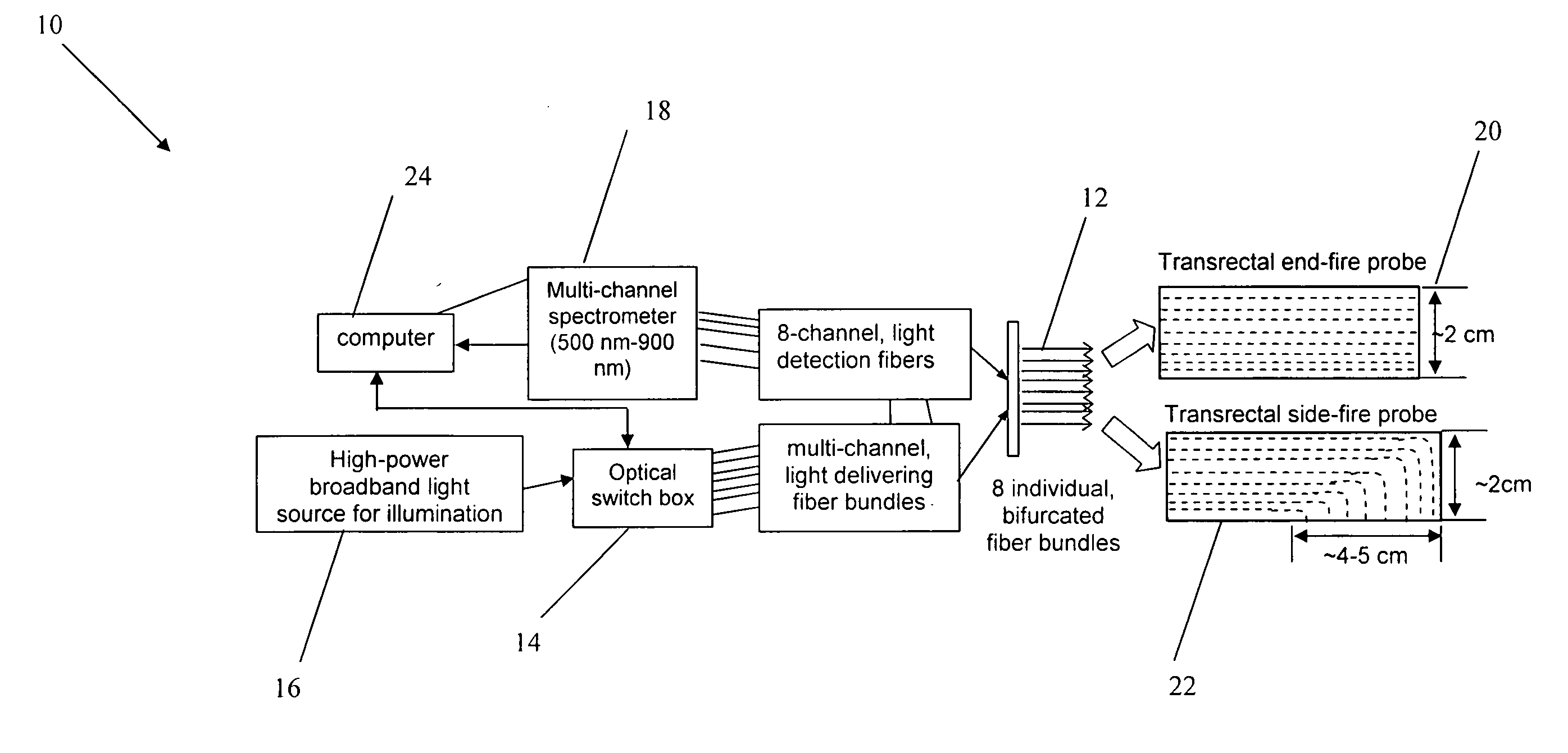
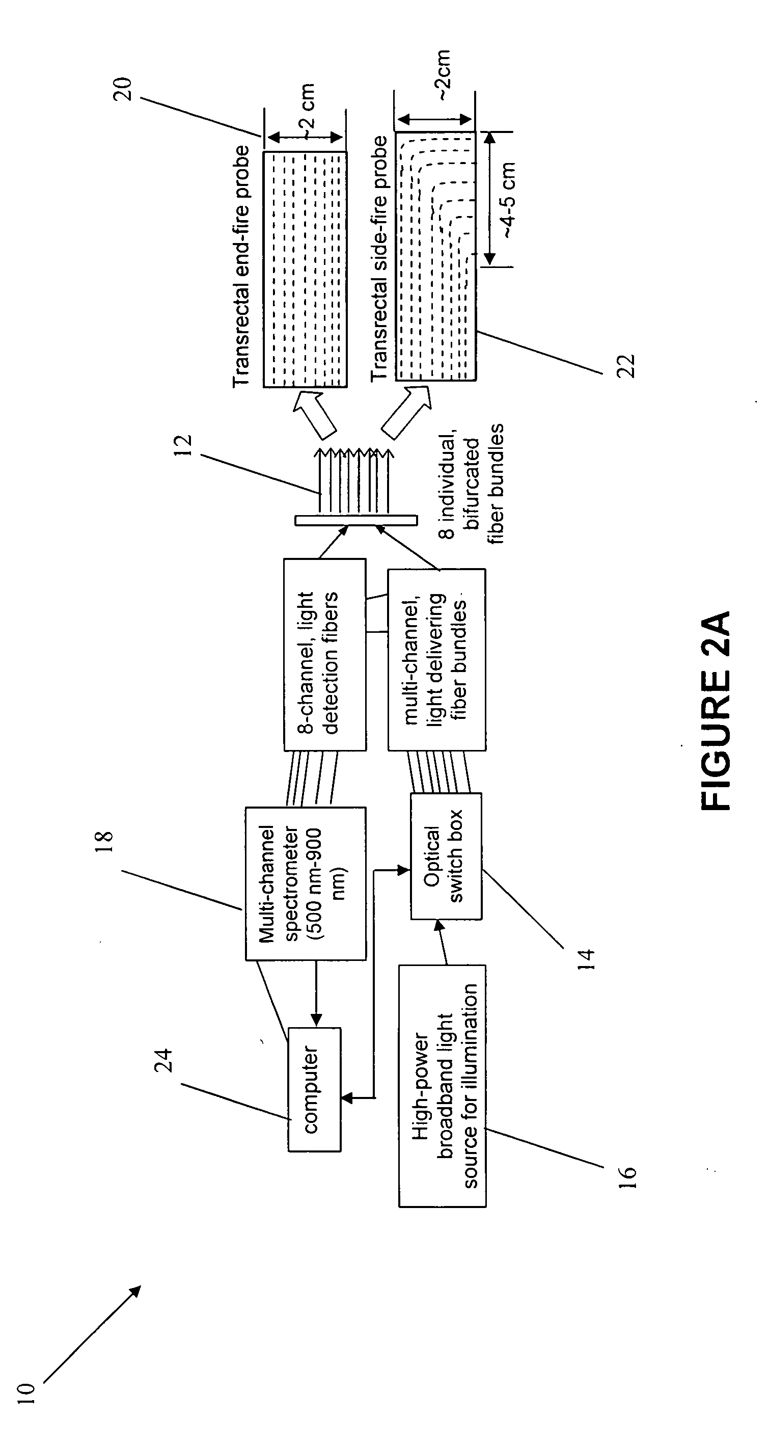
[0033] The present invention provides a broadband, multi-channel, tomographic NIR imaging system 10, particularly designed for the prostate cancer studies. While it is simpler to use just 2-4 wavelengths to obtain hemodynamic parameters, the present invention derives accurate concentrations of several intrinsic chromophores presented in tissue. A system in accordance with the present invention, therefore, can be used to efficiently separate both absorption and reduced scattering coefficients without any non-uniqueness problem. Thus, the present invention unique...


PUM


Abstract

A multi-channel, near infrared (NIR) imaging system comprising one or more optical fiber bundles; and a transrectal NIR probe having an outer material, wherein the optical fiber bundle is integrated into the transrectal probe (an end-fire or a side-fire NIR probe). The outer material is preferably rubber-like silicon or made of two translucent materials exhibiting low viscosity and requires curing at room temperature. The transrectal NIR probe comprises a curvature to match the curvature of a human rectum and may have a circular or oval-shaped silicone holder. The imaging system further comprises a broad-band light source adapted to deliver light to the fiber channels, an optical switch box adapted to allow only one of the fiber channels to pass through at a time; and a multi-channel spectrometer to capture tomographic images of changes in HbO, HbT, light scattering patterns and hemodynamic response times from human prostate.

Description

BACKGROUND [0001] The present invention relates to the general field of cancer detection, and in particular, to transrectal probes for prostate cancer detection and prognosis. [0002] Prostate cancer is the most common male cancer and the second leading cause of cancer death in men in the United States. Effective treatments are highly associated with accurate and early detection of prostate cancer. It is currently difficult, however, to detect early stages of prostate cancer because of lack of any obvious symptoms. There are four common examination and screening methods known in the art for screening prostate cancer: Digital Rectal Examination (DRE), Transrectal Ultrasonography (TRUS), Prostate-Specific Antigen (PSA) test and Magnetic Resonance Imaging (MRI) and Magnetic Resonance Spectroscopic Imaging (MRSI). All of these early detection and screening tests have serious shortcomings as discussed below. [0003] DRE is the most established method of examining the prostate gland. The cl...

Claims


Application Information

Patent Timeline
Patent Type & Authority Applications(United States)
IPC IPC(8): A61B6/00
CPCA61B5/0075A61B5/4381A61B5/0086
Inventor LIU, HANLITRUELSON, THOMAS C.
Owner BOARD OF RGT THE UNIV OF TEXAS SYST
Why Eureka
Try Eureka