Modalities for the treatment of degenerative diseases of the retina

a retinal degenerative disease and retinal technology, applied in the direction of biocide, cardiovascular disorder, drug composition, etc., can solve the problems of increased photoreceptor loss, reduced neovascularization ability, and reduced hla antigen complex, so as to improve the ability to prevent neovascularization and reduce the complexity of hla antigens

Active Publication Date: 2007-02-08
ADVANCED CELL TECH INC
View PDF7 Cites 48 Cited by
  • Summary
  • Abstract
  • Description
  • Claims
  • Application Information

AI Technical Summary

Benefits of technology

[0017] Another embodiment of the present invention is a method for the derivation of new RPE lines and progenitor cells from existing and new ES cell lines. There can be variations in the properties, such as growth rate, expression of pigment, or de-differentiation and re-differentiation in culture, of RPE-like cells when they are derived from different ES cell lines. There can be certain variations in their functionality and karyotypic stability, so it is desirable to provide methods for the derivation of new RPE lines and new ES cell lines which would allow choosing the lines with desired properties that can be clonally selected to produce a pure population of high quality RPE-like cells.
[0020] Another embodiment of the present invention utilized a bank of ES or embryo-derived cells with homozygosity in the HLA region such that said cells have reduced complexity of their HLA antigens.
[0024] Embryonic stem cells (ES) can be indefinitely maintained in vitro in an undifferentiated state and yet are capable of differentiating into virtually any cell type. Thus human embryonic stem (hES) cells are useful for studies on the differentiation of human cells and can be considered as a potential source for transplantation therapies. To date, the differentiation of human and mouse ES cells into numerous cell types have been reported (reviewed by Smith, 2001) including cardiomyocytes [Kehat et. al. 2001, Mummery et. al., 2003 Carpenter et. al., 2002], neurons and neural precursors (Reubinoff et. al.2000, Carpenter et. al.2001, Schuldiner et. al., 2001), adipocytes (Bost et. al., 2002, Aubert et. al., 1999), hepatocyte-like cells (Rambhatla et. al., 2003), hematopoetic cells (Chadwick et. al., 2003). oocytes (Hubner et. al., 2003), thymocyte-like cells (Lin RY et. al., 2003), pancreatic islet cells (Kahan, 2003), and osteoblasts (Zur Nieden et. al., 2003). Another embodiment of the present invention is a method of identifying cells such as RPE cells, hematopoietic cells, muscle cells, liver cells, pancreatic beta cells, neurons, endothelium, progenitor cells or other cells useful in cell therapy or research, derived from embryos, embryonic stem cell lines, or other embryonic cells with the capacity to differentiate into useful cell types by comparing the messenger RNA transcripts of such cells with cells derived in-vivo. This method facilitates the identification of cells with a normal phenotype and for deriving cells optimized for cell therapy for research.

Problems solved by technology

Diseases of the CNS frequently include neuronal cell loss, and, because of the absence of endogenous repopulation, effective recovery of function following CNS-related disease is either extremely limited or absent.
This neovascular (“wet”) form of AMD is particularly destructive and seems to result from a loss of proper regulation of angiogenesis.
Breaks in Bruch's membrane as a result of RPE dysfunction allows new vessels from the choroidal circulation access to the subretinal space, where they can physically disrupt outer-segment organization and cause vascular leakage or hemorrhage leading to additional photoreceptor loss.
There are often undesirable side effects, however, and therefore patient dissatisfaction with treatment outcome.
In addition, laser treatment does not fix the underlying predisposition towards developing CNV.
Since RPE plays an important role in photoreceptor maintenance, and regulation of angiogenesis, various RPE malfunctions in vivo are associated with vision-altering ailments, such as retinitis pigmentosa, RPE detachment, displasia, athrophy, retinopathy, macular dystrophy or degeneration, including age-related macular degeneration, which can result in photoreceptor damage and blindness.
All of the above conditions involve loss of photoreceptors and, therefore, treatment options are few and insufficient.
tion. However, even in an immune-privileged site such as the eye, there is a problem with graft rejection, hindering the progress of this approach if allogenic transplantation is
used. Although new photoreceptors (PRCs) have been introduced experimentally by transplantation, grafted PRCs show a marked reluctance to link up with surviving neurons of the host
retina. Reliance on RPE cells derived from fetal tissue is another problem, as these cells have shown a very low proliferative po
Disease. Although a 30-50% decrease in symptoms was found one year after transplantation, there is a shortage of eye donors, this is not yet FDA approved, and there would still exist a need beyond what could be met by donated e
This results in poor effective coverage of photoreceptors as well as a multilayered RPE with incorrect polarity, possibly resulting in cyst formation or macular edema.
A serious problem exists in that the neural retinal grafts typically do not functionally integrate with the host retina.
In addition, the absence of an intact RPE monolayer means that RPE dysfunction or disruption of Bruch's membrane has not been rectified.
Thus, there exists no effective means for reconstituting RPE in any of the current therapies and there remain deficiencies in each, particularly the essential problem of a functional disconnection between the graft and the host retina.

Method used


Image

  • Modalities for the treatment of degenerative diseases of the retina
  • Modalities for the treatment of degenerative diseases of the retina
  • Modalities for the treatment of degenerative diseases of the retina

Examples


example 1

Spontaneous differentiation into Pigmented Epithelial Cells in Long Term Cultures

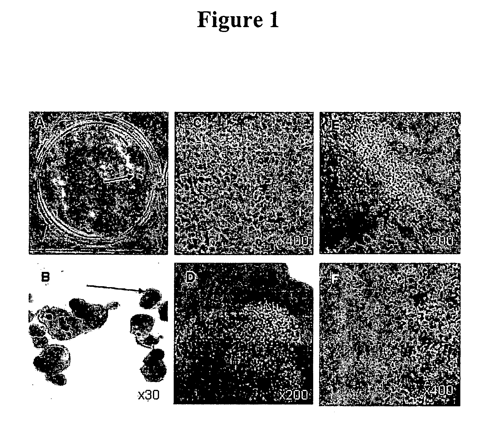
[0060] When hES cell cultures are allowed to overgrow on MEF in the absence of LIF, FGF and Plasmanate, they form a thick multilayer of cells. About 6 weeks later, dark islands of cells appear within the larger clusters (FIG 1). These dark cells are easily seen with the naked eye and looked like “freckles” in a plate of cells as shown in FIG. 1A. At higher magnification these islands appear as tightly packed polygonal cells in a cobblestone monolayer, typical of epithelial cells, with brown pigment in the cytoplasm (FIG. 1C). There are differences in the amount of pigment in the cells with cells in the central part of the islands having the most pigment and those near the edges the least. (FIG. 1, E,F).

[0061] When hES cells form embryoid bodies (EB)—pigmented epithelial cells appear in about 1-2% of EBs in the first 6-8 weeks (FIG. 1B) . Over time more and more EBs develop pigmented cells, and by 3 mo...

example 2

Isolation and Culture of Pigmented Epithelial Cells

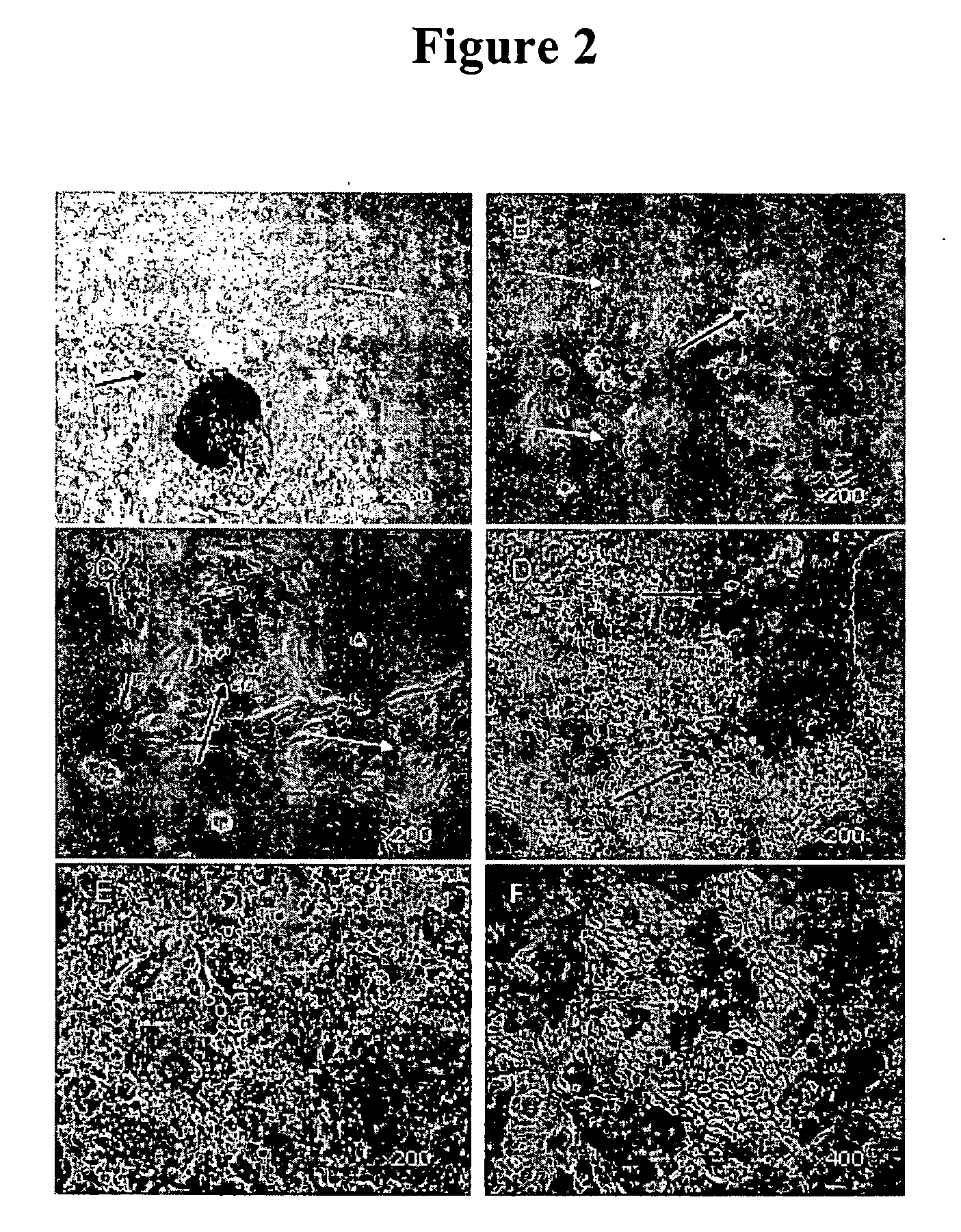
[0062] The inventors isolated pigmented epithelial cells from both adherent hES cell cultures and from EBs. Pigmented polygonal cells were digested with enzymes (trypsin, and / or collagenase, and / or dispase), and the cells from these pigmented islands were selectively picked with a glass capillary. Although care was taken to pick only pigmented cells, the population of isolated cells invariably contained some non-pigmented cells. After plating cells on gelatin or laminin for 1-2 days, the cells were considered to be primary cultures (P0).

[0063] Primary cultures contained islands of pigmented polygonal cells as well as some single pigmented cells. After 3-4 days in culture, non-pigmented cells that seemed to have lost epithelial morphology (flatter and cells with lamellipodia) appeared at the periphery of some islands (FIG. 2). The number of such peripheral cells increased over time, suggesting that these cells were proliferating, a...

example 3

Detection of RPE Markers

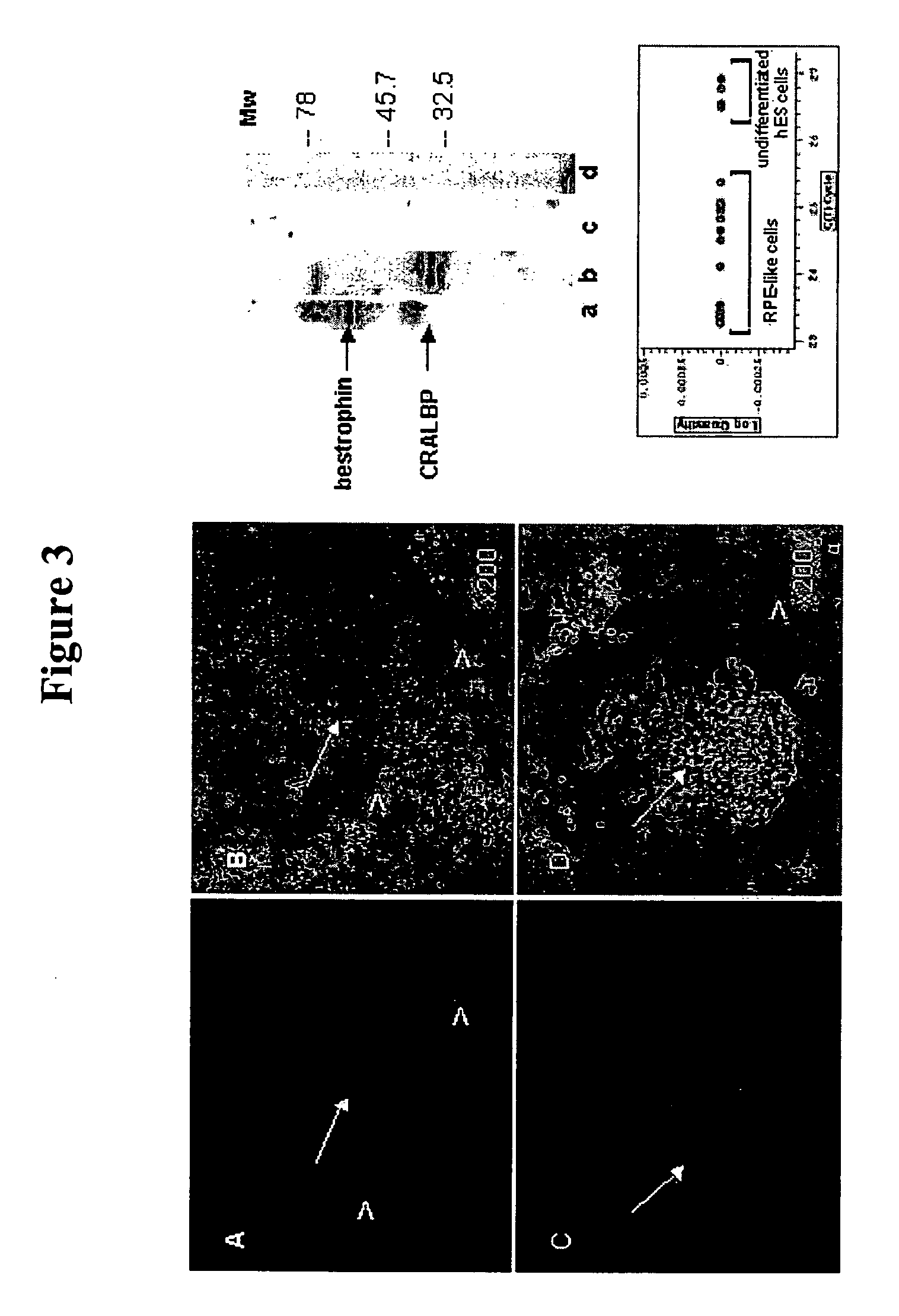
[0064] The preliminary characterization of these differentiated human cells as RPE is based on their similarity to RPE cultures previously described; principally, their epithelial morphology and possession of pigment. There are three types of pigmented epithelial cells in human body: retinal and iris pigmented epithelium and keratinocytes, but the latter don't secrete pigment. The epithelial structure and cobblestone morphology are not shared by other pigmented cells, e.g. melanocytes. It is also noteworthy that RPE cells have been shown to lose and regain their pigment and epithelial morphology when grown in culture (Zhao 1997, Opas and Dziak, 1994), and the pigmented cells behaved in a similar manner, so to test the hypothesis that the ES derived cells may be RPE, they were stained with antibodies to known markers for RPE: bestrophin and CRALBP. FIG. 4 (left panel) shows membrane localization of bestrophin (A) and CRALBP (C), both are found in pigmented ep...


PUM

PropertyMeasurementUnit
distanceaaaaaaaaaa
timeaaaaaaaaaa
fluorescent angiographyaaaaaaaaaa

Abstract

This invention relates to methods for improved cell-based therapies for retinal degeneration and for differentiating human embryonic stem cells and human embryo-derived into retinal pigment epithelium (RPE) cells and other retinal progenitor cells.

Description

[0001] This application is being filed as a PCT International Patent application on 24 Jan. 2005, in the name of Advanced Cell Technology, Inc, a U.S. national corporation, applicant for the designation of all countries except the U.S., and Irina V. Klimanskaya, a U.S. citizen, applicant for the designation of the US only, and claims priority to U.S. Provisional Application Ser. No. 60 / 538,964, filed Jan. 23, 2004.FIELD OF THE INVENTION [0002] This invention relates generally to methods for improved cell-based therapies for retinal degeneration and other visual disorders as well as treatment of Parkinson's disease and for differentiating mammalian embryonic stem cells and mammalian embryo-derived cells into retinal pigment epithelium (RPE) cells and other eye tissue including, but not limited to) rods, cones, bipolar, corneal, neural, iris epithelium, and progenitor cells. BACKGROUND OF THE INVENTION [0003] Many parts of the central nervous system (CNS) exhibit laminar organization,...

Claims


Application Information

Patent Timeline
Patent Type & Authority Applications(United States)
IPC IPC(8): A61K35/30A01N63/00A61K35/12A61K35/44A61K48/00C12N5/071C12N5/0793
CPCA61K35/12A61K35/44C12N5/062C12N5/0621C12N2506/02C12N2501/115C12N2501/15C12N2501/155C12N2501/33C12N2501/01A61P25/16A61P27/00A61P27/02A61P43/00A61P9/10A61P9/14A61K35/30A61K9/0048A61K48/00
Inventor KLIMANSKAYA, IRINA V.
Owner ADVANCED CELL TECH INC
Why Eureka
Try Eureka