

Bioactive Material Delivery Systems Comprising Sol-Gel Compositions
a bioactive material and composition technology, applied in the direction of prosthesis, surgery, coatings, etc., can solve the problems of difficult adhesion of a polymeric coating to a substantially different substrate, such as the metallic substrate of the stent, and the hydrophobic organic polymer cannot spontaneously adhere to the surface of the implantable medical device, etc., to achieve enhanced adhesion, enhanced bioactive material incorporation, and enhanced adhesion
- Summary
- Abstract
- Description
- Claims
- Application Information
AI Technical Summary
Benefits of technology
Problems solved by technology
Method used
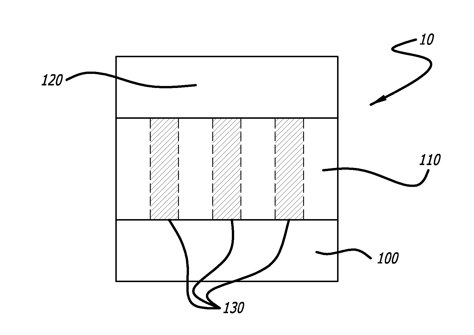
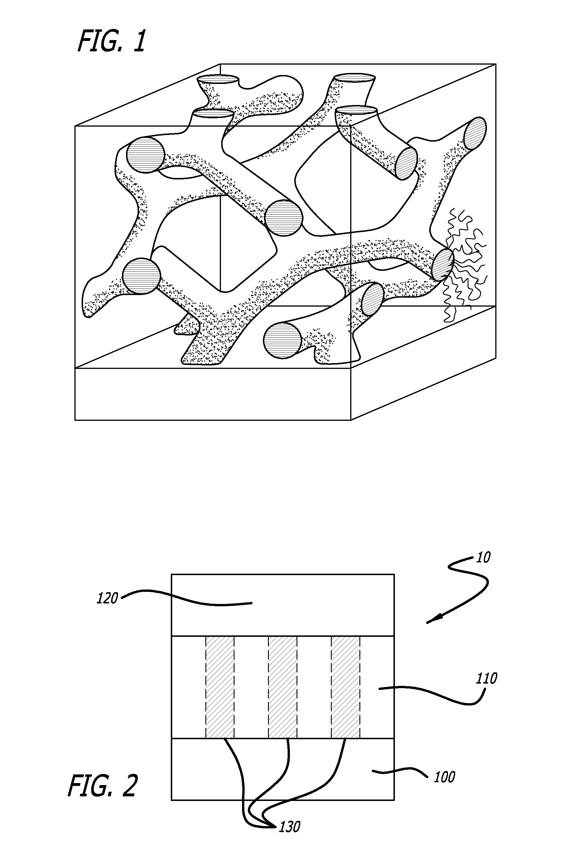
Image

Examples
example i
[0069] A 0.1M tetraethoxy silane (TEOS) sol-gel solution was prepared by first making 0.3M ethanolic hydrochloric acid in a 20 mL glass scintillation vial by mixing 25 μL of concentrated (12M) HCl with 860 μL deionized water and 3 mL of absolute ethanol. In a 1.5-mL microcentrifuge tube 1 mL of ethanol and 112 μL TEOS were combined. The TEOS solution was added dropwise to the acidified ethanol over 30 seconds and the resulting TEOS solution was allowed to hydrolyze for 45 minutes. At the end of the hydrolysis period, 2 mL of the hydrolyzed TEOS solution were added to des-aspartate angiotensin I (DAA-I) (2.5 mg) in a glass vial (1 dram). Brief sonication was used to ensure both dissolution and mixing of the peptide. The resultant solution was transferred to a 5 mL gas tight Hamilton syringe after passage through a 5 micron filter. The syringe was placed in a Harvard Scientific syringe pump and connected to the ultrasonic sprayer.
[0070] Either two cleaned 12 mm or four cleaned 6 mm s...
example ii
[0073] To explore the effects of increasing the amount of TEOS on the rate of elution of DAA-I, increasing amounts, from 0.1-0.5M of TEOS were added to a constant amount of DAA-I (1.25 mg / mL) according to the methods and protocols described above. The stents (9 mm, n=4) were sprayed with each of the solutions as described above, weighed, and the amounts DAA-I eluted over a 24 hour period were compared. FIG. 5A shows the total amount of bioactive material released over 72 hours while FIG. 5B shows elution curves as a function of time. As seen in these FIGS. 5A-5B, increasing the molar ratio of TEOS to the drug significantly delayed the rate of release of the drug into aqueous solution.
example iii
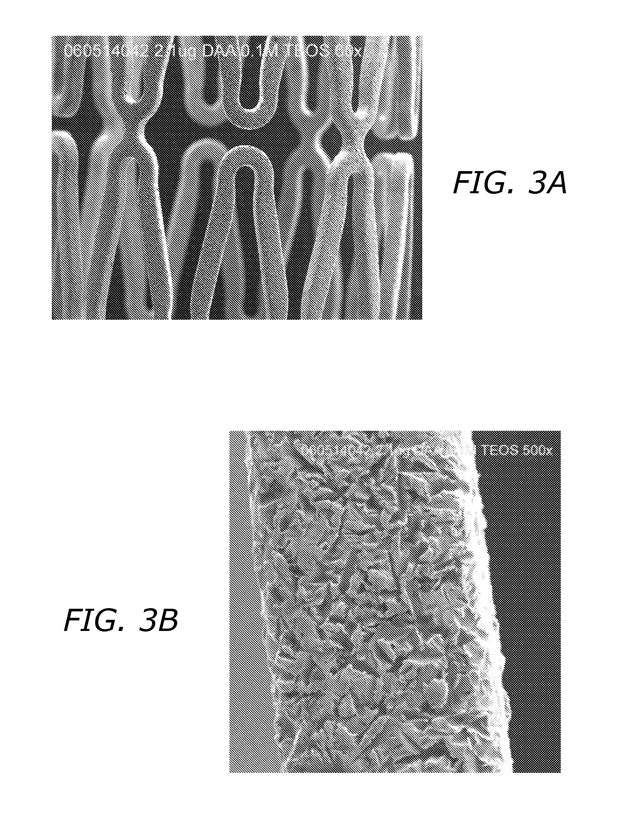
[0074] Sol-gels composed solely of hydrolyzed TEOS or tetramethoxy silane (MEOS) are relatively hydrophilic and even though they more effectively entrap bioactive materials, they do not provide a chemically compatible environment for most hydrophobic drugs such as paclitaxel, rapamycin, cyclosporin, and other compounds with limited water solubility. To increase the hydrophobic character of the resulting sol-gel a variety of alkylated ethoxy silanes can be added to the sol-gel forming solution. Compounds such as methyl, t-butyl, iso-butyl, hexyl, phenyl, octyl, dodecyl, and octadecyl triethoxy silane can be added to the mixtures at different molar ratios to result in dramatically different sol-gel coatings. The inclusion of such compounds results in significant differences in the ability to incorporate a spectrum of compounds into the gel, and will also affect the rate of their release from the gel into aqueous solutions. An example of such an effect is shown in FIG. 6. Sol-gel solut...
PUM
| Property | Measurement | Unit |
|---|---|---|
| pore size | aaaaa | aaaaa |
| pore size | aaaaa | aaaaa |
| size | aaaaa | aaaaa |
Abstract
Description
Claims
Application Information