Methods and devices for intramuscular stimulation of upper airway and swallowing muscle groups

a muscle group and intramuscular stimulation technology, applied in the field of muscular function modification devices and procedures, can solve the problems of pneumonia risk, high treatment cost and the commensurate value of remediation technology if one were available, and underestimated cost, so as to prevent pneumonia risk and prevent aspiration in chronic dysphagia

Inactive Publication Date: 2007-05-31
UNITED STATES OF AMERICA
View PDF13 Cites 53 Cited by
  • Summary
  • Abstract
  • Description
  • Claims
  • Application Information

AI Technical Summary

Benefits of technology

[0010] The shortcomings of electrode control of internal solid body part movements summarized above are alleviated by embodiments of the invention. One such embodiment provides a method of synergistic production of movements during speech, swallowing or voice production, comprising: chronic implantation of at least two intra-muscular stimulators into different muscles involved in the upper airway and vocal tract, chronic implantation of a signal generator that generates electrical pulses to at least two intra-muscular stimulators; wherein electrical pulses from the signal generator activate at least two muscles to produce the synergistic movement control during the activity. Another embodiment provides a method of moving the hyoid bone, and/or parts of the upper airway and/or vocal tract within an animal by two or more different controlled muscles, comprising: implanting at least one electrode into each of two or more different muscles; electrically connecting each electrode to a indwelling subcutaneous signal generator capable of generating a pattern of stimulation; and energizing the controlled muscles at the same time by the signal generator to synergistically move the parts of the upper airway, hyoid or vocal tract. Yet another embodiment provides a method of simultaneously moving the hyoid bone and opening the upper esophageal sphincter within an human via at least one muscle attached to the hyoid bone, comprising: implanting at least one electrode into each of two or more said muscles; electrically connecting each electrode to a signal generator capable of generating a complex pattern to activate the muscle attached to the electrode; and energizing electrodes in at least two of the muscles at the same time with the signal generator, thereby synergistically moving the hyoid bone and/or opening the upper esophageal sphincter. Yet another embodiment provides a system for moving a cartilage within an animal, comprising: a first electrode implanted in a first muscle attached to the cartilage; a second electrode implanted in a second different muscle attac...

Problems solved by technology

Dysphagia is a common complication with diseases such as stroke, neurodegenerative diseases, brain tumors, respiratory disorders, and the like wherein insufficient control of muscles needed for swallowing engender a risk of aspiration pneumonia.
The treatment costs and the commensurate value of a remediation technology if one were available, are very high.
Furthermore, this cost is an underestimate because Medicare pays only half of the enteric home feeding costs and approximately 412,000 patients per year receive enteric feedings due to risk of aspiration in the United States alone.
Dysphagia often results from poor control of some muscles in the upper respiratory system.
Patients sometimes lack proper control of muscles used for these other activities and unfortunately remedial efforts leave much to be desired here as well.
These advances are helpful but generally do not address sufficiently the control of specific cartilage, tissue or bone movements, which require the coordinated action of multiple muscles.
However, no suitable description of a chronically implanted (i.e. implanted for multiple stimulations) neuroprosthestic system exists for long term prevention of aspiration during swallowing in patients who have not been able to take food or liquids by mouth following unsuccessful rehabilitation.
The Freed et. al. rehabilitation device is not appropriate for patients with a chronic...

Method used


Image

  • Methods and devices for intramuscular stimulation of upper airway and swallowing muscle groups
  • Methods and devices for intramuscular stimulation of upper airway and swallowing muscle groups
  • Methods and devices for intramuscular stimulation of upper airway and swallowing muscle groups

Examples


example 1

[0065] This example demonstrates intra-muscular stimulation of swallowing and is also detailed in J. Applied Physiology, 94: 128-134, (2003), which is incorporated by reference in its entirety, particularly with respect to methodological details relevant to the present examples.

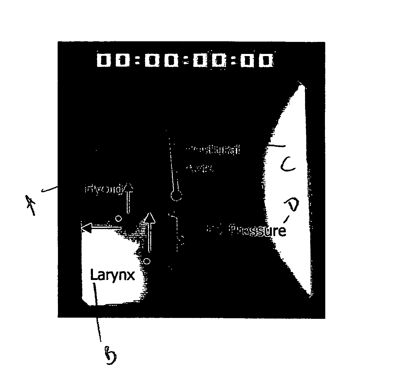
[0066] Fifteen healthy men, each selected for his highly visible thyroid prominence, participated in this study. Average age was 42 years (range 28 to 62). Normal laryngeal structure and function was confirmed on fiberoptic laryngeal examination by an otolaryngologist (E. M.). A lateral view of the neck surface was video recorded during each trial for data analysis. To aid later video data analysis, a 5 cm×3 mm strip of white tape was adhered to the left side of the participant's neck parallel to the direction of movement of the thyroid prominence during swallowing. Placement of the tape was sufficiently lateral to assure that the underlying skin did not move in conjunction with the prominence during swallow...

example 2

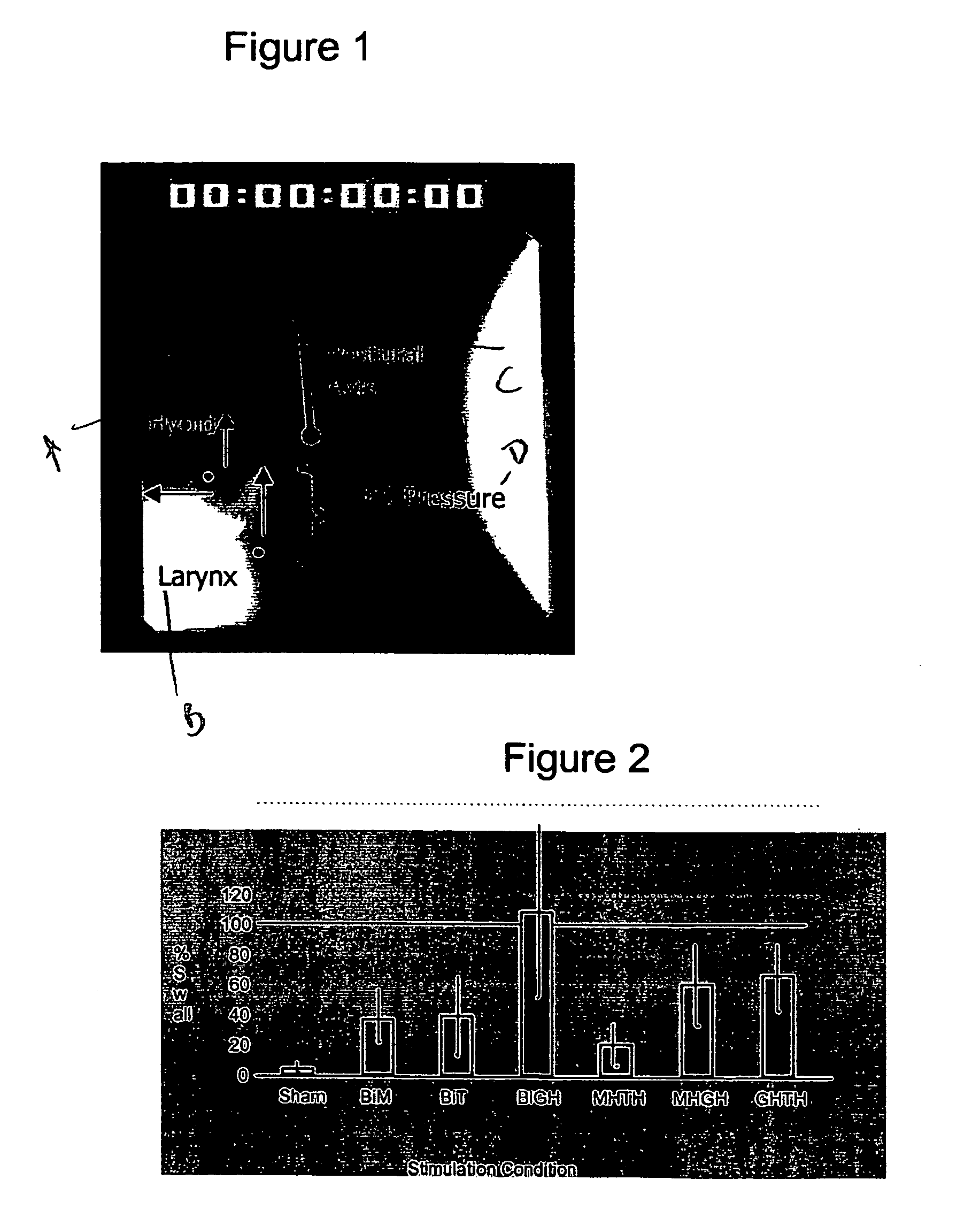
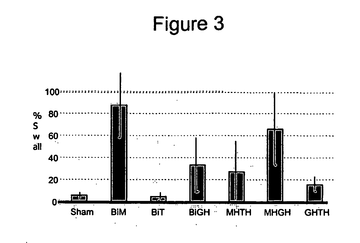
[0077] Unless otherwise described, the procedures described above for Example 1 were used for this example, which demonstrates that combined muscle stimulation can move the hyoid bone in the anterior and superior direction and the larynx in the superior direction to the same or a greater extent than occurs during normal swallowing.

[0078] The skin at each electrode insertion site was anesthetized with injections of 2% lidocaine. For each site, after numbness was reported, a 27 gauge monopolar needle was inserted in a desired muscle region and a train of stimulating pulses (200 μs width, 0.5-5.0 mA amplitude, 30 Hz frequency) was delivered to induce muscle contraction. Needle location was found to be mylohyoid (MH) if the stimulated contraction retracted the submental tissue and moved the thyroid prominence superiorly. A thyrohyoid (TH) location was found if the thyroid prominence moved superiorly and twisted contralaterally. Once a desired insertion site was located, the locating ne...

example 3

[0083] Unless otherwise described, the procedures described above for Example 1 were used for this example, which quantitatively demonstrates decreased pressure in the upper esophageal sphincter in response to anterior motion of the hyoid induced by combined muscle stimulation.

[0084] To record changes in pressure exerted at the upper esophageal sphincter, a manometer was inserted into the upper esophagus. The height was adjusted until the point of maximum resting pressure was identified and recorded with the transducer. The transducer pressure signal was examined before each stimulation or swallow to determine if the transducer had returned to the same position.

[0085] Simultaneous measurements of upper esophageal sphincter pressure both during stimulation at rest and without stimulation during normal swallowing determined whether the decrease in pressure, representing opening of the upper esophageal sphincter (UES) during mylohyoid stimulation, relates to the degree and / or duratio...


PUM


Abstract

Devices and methods were discovered that successfully provided patient autonomous control of both hyolaryngeal elevation, anterior hyoid motion and opening of the upper esophageal sphincter for swallowing by intramuscular stimulation of two muscles. The technology allows patient self stimulation of swallowing and can return oral feeding to dysphagia patients. Indwelling electrode stimulation of only two muscles generated as much as 80 % of normal synergistic movement leading to swallowing. The devices and methods also are useful for control of other upper respiratory muscle groups involved in speech and voice. Calibration techniques may be used in combination for greater freedom in setting and using electrodes over extended implantation time periods. These methods and devices can control complex movements of body solids such as bone and cartilage and tissues by electro stimulation of a minimum set of muscles simultaneously.

Description

FIELD OF THE INVENTION [0001] The invention relates generally to devices and procedures for modifying muscular function and more specifically to control of muscular activity by electrode stimulation. BACKGROUND OF THE INVENTION [0002] Dysphagia is a common complication with diseases such as stroke, neurodegenerative diseases, brain tumors, respiratory disorders, and the like wherein insufficient control of muscles needed for swallowing engender a risk of aspiration pneumonia. Aspiration pneumonia has been estimated to inflict a 20% death rate in the first year after a stroke and 10-15% each year thereafter. Treatment for this disorder requires either feeding through a nasogastric tube on a temporary basis or enteric feeding through a stoma to the stomach in chronically affected cases. The treatment costs and the commensurate value of a remediation technology if one were available, are very high. For example, in 1992 Medicare paid for enteral feedings of 206,000 patients at a cost of...

Claims


Application Information

Patent Timeline
IPC IPC(8): A61N1/36A61HA61H1/00A61N1/18
CPCA61N1/36007
Inventor LUDLOW, CHRISTY L.MANN, ERICBURNETT, THERESABIELAMOWICZ, STEVEN
Owner UNITED STATES OF AMERICA
Why Eureka
Try Eureka