Eureka AIR delivers breakthrough ideas for toughest innovation challenges, trusted by R&D personnel around the world.

Brain tissue classification

Active Publication Date: 2007-07-19
BRAINLAB
View PDF8 Cites 19 Cited by
  • Summary
  • Abstract
  • Description
  • Claims
  • Application Information

AI Technical Summary

Benefits of technology

[0018] RF inhomogeneities can be minimized by using at least two related image sequences in order to estimate and remove a bias term arising from the inhomogeneity. Multiple imaging modalities can be employed in order to identify white matter, even in the presence of pathological conditions. MR diffusion tensor imaging may be used, as it is particularly sensitive to variations in cell topology and is thus preferred for this task.
[0019] Preferably, medical images are used that show contrast between the white matter, gray matter, and CSF. In previous methods, this usually is taken to be a single three-dimensional MRI volume. Advantage is gained by using two similar MRI volumes with preferably only a single, but optionally two or more varied acquisition parameters to obtain different WM / GM / CSF contrast levels. It may be assumed that the gain field arising from magnetic susceptibility and RF field inhomogeneity will be similar in the two volumes. By taking the ratio of the two volumes, the common gain factor can be eliminated. Furthermore, by carefully selecting the acquisition parameters, the WM / GM / CSF contrast can be increased with respect to the individual volumes. This methodology can improve the performance of the remainder of the overall classifying method, but is not required. Some low-frequency distortion may remain, but it can be addressed through further processing as described below.

Problems solved by technology

Locating these surfaces is beneficial for some neurosurgical procedures, such as convection-enhanced drug delivery, since placement of infusion catheters too close to CSF boundaries is known to lead to poor dispersion of infusate in tissue, as the infusate flows into the less-resistive CSF-filled regions.
At the cortical surface, however, thin sulci are often narrower than the resolution provided by MR imagery and, thus, are not reliably detected.
Furthermore, the problem can be complicated by brain pathologies.
In particular, edema in white matter can greatly alter signal levels, making it difficult to separate white matter from other tissues.
However, it can be difficult to differentiate gray matter from white matter.
This difficulty is often compounded by inhomogeneities in the RF fields, making it difficult or impossible to separate gray and white matter with a constant threshold value.
Deformable surface models can represent partial voxels, but are generally complicated to implement and computationally intensive to run.
Brain tissue classification methods that use MRI data as input can encounter difficulties.
These difficulties include a variation in signal response, and structures that are smaller than the sampling rate.
MRI data used for brain tissue classification often contain some amount of artifact or distortion due to inhomogeneities in the radio frequency (RF) fields and magnetic susceptibility variations in the tissue being imaged.
The difficulties with this method are that they are complicated to implement and relatively time-consuming to perform.
The second difficulty, structures too small to be detected at MRI resolution, occurs frequently at the cortical surface.
This method performs well at locating the cortical surface, but the level set approach for locating the GM-WM boundary is compute-intensive, considering that the WM-GM interface is amenable to simpler classification methods, such as thresholding.

Method used


Image

  • Brain tissue classification
  • Brain tissue classification
  • Brain tissue classification

Examples


Embodiment Construction

[0027] A CSF boundary detection procedure includes the steps shown below.

[0028] 1. Image pair pre-processing to enhance contrast and reduce bias (optional).

[0029] 2. WM / GM / CSF thresholded classification / inhomogeneity correction.

[0030] 3. Thin sulcus detection using the gray matter skeleton.

1. Image Pair Pre-Processing

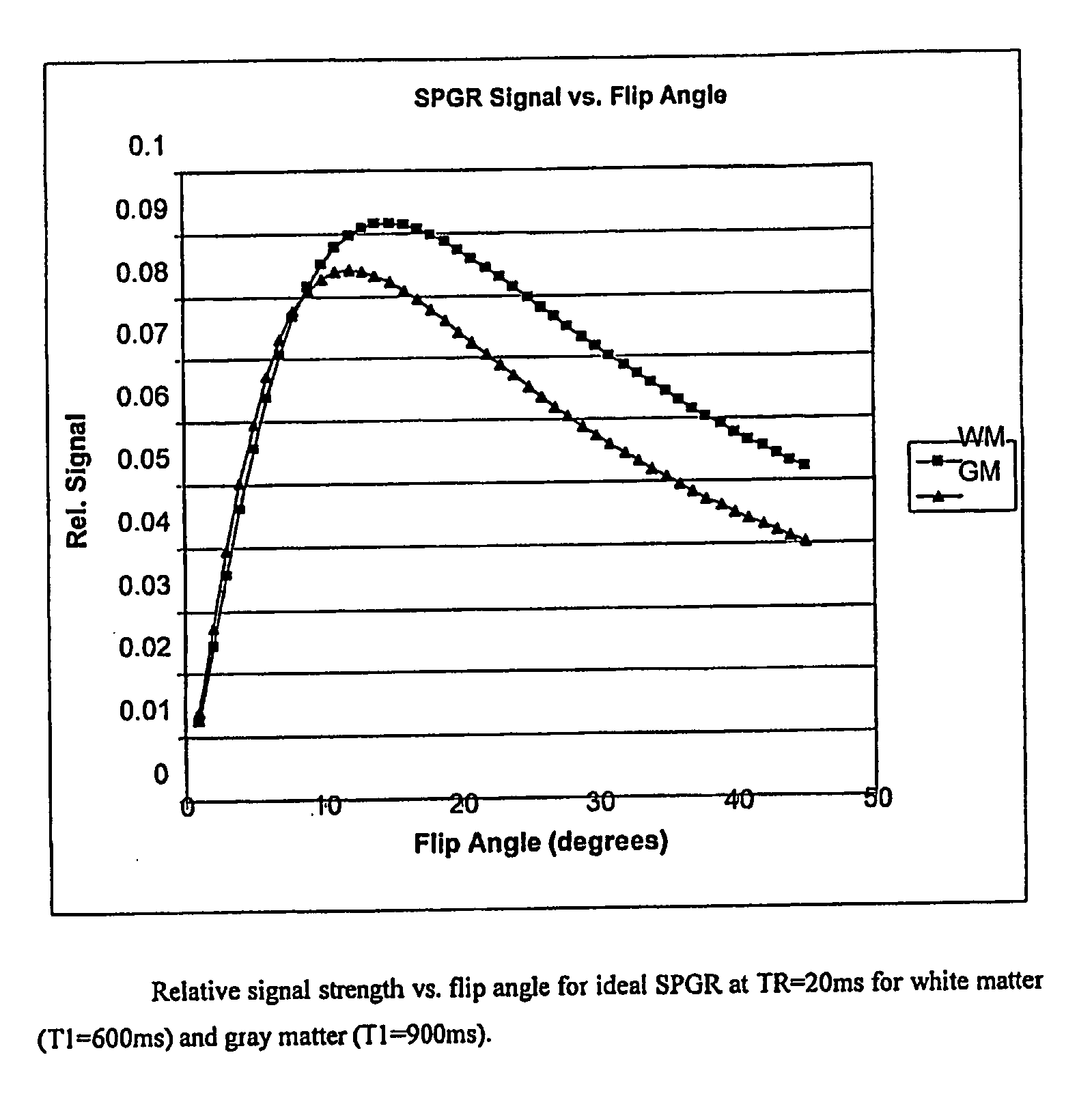
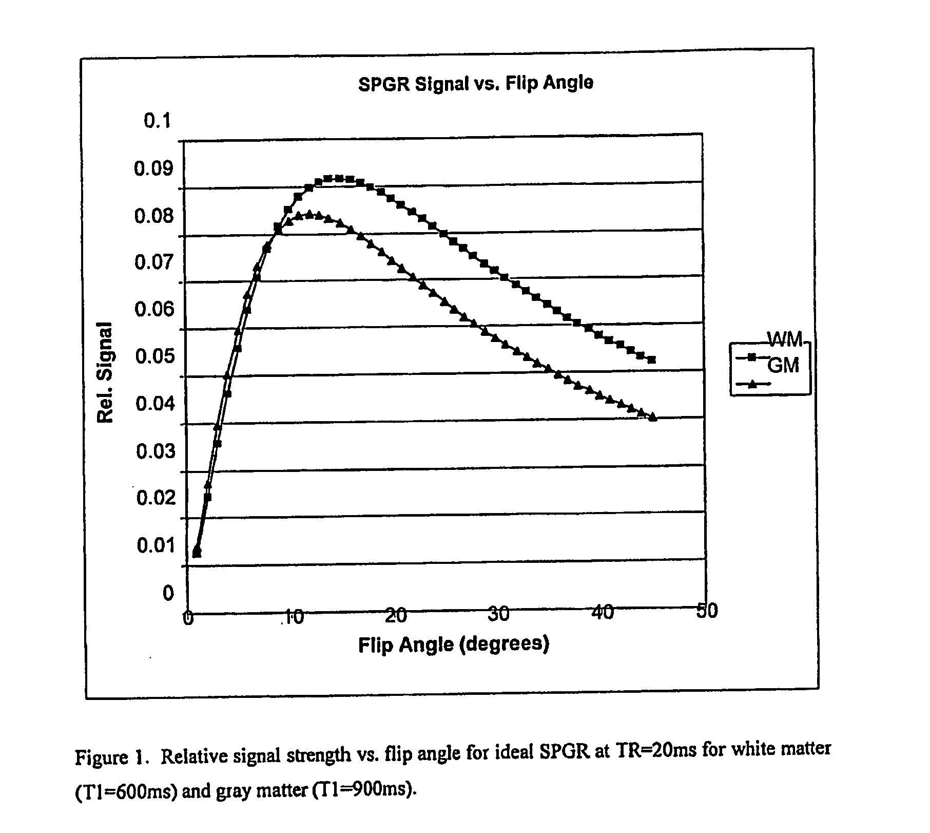
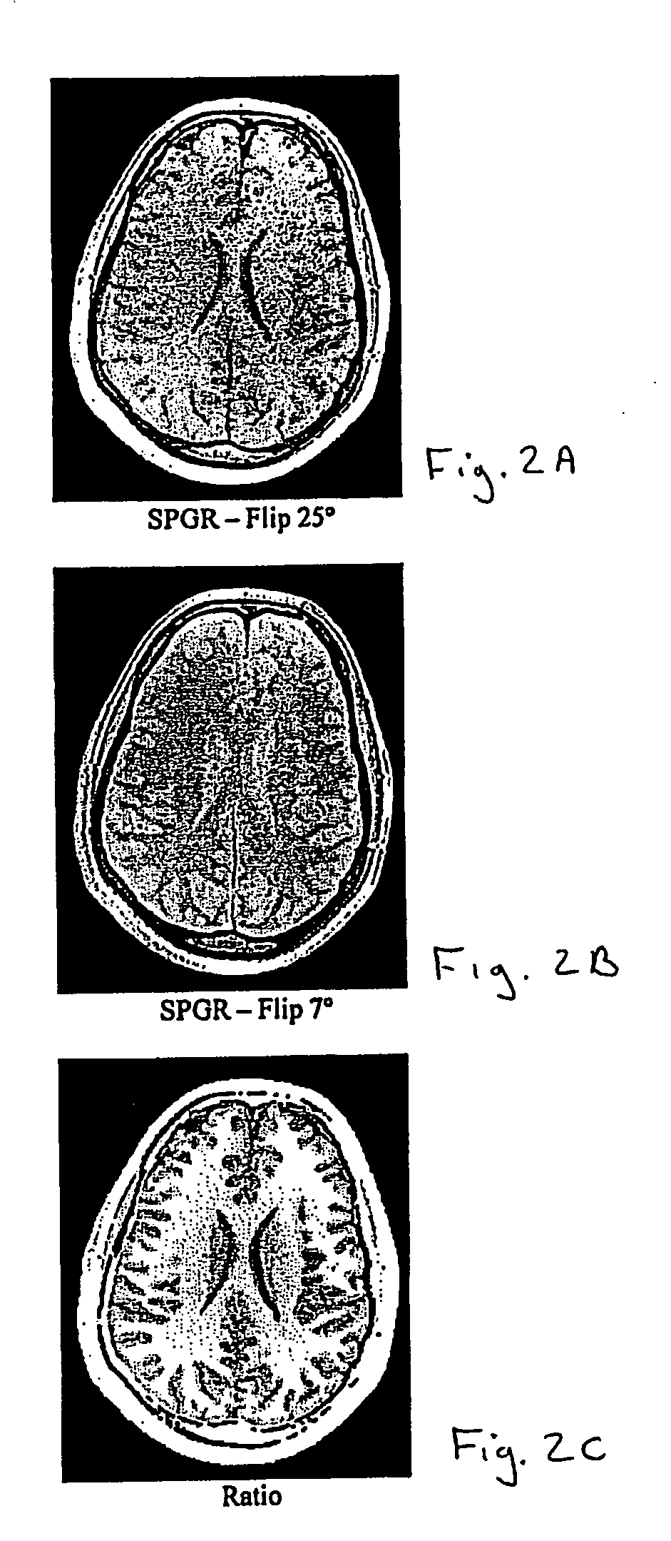
[0031] Two MRI volumes can be acquired using identical acquisition parameters, varying, for example, one acquisition parameter. These two volumes can be co-registered, if necessary, using known methods. Then the ratio of the two volumes is determined (any division by zero is replaced with the value zero). The ratio, for example, may be based on a voxel by voxel or data point by data point calculation. Determining the ratio of the two volumes has two advantages. First, intensity variations due to field inhomogeneities tend to be similar in both images, so the division tends to normalize this undesirable effect such that it is insignificant. Second, when the acquisi...


PUM


Abstract

A medical imaging processing method includes: using an imaging method to acquire at least first and second data sets of a region of interest of a patient's body, with at least one image acquisition parameter being changed so that first and second data sets yield different contrast levels relating to different substance and / or tissue types, and wherein the at least one acquisition parameter used to obtain the first data set is selected to enhance the contrast between one of the substance and / or tissue types relative to other substance and / or tissue types, and the at least one acquisition parameter used to obtain the second data set is selected to enhance the contrast between a different one of the substance and / or tissue types relative other substance and / or tissue types, thereby to optimize the contrast between at least three different substance and / or tissue types: and processing the two data sets to identify the different tissue types and / or boundaries therebetween.

Description

RELATED APPLICATION DATA [0001] This application claims priority of U.S. Provisional Application No. 60 / 724,231 filed on Oct. 6, 2005, which is incorporated herein by reference in its entirety.FIELD OF THE INVENTION [0002] The present invention relates to image processing for surface delineation and, more particularly, to a method and apparatus for detecting boundaries between brain tissue and cerebrospinal fluid (CSF) filled regions to classify brain tissue. BACKGROUND OF THE INVENTION [0003] Tissue-CSF boundaries between brain tissue and cerebrospinal fluid (CSF) occur primarily at an outer cortical surface and in interrior regions of the brain at CSF-filled ventricles. Locating these surfaces is beneficial for some neurosurgical procedures, such as convection-enhanced drug delivery, since placement of infusion catheters too close to CSF boundaries is known to lead to poor dispersion of infusate in tissue, as the infusate flows into the less-resistive CSF-filled regions. [0004] La...

Claims


Application Information

Patent Timeline
IPC IPC(8): A61B8/00
CPCA61B5/7264A61B5/055G06T2207/30016G06T7/12G06T2207/10088G01R33/5608A61B2576/026A61B5/4064A61B5/0042
Inventor HARTLEP, ANDREASPEDAIN, CHRISTOPHBRADY, MARTINRAGHAVAN, RAGHU
Owner BRAINLAB
Why Eureka
Eureka Blog
Learn More