Method for fluorescently staining tissue

a fluorescent staining and tissue technology, applied in the field of fluorescent staining tissue, can solve the problems of sample having a different state from that within the living body, sample surface being quite delicate, decoloration of dye from stained tissue, etc., and achieves enhanced fluorescent properties, clear and accurate stained images, and high ph dependence

Inactive Publication Date: 2007-07-26
ASAHI KOGAKU KOGYO KK
View PDF3 Cites 7 Cited by
  • Summary
  • Abstract
  • Description
  • Claims
  • Application Information

AI Technical Summary

Benefits of technology

[0016]According to the staining method of the present invention, a clear and accurate stained image can be obtained because only fluorescein bound with a tissue is selectively capable of fluorescent staining. Namely, fluorescein is highly dependent on pH in general, and its fluorescent property tends to be enhanced more at higher pH values. Only a fluorescein molecule that is permeated into the internal region of a living body tissue and bound wit

Problems solved by technology

However, the sample to be observed has a quite delicate surface and must be washed with care in avoiding the deformation or damage of the sample and the denaturation of the tissue.
Moreover, excessive washing also causes the detachment of the dye (decoloring) from the stained tissue.
On the other hand, when an extr

Method used


Image

  • Method for fluorescently staining tissue
  • Method for fluorescently staining tissue

Examples


example 1

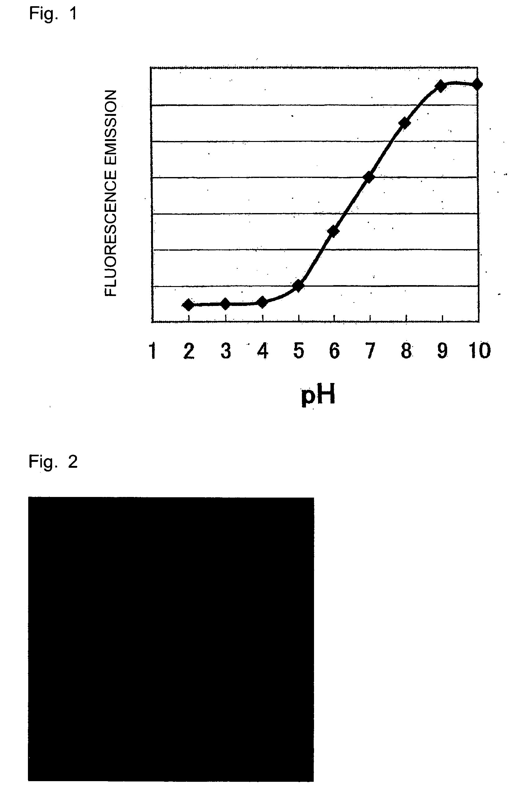
[0031]pH-Dependent Fluorescence Emission of Fluorescein

[0032]The pH of an aqueous solution of fluorescein sodium (1 mg / mL) was adjusted with 0.1 M sodium phosphate buffer solution, followed by measurement at an excitation wavelength of 490 nm and a fluorescence wavelength of 530 nm.

[0033]A microplate reader (manufactured by Corona, MTP-800AFC) was used in the fluorescent measurement.

[0034]As seen from the result shown in FIG. 1, the fluorescence intensity of fluorescein gets stronger at higher pH values.

example 2

[0035]Fluorescein Sodium Staining Test on Rat Large Intestine

[0036]The formalin-fixed large intestines of rats (8-week-old, male) were used to observe a difference in staining property against pH. The large intestines were washed for 10 seconds with a phosphate buffered saline (137 mmol / L NaCl, 8.1 mmol / L Na2HPO4, 2.7 mmol / L KCl, 1.57 mmol / L KH2PO4, hereinafter referred to as PBS (−)) and then immersed into solutions of fluorescein sodium (manufactured by Sigma, F6377, hereinafter referred to as F—Na) adjusted and diluted to 1 mg / mL with acidic, alkaline, and neutral buffer solutions. The neutral, alkaline, and acidic buffer solutions used were 0.1 M sodium phosphate buffer solution (pH 7.0), 0.1 M borate buffer solution (pH 9.1), and 0.4 M NaH2PO4 buffer solution (pH 4.65), respectively. After further washing for 10 seconds with PBS (−), the resulting large intestines were fixed with 10% formalin-acid buffer solution and observed with a confocal scanning microscope (manufactured by...

example 3

[0038]Difference in Permeation and Fluorescent Property of Dye Depending on Changes in Solvent pH

[0039]The large intestines of rats (8-week-old, male) were extracted and subjected to a staining test.

[0040]F—Na (manufactured by Sigma, F6377) was adjusted to 1 mg / mL and diluted with 0.1 M sodium phosphate buffer solution (pH 7.0), 0.1 M borate buffer solution (pH 9.1), and 0.4 M NaH2PO4 buffer solution (pH 4.65) to prepare their respective 0.1 mg / mL solutions, into which the tissues were then immersed for 1 minute. After further washing for 10 seconds with PBS (−), the resulting tissues were observed with a confocal scanning microscope (manufactured by Leica Microsystems, TCS-SP2). The results are shown in Table 1.

TABLE 1Fluorescence intensityexamined visually(indicated in five tiers)FreeFluoresceinState of observedfluoresceinin internalimage (clarityinregion ofof fluorescentSolventpHliquid pooltissueimage)0.4 M NaH2PO44.6513Clearsolution0.1 M Na7.053Not clear butphosphatetissue isbuf...


PUM


Abstract

It is intended to provide a method for fluorescently staining a tissue accurately and clearly by convenient procedures and a staining agent used in this method. The present invention provides a method for fluorescently staining a tissue, comprising treating a tissue with a solution comprising fluorescein or a salt thereof and then fluorescently observing the treated portion under an acidic condition of lower than pH 7.

Description

BACKGROUND OF THE INVENTION[0001]1. Field of the Invention[0002]The present invention relates to method for fluorescently staining a living body tissue or a living body-derived tissue conveniently and clearly and to a staining agent used in this method.[0003]2. Description of the Related Art[0004]The acquisition of cross-sectional images of living body tissues is of extreme importance in the medicobiological field. Cross-sectional images of tissues extracted from living bodies could previously be obtained by chemical fixation, dehydration, slicing, and staining.[0005]The development of a confocal imaging system and the prevalence of a confocal laser scanning microscope enabled noninvasive observation of cells and tissues in the observation of sections of biological samples with intricately multi-layered cells and connective tissues. Medical endoscopes equipped with a built-in confocal imaging system have recently been developed.[0006]The observation of fluorescent images in a confoc...

Claims


Application Information

Patent Timeline
IPC IPC(8): G01N1/30G01N33/48
CPCG01N1/30A61K49/0043A61K49/0017G01N33/52
Inventor YAMAMOTO, AKIRAIIMORI, YUSUKESAZE, MIZUEISHIGURO, MARIKO
Owner ASAHI KOGAKU KOGYO KK
Why Eureka
Try Eureka