
Immune diagnostic assay to diagnose and monitor tuberculosis infection

a tuberculosis and immuno-diagnosis technology, applied in the field of immuno-diagnosis assay to diagnose and monitor tuberculosis infection, can solve the problems of inability to culture virulent m. tuberculosis, and inability to monitor the infection of tuberculosis, etc., to achieve the highest sensitivity of active tuberculosis and the high sensitivity of assay

Inactive Publication Date: 2007-08-23
INST NAT PER LE MALATTIE INFETTIVE LAZZARO SPALLANZANI IRCCS
View PDF0 Cites 13 Cited by
  • Summary
  • Abstract
  • Description
  • Claims
  • Application Information

AI Technical Summary

Benefits of technology

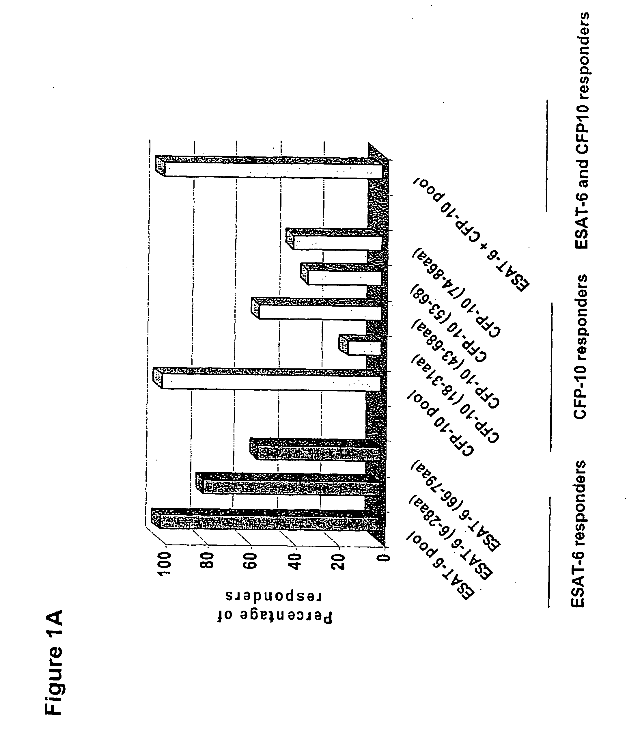
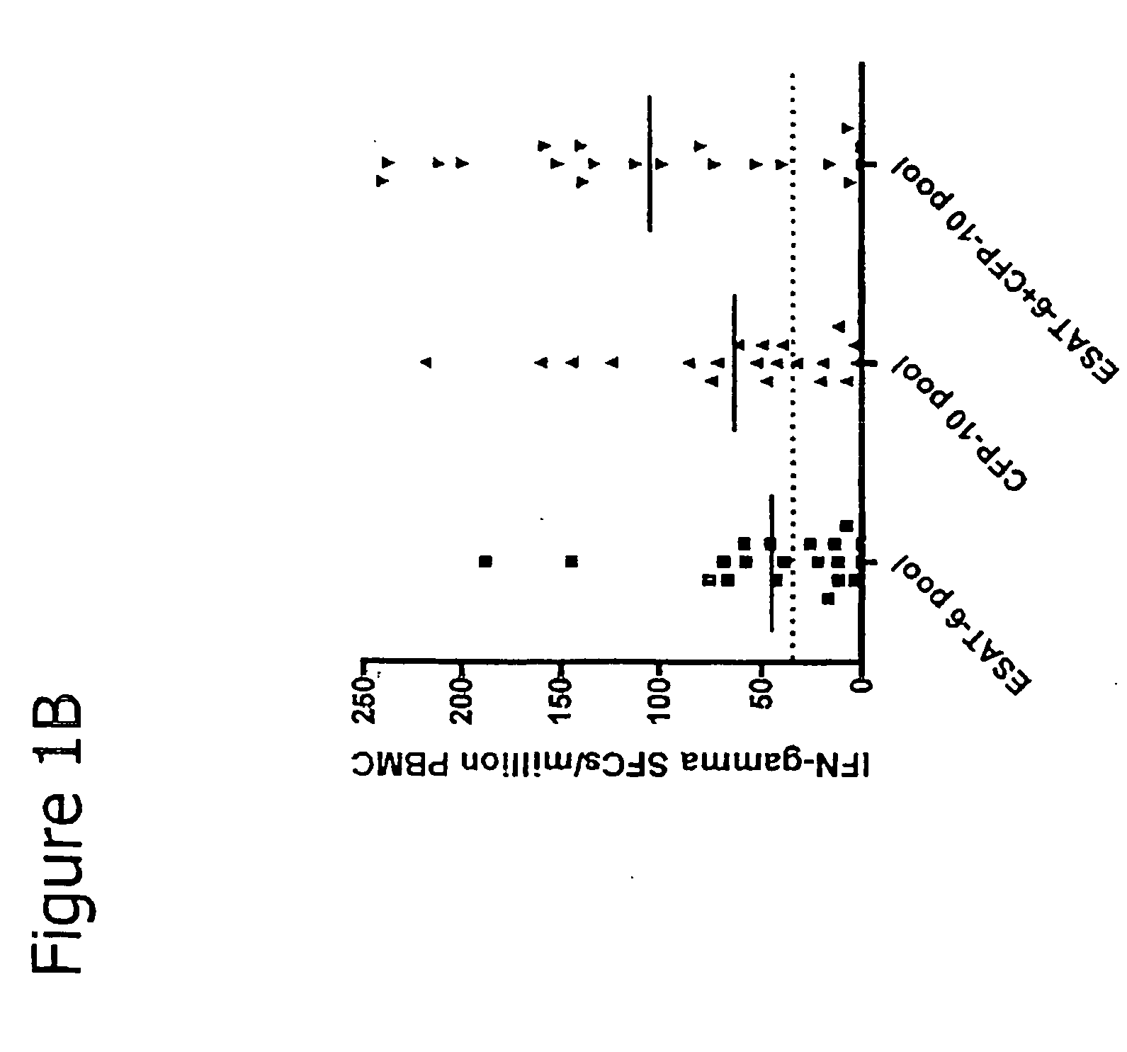
[0029] The novelty of our assay is the use of specifically selected CFP-10 peptides separately (FIG. 1A) and in combination with ESAT-6 specifically selected peptides (Vincenti et al 2003). As seen in FIG. 1B among patents with microbiologically confirmed active tuberculosis, the use of selected peptides from CFP-10 leads to a higher sensitivity of the assay compared to the use of only ESAT-6 selected peptides. In FIG. 1B it is also noticeable that the combination of ESAT-6 and CFP-10 has the highest sensitivity for active tuberculosis. Overtapping ESAT-6 and CFP-10 peptides were used in the previous patent applications and scientific articles (WO0026248; US2004058399; WO 20041099771, EP 1 350 839 A; van Pinxteren 2000; Cockle 2002; Andersen 2001; WO 99 / 04005; WO 99 / 42076 A: WO 97 / 09428 A; WO 01 / 04151 A; WO 01 / 79274 A; WO 03 / 093307 A). In addition we report that these selected peptides may be used with different experimental methods to achieve the same result: such as ELISPOT assay, FACS analysis and whole blood ELISA. In addition, in the present invention we used a rapid method to separate circulating mononuclear cells in the blood sample (LeucoSep, ARNIKA, Milan, Italy) which is different from what has been used in other patents (for example Par. 18 of EP1152012). This method, compared to traditional methods, allows processing several samples at once.

Problems solved by technology

“patient with tuberculosis infection” either a) an individual with a recent or old contact with tuberculosis and in these cases it is not possible to culture virulent M. tuberculosis; b) an individual with culture proven infection with virulent M. tuberculosis.
It is not possible to culture virulent M. tuberculosis.
Therefore tuberculosis remains a global health problem because of the long time needed for microbiological diagnosis and because of the difficulty in monitoring the efficacy of anti-tuberculosis therapy.
However, this test has significant limitations as purified protein derivative (PPD) used for TST is a crude precipitate of filtered MTB containing more than 200 antigens widely shared among environmental mycobacteria (Fine et al 1999, Huebner et al 1993, American Thoracic Society 2000.
However, such response does not allow discriminating between subjects with active tuberculosis and subjects with latent infection.
These results can lead to an incorrect diagnosis, because the subject may have a disease that mimics active tuberculosis, but different from tuberculosis (such as a bacterial pneumonia, a viral fever, a bronchitis) and thus he may start an incorrect treatment, he may be isolated from the community if the suspect is a pulmonary tuberculosis, with relevant human and economic costs.

Method used


Image

  • Immune diagnostic assay to diagnose and monitor tuberculosis infection
  • Immune diagnostic assay to diagnose and monitor tuberculosis infection
  • Immune diagnostic assay to diagnose and monitor tuberculosis infection

Examples


example 2

Procedure of the Immune Diagnostic Assay Performed on Whole Blood by ELISA (see FIG. 3)

[0174] The ELISA method on whole blood is performed by the use of the QuantiFERON-CMI (QF-CMI, manufactured by Cellestis Limited. South Melbourne, Australia), modified by us. In detail, we use 0.5 ml of blood instead of 1 ml, and therefore we use a 48-well plate instead of a 24-well. The whole procedure of the test requires the use of the following materials and reagents: IFN-gamma QuantiFERON-CMI kit; 48-well plate and 7 tubes, each containing the different stimuli at the desired concentration. The ELISA technique, performed on whole blood, is composed of the following steps:

1. Culture of Whole Blood

[0175] 1. mix the tubes containing heparinised whole blood [0176] 2. distribute blood (500 μl / well) into sterile 48-well plaies. For the children similar results can be obtained using 250 μ / well of blood. Consequently the volume of the Reagents 1-7 to be added will be 50% of the volume indicated. ...

example 3

Procedure of the Immune Diagnostic Assay Performed by FACS (see FIG. 4)

[0197] The whole procedure of the test requires the use of an IFN-gamma flow cytometer and the following materials and reagents: 24-well plate; mixture of antibodies (Becton-Dickinson, CA); brefeldin-A (Sigma, St. Louis, Mo.) and 7 tubes, each containing the different stimuli at the desired concentration.

[0198] In particular the FACS analysis method is composed of the following steps: [0199] i. isolation of mononuclear cells (PBMC) from venous blood [0200] ii. stimulation of Ag-Sp and incubation [0201] iii. immunofluorescence staining [0202] iv. acquisition and FACS analysis [0203] v. diagnostic response.

[0204] In step (i), peripheral blood mononuclear cells (PBMC) are isolated from a small aliquot of venous blood (5 ml) by density gradient centrifugation (Ficoll-Hypaque, Pharmacia; Uppsala; Sweden) using a rapid test based on the use of special tubes equipped with a filter for the separation of leukocytes (Le...


PUM

PropertyMeasurementUnit
Timeaaaaaaaaaa
Timeaaaaaaaaaa
Timeaaaaaaaaaa

Abstract

The present invention relates to a method of diagnosing and monitoring various distinct presentations of tuberculosis: active tuberculosis disease, latent tuberculosis infection and recent tuberculosis infection. The rapid immune assay is based on the evaluation of the frequency of Interferon (IFN) gamma-producing antigen-specific T lymphocytes responding to selected peptide sequences from Mycobacterium tuberculosis, selected for their immunogenicity. The invention concerns also immunogenic and vaccine compositions based on these specific peptide sequences.

Description

FIELD OF INVENTION [0001] The present invention relates to a method of diagnosing and monitoring various distinct presentations of tuberculosis: active tuberculosis disease, latent tuberculosis infection and recent tuberculosis infection. The rapid immune assay is based on the evaluation of the production of Interferon (IFN) gamma by antigen-specific T lymphocytes responding to selected peptide sequences from secretory proteins of Mycobacterium tuberculosis. These peptide sequences have been selected for their immunogenicity. The test may be performed by ELISPOT, ELISA or FACS techniques and the eventual choice of methodology to carry out the lest is done according to the resources and the requirements of the laboratory. Definitions [0002] In the present invention, by using the following terms, we indicate by. [0003]“virulent M. tuberculosis” a bacterium capable of causing the tuberculosis disease in a mammal including a human being. [0004]“patient with tuberculosis infection” eith...

Claims


Application Information

Patent Timeline
IPC IPC(8): G01N33/554C07H21/04C07K14/35G01N33/569
CPCG01N33/5695C07K14/35Y10S435/975
Inventor GOLETTI, DELIAVINCENTI, DONATELLACARRARA, STEFANIAGIRARDI, ENRICOPOCCIA, FRABRIZIOCASETTI, RITAAMICOSANTE, MASSIMO
Owner INST NAT PER LE MALATTIE INFETTIVE LAZZARO SPALLANZANI IRCCS
Learn More