Systems and methods for generating data using one or more endoscopic microscopy techniques

a technology of endoscopic microscopy and system, applied in the field of system and method for generating data using one or more endoscopic microscopy techniques, can solve the problems of difficult implementation of endoscopic cm systems, difficult to achieve endoscopic subcellular imaging, and difficulty in achieving endoscopic subcellular implementation, so as to reduce spatial coherence mismatch in spatial modes, prevent a desired level of interference, and improve the effect of temporal and spatial coherence match

Inactive Publication Date: 2007-10-11
THE GENERAL HOSPITAL CORP
View PDF99 Cites 121 Cited by
  • Summary
  • Abstract
  • Description
  • Claims
  • Application Information

AI Technical Summary

Benefits of technology

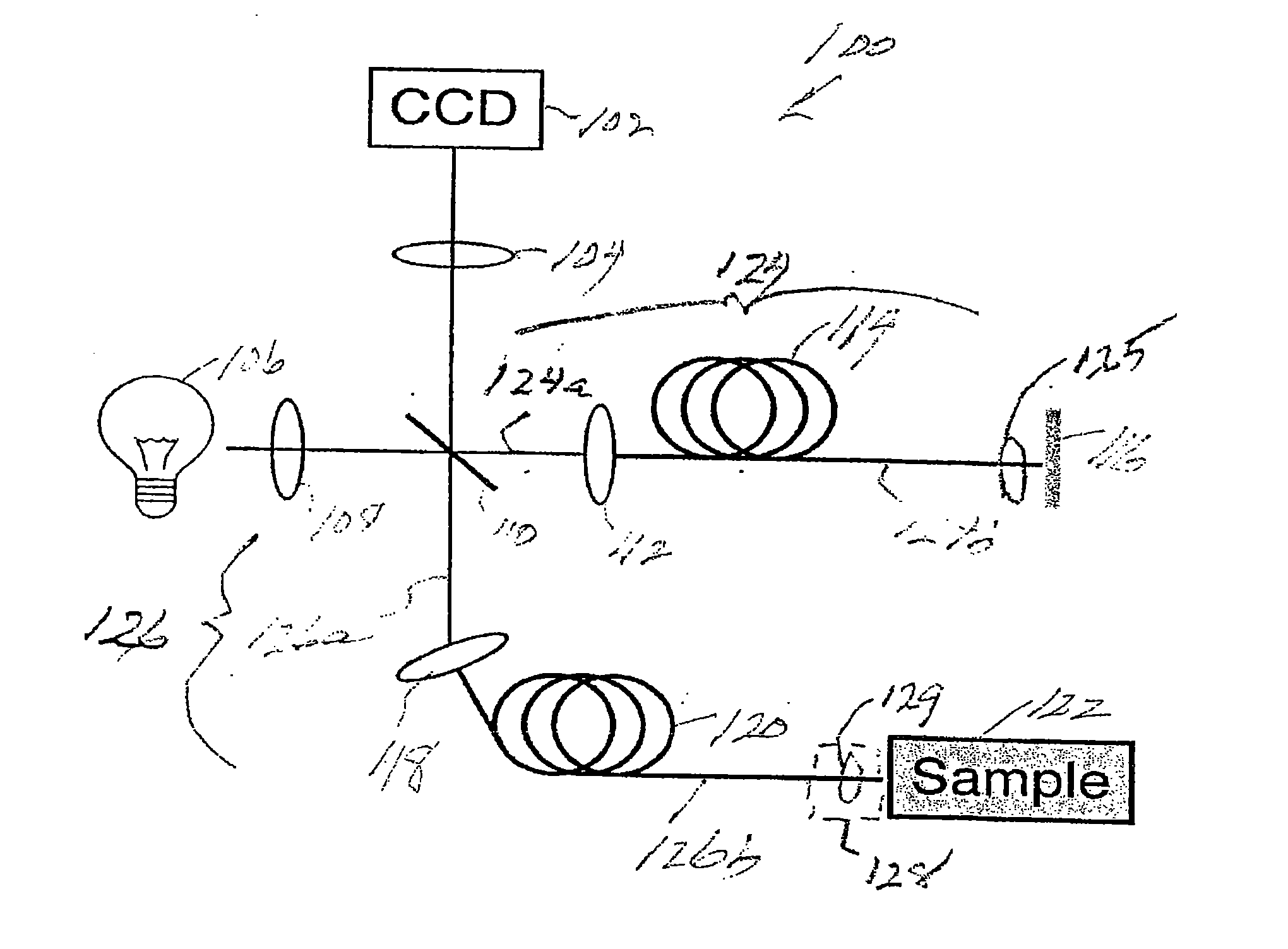
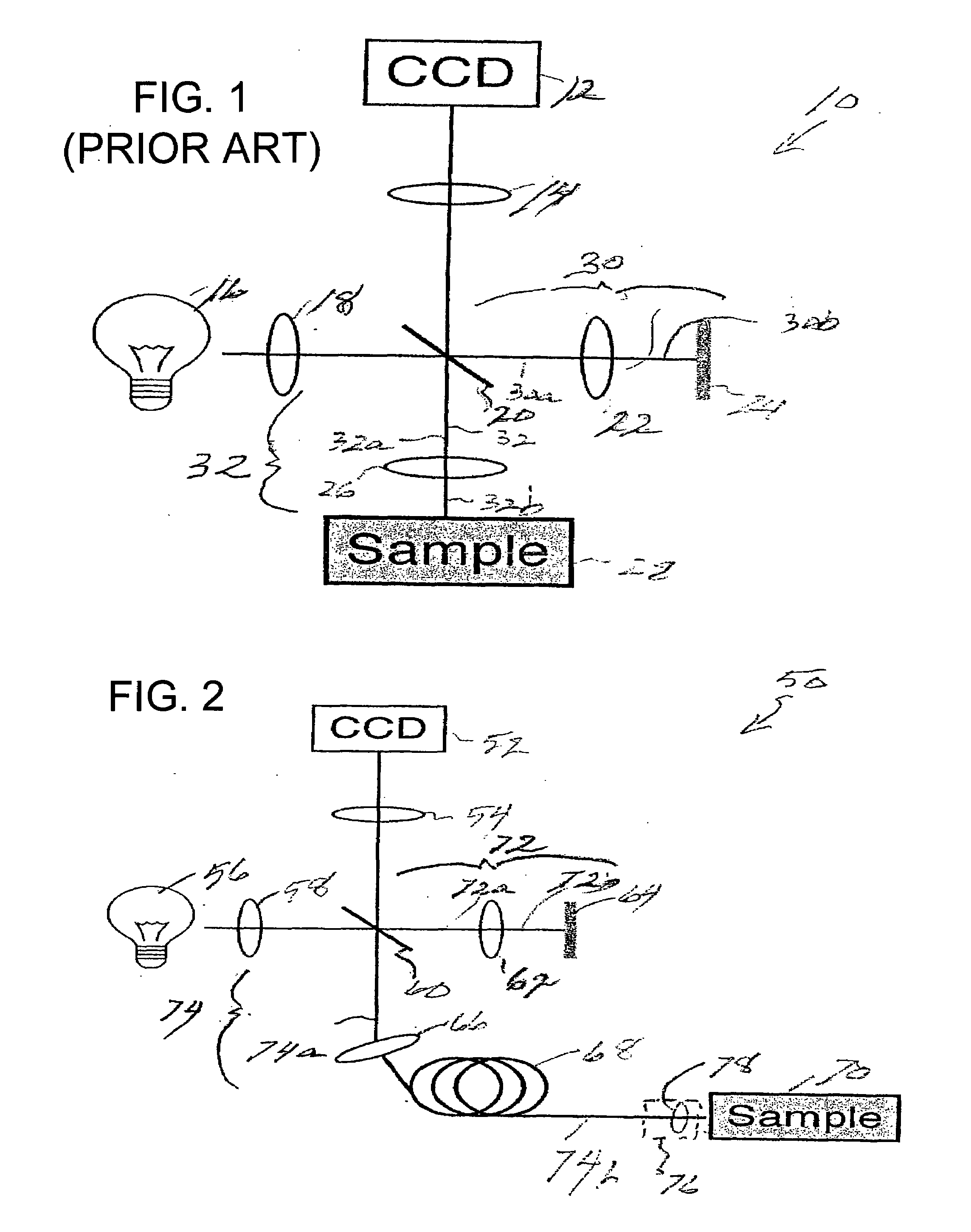
[0025]In another exemplary embodiment of the present invention, a method and system can be provided for performing endoscopic full-field optical coherence microscopy (“E-FFOCM”). Certain variants of the exemplary embodiments of the present invention can utilize an endoscopic probe having a fiber-optic bundle arranged in a Linnik interferometer, which can provide light to the endoscopic probe. The fiber-optic bundle can be single- or multi-mode, but preferably multimode for optimal coupling of the source light and detection of light remitted by the sample. By allowing light delivery through the fiber-optic bundle, the system can facilitate use of the E-FFOCM techniques in a catheter or endoscope. This exemplary embodiment can therefore enables, e.g., a high-resolution microscopy of surfaces of the body accessible by endoscope.
[0027]According to yet another exemplary embodiment of the present invention, an optical-fiber imaging bundle can be used in both the sample and in the reference arms, which should be substantially identical in order to provide spatial and temporal coherence. Even though this exemplary configuration can reduce the spatial coherence mismatch in spatial modes between the arms, the sample arm fiber-optic bundle may change with respect to the reference arm fiber-optic bundle during the diagnostic procedure. As a result, the reference and sample arms can both be spatially and temporally mismatched, possibly preventing a desired level of interference.
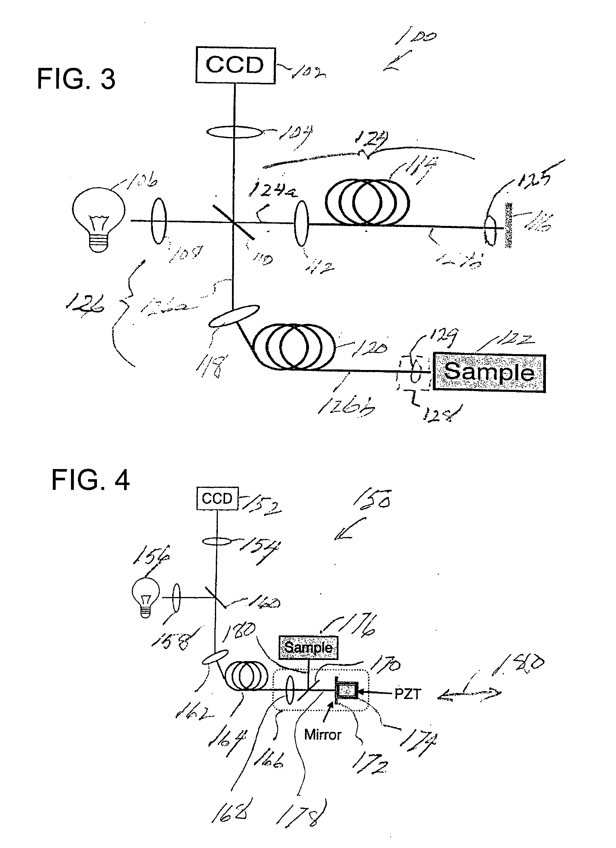
[0028]In still another exemplary embodiment of the present invention, to further improve the temporal and spatial coherence match between reference and sample arms, one fiber-optic bundle can be used to transmit and / or receive the both reference and sample arm light. In such exemplary embodiment, the interferometer can be placed distal to the fiber-optic bundle. The reference arm and sample arm illumination light can travel through the same bundle. At the distal end of the endoscope, the reference arm path can be incident on a mirror mounted to a small linear translator such as a piezoelectric stack. The sample and reference arm light may be combined at the distal beam splitter and transmitted back through the fiber bundle. Since the sample and reference arm paths can traverse the same bundle, they generally remain spatially and temporally coherent with respect to each other, thus facilitating a high contrast interference at the CCD. Furthermore, a dispersion mismatch caused by the bundle can be balanced due to the common paths of the reference and sample arms.

Problems solved by technology

While the OCT and CM systems and methods show potential for solving several important diagnostic problems, these techniques have certain technical requirements that can make endoscopic subcellular imaging difficult.
Further, while the CM systems and methods can provide images in the human tissue with 1 μm transverse resolution, endoscopic implementation of CM may be difficult to achieve.
Endoscopic CM systems, which generally use a small-diameter endoscopic probe, are difficult to implement due to certain endoscopic probe size constraints resulting from a requirement for a high numerical aperture (NA) objective lenses (NA≧0.7) and rapid beam scanning arrangements.
In addition, since both OCT and CM methods and systems generally use lasers to illuminate a sample, the OCT and CM images likely contain significant coherent interference or speckle noise, which can degrade the resolution of the resulting images, e.g., by up to a factor of four.
However, similar to CM principles, the OCM systems and methods likely utilize a rapid scanning of a focused beam by way of a rapid beam scanning mechanism, and thus may also be difficult to implement in a small diameter endoscopic probe.
It may be difficult to miniaturize the components of FIG. 1 within the confines of an endoscopic probe having a small diameter, e.g., less than 5 mm.
However, due to the complexities of miniaturizing the FFOCM system, it has been difficult to realize an endoscopic FFOCM system, which requires a small probe diameter.

Method used


Image

  • Systems and methods for generating data using one or more endoscopic microscopy techniques
  • Systems and methods for generating data using one or more endoscopic microscopy techniques
  • Systems and methods for generating data using one or more endoscopic microscopy techniques

Examples


Embodiment Construction

[0070]Prior to providing a detailed description of the various exemplary embodiments of the methods and systems for endoscopic microscopy according to the present invention, some introductory concepts and terminology are provided below. As used herein, the term “endoscopic probe” can be used to describe one or more portions of an exemplary embodiment of an endoscopic system, which can be inserted into a human or animal body in order to obtain an image of tissue within the body.

[0071]As used herein, the term “monolithic” can be used to describe a structure formed as a single piece, which may have more than one optical function. As used herein, the term “hybrid” can be used to describe a structure formed as a plurality of pieces, each piece having one optical function.

[0072]The exemplary embodiments of the methods and systems according to the present invention described below can be used with any wavelength of light or electro-magnetic radiation, including but not limited to visible l...


PUM


Abstract

Exemplary systems and methods for imaging at least one portion of a sample can be provided. For example, according to one exemplary embodiment of such systems and methods, it is possible to receive at least one first electro-magnetic radiation from the sample and at least one second electro-magnetic radiation from a reference using at least one arrangement. Such arrangement and the reference can be provided in an endoscope enclosure. The image data associated with the portion can be generated as a function of the first and second electro-magnetic radiations. In another exemplary embodiment, an endoscope arrangement can be provided for imaging such portion of the sample. The endoscope arrangement can include at least one interferometric arrangement configured to receive at least one electro-magnetic radiation from the sample, and situated within and at one end of an endoscope enclosure of the endoscope arrangement. According to yet another exemplary embodiment, at least one first Linnik interferometric arrangement at least one second fiber arrangement being in optical communication with the at least one first arrangement can be provided. The second arrangement can be configured to transmit an electro-magnetic radiation to the first arrangement. The first arrangement can be configured to receive an additional electro-magnetic radiation from the sample which can be associated with the first electro-magnetic radiation. The first arrangement can be configured to forward at least one third electro-magnetic radiation which is associated with the at least one second electro-magnetic radiation to the at least one second arrangement.

Description

CROSS-REFERENCE TO RELATED APPLICATION(S)[0001]This application is based upon and claims the benefit of priority from U.S. Patent Application Ser. No. 60 / 759,936, filed Jan. 18, 2006, the entire disclosure of which is incorporated herein by reference.STATEMENT REGARDING FEDERALLY SPONSORED RESEARCH[0002]The invention was made with the U.S. Government support under Contract No. BES-0086709 awarded by the National Science Foundation. Thus, the U.S. Government has certain rights in the invention.FIELD OF THE INVENTION[0003]The present invention generally relates to systems and methods for generating data using one or more endoscopic microscopy techniques and, more particularly to e.g., generating such data using one or more high-resolution endoscopic microscopy techniques.BACKGROUND OF THE INVENTION[0004]Medical imaging technology has advanced to provide physicians with important information regarding the macroscopic anatomy of patients. Imaging modalities such as radiography, magnetic...

Claims


Application Information

Patent Timeline
Patent Type & Authority Applications(United States)
IPC IPC(8): A61B5/05
CPCA61B1/00096G01B9/02091A61B1/00172A61B1/06A61B1/07A61B5/0066A61B5/0068A61B5/0075A61B5/0084G01N21/4795G02B23/2446G01B9/02079G01B9/02057G01B9/0205A61B1/00167
Inventor TEARNEY, GUILLERMO J.IFTIMIA, NICUSORBOUMA, BRETT EUGENEOH, WANG-YUHL
Owner THE GENERAL HOSPITAL CORP
Why Eureka
Try Eureka