Bioactive Coating of Biomedical Implants

a bioactive coating and implant technology, applied in the field of bioactive coating of biomedical implants, can solve the problems of metal surface providing a relatively poor interface for bone or other tissue attachment, propensity for the coating to erode or detach from the metal implant substrate, etc., and achieves the effect of promoting bone regrowth and attachment, and high silanol group density

Inactive Publication Date: 2008-03-20
UNIV OF SOUTH AUSTRALIA
View PDF7 Cites 21 Cited by
  • Summary
  • Abstract
  • Description
  • Claims
  • Application Information

AI Technical Summary

Benefits of technology

[0010]The present invention also provides a method of improving the fixation of an implanted biomedical implant, the method including the step of contacting at least part of the surface of the implant prior to implantation with a plasma gas containing a reactive hydroxylating oxidant species.
[0046]An advantage of the method of the present invention is that the step of contacting at least part of the surface of the implant (with or without a composite silica coating) with a plasma gas containing the reactive hydroxylating oxidant species is that it is possible to form a higher density of silanol groups on the treated surface than with other known methods. As discussed previously, it has previously been suggested that it is the presence of hydroxyl groups on the surface of the treated implant that induces calcium hydroxy phosphate nucleation and hence promotes bone regrowth and attachment to the surface. Therefore the present invention also provides a metal implant having a bioactive coating that has a surface concentration of hydroxyl (OH) groups that is about 3 times the surface OH group concentration of a coating that is formed using methods other than the methods of the present invention.

Problems solved by technology

A common problem with uncoated metallic implants is that the metal surface provides a relatively poor interface for attachment of bone or other tissue.
Whilst coated metallic implants provide an interface that is more suitable for bone regrowth, there is frequently a propensity for the coating to erode or detach from the metal implant substrate.

Method used


Image

  • Bioactive Coating of Biomedical Implants
  • Bioactive Coating of Biomedical Implants
  • Bioactive Coating of Biomedical Implants

Examples


example 1

Treatment of Implant Substrates with Plasma containing a Reactive Hydroxylating Oxidant Species

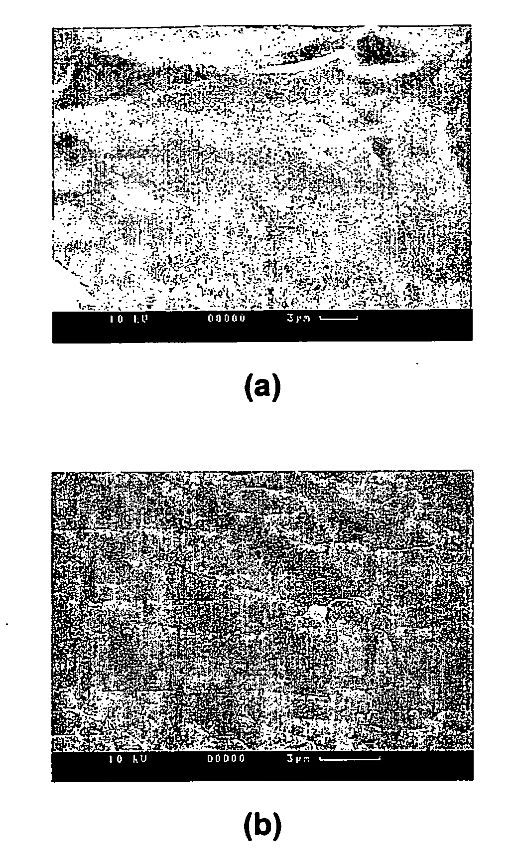
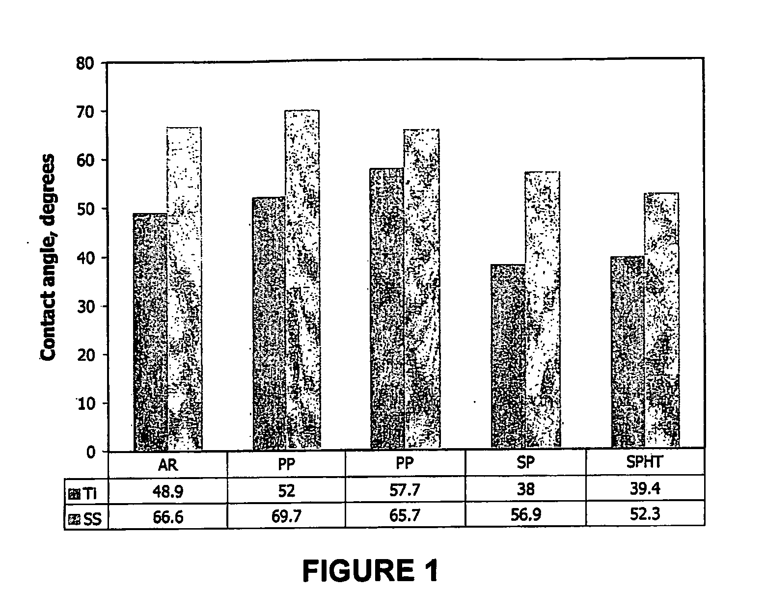
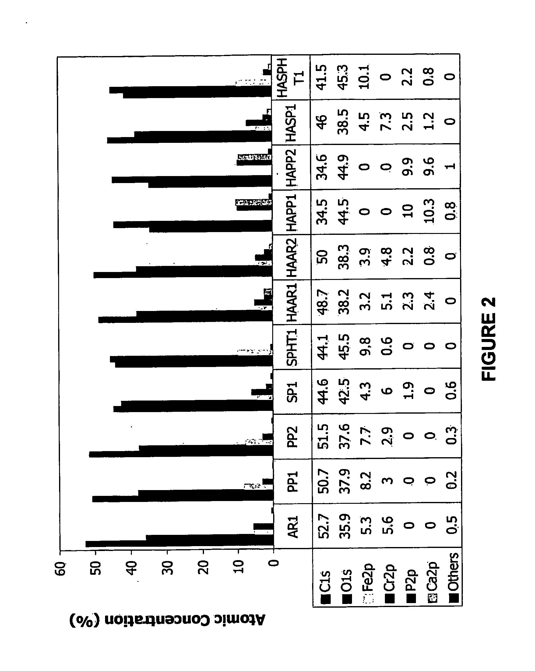
[0070]Commercially available titanium or stainless steel self-tapping cortex screws, 3.5 mm diameter and 30 mm long were treated using the methods of the invention and were compared with untreated samples and also with samples that had been treated using published methods. Brief sample preparation details are:[0071]AR (As-Received) samples: titanium and stainless steel screws as-received, in polyethylene bags.[0072]PP (Plasma Processed) samples: Using the methods of the present invention, the samples were subjected to a two-step plasma treatment using H2O2 as the reactive hydroxylating oxidant species (5 min air plasma, followed by 5 min air / 30% H2O2 plasma). The system used to prepare these samples was a Harrick radio frequency plasma unit (model PDC-32G). The flow rates of air were controlled by using two mass flow controllers (MFCs). The flow rate range of the MFCs was 2-100 sccm based ...

example 2

Treatment of Implant Substrates with Organosilanes Followed by Plasma Treatment with a Reactive Hydoxylating Oxidant Species

[0094]Titanium sheet (99.7% purity) and tetraethoxysilane (TEOS) (98% purity) were purchased from Aldrich Chemical Co. and hydrogen peroxide (H2O2) 30% (Univar) analytical reagent was obtained from APS Finechem (Australia). The titanium substrates were prepared by cuffing the 0.25 mm thick sheet into 10×10 mm2 pieces. Prior to plasma deposition, the metal pieces were rinsed in 5 wt. % KOH solution followed by a Milli-Q water wash, then immersed in a warm solution containing 1.6 wt. % HF, 33 wt % HNO3 for 2-3 min followed by copious Milli-Q water rinse.

[0095]As mentioned above, the technique of plasma assisted chemical vapour deposition (PACVD) was employed for preparing thin-film coatings of hydroxylated silica onto titanium substrates. The configuration and use of a PACVD apparatus is known to a person skilled in the art. A radio frequency (13.56 MHz) plasma r...


PUM

PropertyMeasurementUnit
pressureaaaaaaaaaa
thicknessaaaaaaaaaa
thicknessaaaaaaaaaa

Abstract

A method of forming a bioactive coating on a biomedical implant, the method including the step of contacting at least part of the surface of the implant prior to implantation with a plasma gas containing a reactive hydroxylating oxidant species. The step of contacting the surface of the implant prior to implantation with a plasma gas containing a reactive hydroxylating oxidant species may be preceded by a step of exposing the implant to an organosilane or organosilicate species to form a silica coating on at least part of the implant prior to contact with the oxidant species.

Description

FIELD OF THE INVENTION[0001]The present invention relates to methods that can be used to modify the surfaces of a metallic biomedical implant such as an artificial bone, artificial joint or an artificial tooth root used in reconstructive surgery, to accelerate or improve in situ fixation of the implant to bone. The invention also relates to biomedical implants that are produced using the methods of the invention.BACKGROUND OF THE INVENTION[0002]Medical and dental implants (‘biomedical implants’) such as hip, knee, dental and other bone implants are widely used in restorative surgery. For example, artificial joint implants are widely used in the field of orthopedics and artificial tooth root implants are attracting much attention in the field of dentistry. The implants are fixed in bone either by bonding to the bone by means of a suitable adhesive resin or by making use of the regrowth of a patient's bone tissue to fix the implant to the existing bone structure. The latter method is ...

Claims


Application Information

Patent Timeline
Patent Type & Authority Applications(United States)
IPC IPC(8): A61F2/28B05D1/00A61F2/00A61L27/04A61L27/50
CPCA61F2250/0067A61L2420/02A61L27/50A61L27/04
Inventor KUMAR, SUNILZHANG, HAILONGSIMPSON, DARREN JOHNSMART, ROGER ST. CLAIR
Owner UNIV OF SOUTH AUSTRALIA
Why Eureka
Try Eureka