Eureka AIR delivers breakthrough ideas for toughest innovation challenges, trusted by R&D personnel around the world.

Prevention of Cell Proliferation by Inhibiting Myosin Light Chain Kinase

a technology of myosin light chain kinase and cell proliferation, which is applied in the direction of nitro compound active ingredients, drug compositions, peptides, etc., can solve the problems of inability to treat tumors located in other areas such as the backbone, inability to control cell growth, and inability to use in the treatment of disseminated neoplastic conditions such as leukemia, so as to reduce the number of tumor cells, inhibit growth or proliferation, and reduce mlc-p

Inactive Publication Date: 2008-04-03
THE BOARD OF TRUSTEES OF THE UNIV OF ILLINOIS
View PDF0 Cites 7 Cited by
  • Summary
  • Abstract
  • Description
  • Claims
  • Application Information

AI Technical Summary

Benefits of technology

[0023]In a further embodiment, the present invention is a method of inhibiting growth or proliferation of, or inducing reduction in the number of tumor cells in a subject, comprising administering to the subject at least one cytotoxic agent and at least one myosin light chain kinase (MLCK) inhibitor, in an amount which, together, is effective to cause a reduction in MLC-P and inhibit growth or proliferation of the tumor cells.
[0024]Also, in another embodiment, the present invention is a method of inhibiting unwanted growth or proliferation of, or reducing the number of tumor cells in a human subject. The method includes administering to the human subject at least one cytotoxic agent and at least one MLCK inhibitor, in an amount, which together, is effective to cause a reduction in MLC-P and reduce or inhibit the growth or proliferation of the established tumor, induce cell death of the established tumors, or to reduce the size of the established tumors.
[0025]In yet another embodiment, the present invention is a method of inhibiting unwanted proliferation of, or reducing the size of, an established tumor in a subject. The method comprises administering to the subject at least one cytotoxic agent and at least one MLCK inhibitor, in an amount, which together, is effective to cause a reduction in MLC-P and reduce or inhibit the growth or proliferation of the established tumor, induce cell death of the established tumors, or to reduce the size of the established tumors. The established tumor is breast cancer, prostate cancer, lung carcinoma, renal cell carcinoma, glioma, melanoma, chemotherapy resistant tumors or metastatic tumors.

Problems solved by technology

Suppressive genes are growth regulatory genes, which upon mutation, can no longer control cell growth.
The products of transforming genes cause inappropriate cell growth.
While surgery is sometimes effective in removing tumors located at certain sites, for example, in the breast, colon, and skin, it cannot be used in the treatment of tumors located in other areas, such as the backbone, nor in the treatment of disseminated neoplastic conditions such as leukemia.
Chemotherapy-induced side effects significantly impact the quality of life of the patient and may dramatically influence patient compliance with treatment.
Additionally, adverse side effects associated with chemotherapeutic agents are generally the major dose-limiting toxicity (DLT) in the administration of these drugs.
Many of these chemotherapy-induced side effects if severe, may lead to hospitalization, or require treatment with analgesics for the treatment of pain.
As set forth above, untreated cancer may result in severe consequences, including death.

Method used


Image

  • Prevention of Cell Proliferation by Inhibiting Myosin Light Chain Kinase
  • Prevention of Cell Proliferation by Inhibiting Myosin Light Chain Kinase
  • Prevention of Cell Proliferation by Inhibiting Myosin Light Chain Kinase

Examples


example 1

Myosin Light Chain Dephosphorylation and Apoptosis

[0183]A. Studies with Actinomycin D

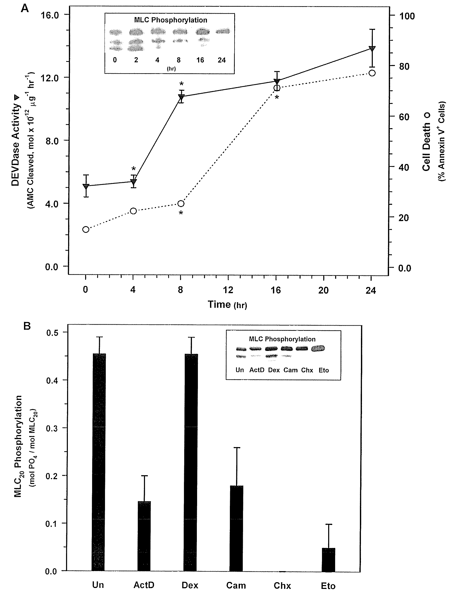
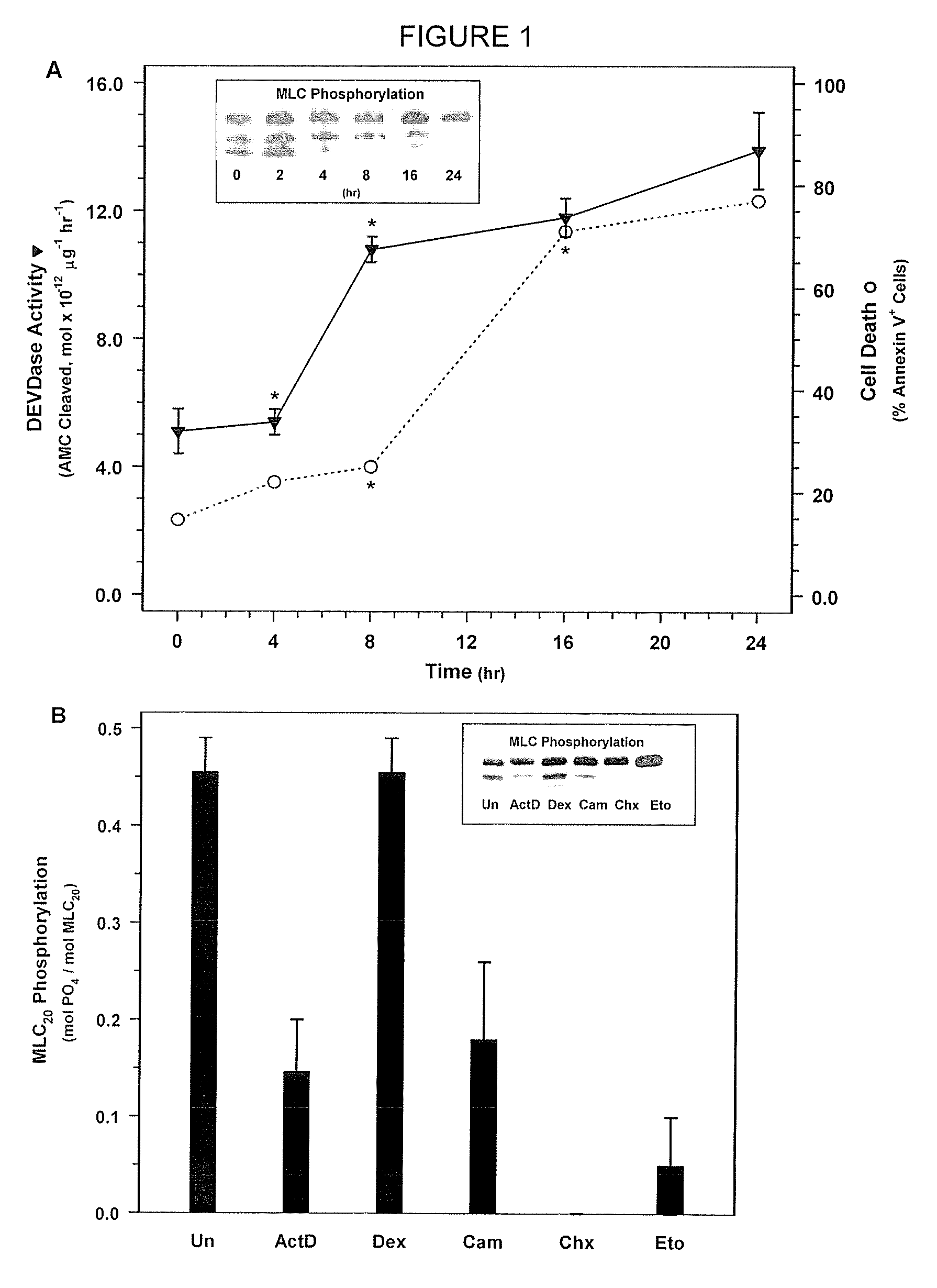
[0184]NIH 3T3 fibroblasts (ATCC Accession No. CRL-1658) were treated with actinomycin D, which is a compound known to rapidly induce cell death, to determine its effect on MLC-P.

[0185]NIH 3T3 fibroblasts were obtained from ATCC and were grown in Dulbecco's Modified Eagle Medium (DMEM) (Gibco BRL, Gaithersburg, Md.) supplemented with 10% FBS and 1% penicillin and streptomycin in a 37° C. incubator. 3T3 cells were treated with 500 nM actinomycin D in DMEM containing 0.5% FBS without antibiotics for the 0, 2, 4, 8, 16, or 24 hours. Cells were then washed with phosphate buffered saline (PBS). Floating cells were collected by centrifugation and combined with attached cells that were harvested by trypsinization. MLC20 phosphorylation was quantified be Western blotting with an anti-MLC20 antibody (FIG. 1). In short, the harvested cells were treated with ice cold 10% TCA, 10 mM DTT to precipitate the cellul...

example 2

MLC-DP Triggers Cell Death

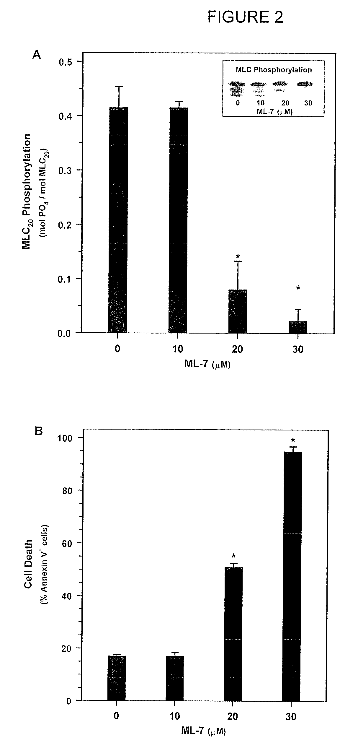
[0190]Next, in order to determine whether MLC-DP can trigger cell death, SMC were treated with ML-7 or KT5926, two inhibitors of MLCK (Nakanishi et al., 1990; Garcia, 1998; and Bain et al., 2003). Treatment with increasing doses of ML-7 (0, 10, 20, and 30 μM concentrations) for 16 hours resulted in a dose-dependent decrease in MLC20 phosphorylation (FIG. 2A) and a corresponding increase in cell death (FIG. 2B). Similarly, treatment with KT5926 resulted in cell death, including genome digestion, in NIH 3T3 cells (data not shown). As is the case with non-muscle cells induced with actinomycin D, MLC-DP was an early event in cell death in SMC treated with 20 μM ML-7 (FIG. 3A). Thus, MLC20 dephosphorylation precedes the onset of apoptosis and inhibiting MLCK appears to be sufficient to induce cell death.

example 3

MLCK Inhibitory Antibody Triggers Apoptosis

[0191]The role of MLC-DP in apoptosis was investigated further by microinjecting SMC with an affinity purified, inhibitory antibody to MLCK (de Lanerolle, 1981). An affinity purified, goat anti-human IgG was used as a control. Each antibody was mixed separately with either rhodamine-labeled dextran or fluorescein-labeled dextran as an injection marker. Results were mean±SEM for 3 experiments (*p value <0.01 using a paired T test). FIG. 3B shows confocal images of cells microinjected with a control antibody or an affinity-purified inhibitory antibody to MLCK immediately following microinjection (Panels A, B, E, F) and 4 hrs later (C, D, G, H). The control antibody (affinity-purified goat anti-human IgG) was mixed with FITC-labeled dextran prior to injection and the MLCK antibody was mixed with rhodamine-labeled dextran.

[0192]Three to four hours after injection, the cells were scored for morphological markers of cell death without prior knowl...


PUM

PropertyMeasurementUnit
pHaaaaaaaaaa
pHaaaaaaaaaa
emission wavelengthaaaaaaaaaa

Abstract

Methods of using compounds having certain inhibitory effect to treat cancer are disclosed. More specifically, methods of using myosin light chain kinase inhibitor (MLCK) to treat cancer are disclosed. MLCK inhibitors may cause reduction in MLC-P and induce apoptosis in neoplastic cells and prevent and or inhibit the tumor growth.

Description

STATEMENT OF RELATED CASES[0001]This application claims priority to U.S. Provisional Application Ser. No. 60 / 822,349, filed Aug. 14, 2006, the entirety of which is incorporated by reference herein.FEDERALLY SPONSORED RESEARCH OR DEVELOPMENT[0002]The subject matter of this application was funded by the National Institute of Health (Grant nos. NIH HL 59618 and NIH HL 0241 1). The government may have certain rights in this invention. Any opinions, findings, and conclusions or recommendations expressed in this publication do not necessarily reflect the views of the National Institutes of Health.BACKGROUND OF THE INVENTION[0003]1. Technical Field of the Invention[0004]The present invention relates to the methods of treating cancer and other proliferative diseases. More specifically, the present invention relates to administering compounds having certain inhibitory effects to prevent or inhibit tumor growth.[0005]2. Background of the Related Art[0006]Cancer is one of the leading causes of...

Claims


Application Information

Patent Timeline
Patent Type & Authority Applications(United States)
IPC IPC(8): A61K31/551A61K31/00A61K31/04A61K31/05A61K31/265A61K31/282A61K31/337A61K31/70A61K38/00C12Q1/02A61P35/00A61K38/16A61K33/24A61K31/566A61K31/4164A61K31/44A61K31/47A61K31/497A61K31/505
CPCA61K31/00A61K38/00A61K31/05A61K31/265A61K31/282A61K31/337A61K31/4164A61K31/44A61K31/47A61K31/497A61K31/505A61K31/551A61K31/566A61K31/70A61K45/06G01N33/5011G01N2510/00A61K31/04A61P35/00
Inventor DE LANEROLLE, PRIMAL
Owner THE BOARD OF TRUSTEES OF THE UNIV OF ILLINOIS
Why Eureka
Eureka Blog
Learn More