Coatings for implantable medical devices for liposome delivery

Inactive Publication Date: 2008-07-31
MIV SCI HLDG
View PDF38 Cites 46 Cited by
  • Summary
  • Abstract
  • Description
  • Claims
  • Application Information

AI Technical Summary

Benefits of technology

[0008]One embodiment provides a stent, comprising at least one coating covering at least a portion thereof, the at least

Problems solved by technology

Stents reduce but do not eliminate restenosis.
Recently it has been found that both of these stents, though effective at preventing restenosis, cause potentially fatal thromboses (clots) months or years after implantation.
This is consistent with the possible toxicity of the retained drugs or non-degradable polymer.
T

Method used


Image

  • Coatings for implantable medical devices for liposome delivery
  • Coatings for implantable medical devices for liposome delivery
  • Coatings for implantable medical devices for liposome delivery

Examples


example 1

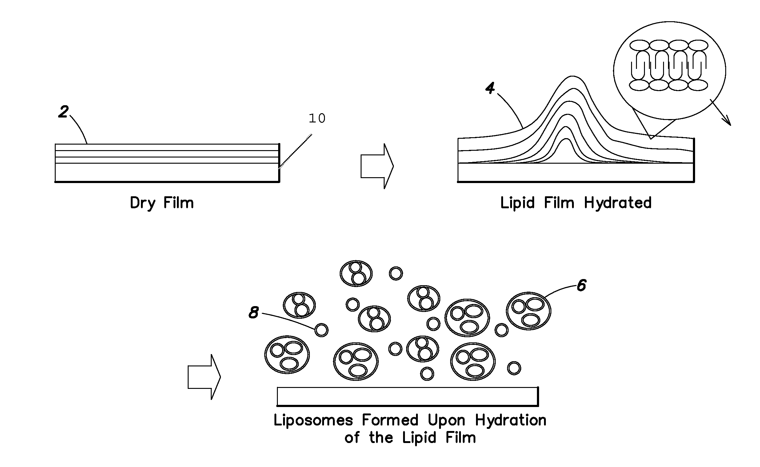
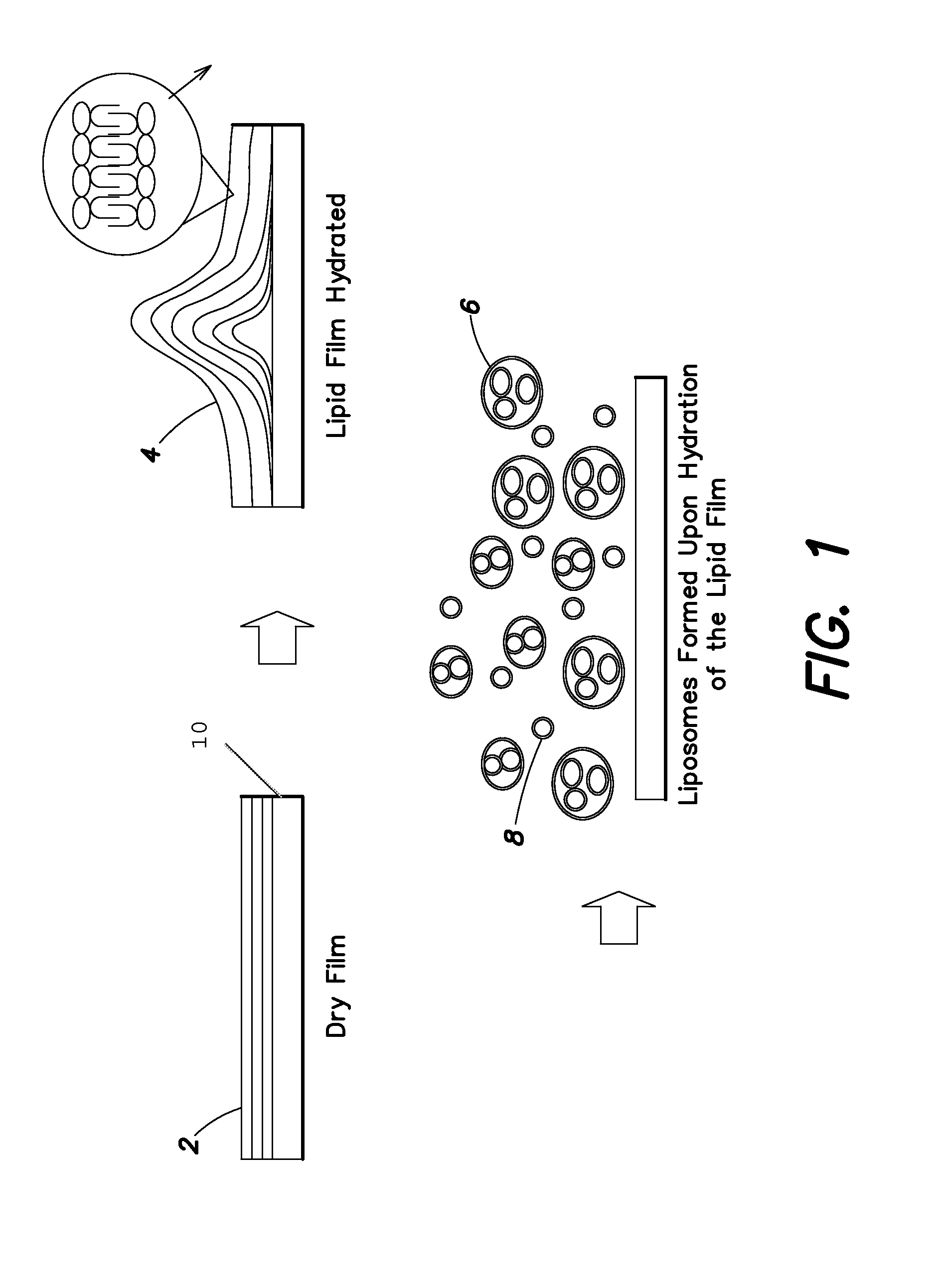
[0177]This Example describes a method for producing a dry phospholipid film. Liposome formation from the film in aqueous solution is observed optically. L-α-Phosphatidylcholine (PC, from soybean) and cholesterol were dissolved in dichloromethane. Paclitaxel (PTX), as a model hydrophobic drug, was added to this solution to produce Formulation B. The weight percent of cholesterol in Formulation B is 10% of the total lipid. The precise amounts of various components are listed in Table 1.

TABLE 1Composition of Formulation BIngredientsFormulation BPC (g)0.18Cholesterol (g)0.02Paclitaxel (g)0.07DCM (mL)10Total of solid0.27phase (g)

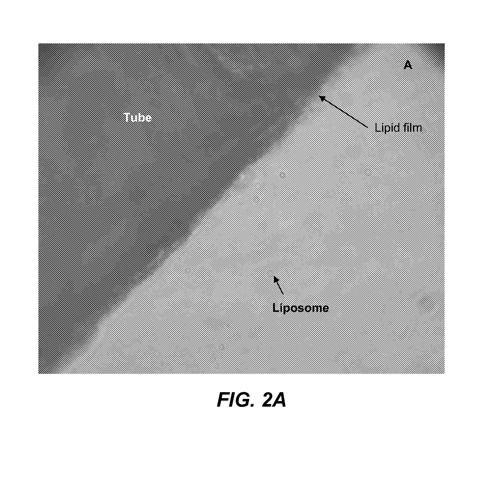
[0178]Formulation B was sprayed on a stainless tube and the tube was placed in a vacuum oven (Napco model 5831, Thermo Electron Corporation) for 12 hours at 30 in. Hg to remove solvent at room temperature. The tube was placed in 1 ml of phosphate buffer solution (PBS). Optical micrographs were obtained from the coating at different time intervals using an inverte...

example 2

[0179]This Example describes the preparation of a lipid film comprising a hydrophobic drug and the ability to tailor the amount of liposome formation by changing the amounts of lipid in the dry film. Two different formulations (Formulations A and B) were prepared comprising a mixture of lipids L-α-phosphatidylcholine (PC, from soybean) and cholesterol dissolved in dichloromethane. The weight percent of cholesterol in Formulation A was 30% of the total lipid and in Formulation B is 10%, and the precise amounts are listed in Table 2. Paclitaxel (PTX), as a model hydrophobic drug, was added to this solution. One hundred μL of the above solution added to a round bottom tube and was dried under vacuum (30 inches Hg) for 12 hours in order to remove solvents, simulating the formation of a lipid film on the surface of a substrate.

TABLE 2Composition of Formulations A and BIngredientsFormulation AFormulation BPC (g)0.140.18Cholesterol (g)0.060.02Paclitaxel (g)0.070.07DCM (mL)55Total of solid0...

example 3

[0186]This Example demonstrates the formation of a lipid film comprising a hydrophilic drug via an emulsion method.

[0187]Lecithin (0.14 g) and cholesterol (0.06 g) are dissolved in 9 mL of dichloromethane (solution 1). Imatinib mesylate (0.0641 g) is dissolved in 200 μL of distilled water (solution 2). Solution 2 is added drop-wise into solution 1 to form an (emulsion 1). Ethanol (2 mL) is added to emulsion 1 to obtain a clear emulsion. The emulsion is then applied on the surface of the medical device.


PUM

PropertyMeasurementUnit
Fractionaaaaaaaaaa
Thicknessaaaaaaaaaa
Thicknessaaaaaaaaaa

Abstract

Disclosed herein are medical devices, such as implantable medical devices (e.g., stents), comprising at least one coating covering at least a portion of the device comprising dry film. The dry film comprises at least one lipid bilayer and at least one pharmaceutically effective agent. Upon exposure to an aqueous fluid, liposomes are released comprising lipids from the dry film encapsulating the pharmaceutically effective agent. The film can contact the device directly or can be coated on a substrate, such as a ceramic.

Description

RELATED APPLICATIONS[0001]This application claims the benefit of priority under 35 U.S.C. § 119(e) to U.S. Provisional Appl. No. 60 / 876,829, filed Dec. 22, 2006, U.S. Prov. App. No. 60 / 942,565, filed Jun. 7, 2007, and U.S. Prov. App. No. 60 / 981,245, filed Oct. 19, 2007, the disclosures of which are incorporated herein by reference.FIELD OF THE INVENTION[0002]Disclosed herein are coatings for medical devices, such as implantable medical devices (e.g., stents), and processes for making the same. The coating comprises and a film containing a lipid bilayer and one or more therapeutic agents.BACKGROUND OF THE INVENTION[0003]Implantable medical devices are used in a wide range of applications including bone and dental replacements and materials, vascular grafts, shunts and stents, and implants designed solely for prolonged release of drugs. The devices may be made of metals, alloys, polymers or ceramics.[0004]Arterial stents have been used for many years to prevent restenosis after balloo...

Claims


Application Information

Patent Timeline
IPC IPC(8): A61F2/04A61K31/337A61K31/506A61P35/00A61P29/00A61P9/00
CPCA61K9/127A61L31/08A61L2300/62A61L2300/00A61L31/16A61P9/00A61P29/00A61P35/00
Inventor HAKIMI-MEHR, DORNALANDY, MARKTSUI, MANUSBUDZYNSKI, VLADTSETKOV, ALEKSY
Owner MIV SCI HLDG
Why Eureka
Try Eureka