Compositions and Method for Multimodal Imaging

a multi-modal imaging and composition technology, applied in the field of medical imaging, can solve the problems of limited size, no available non-invasive methods for distinguishing between lymph nodes, and used in image-guidance applications, and achieve the effect of prolonging contrast imaging

Active Publication Date: 2008-08-28
UNIV HEALTH NETWORK
View PDF6 Cites 24 Cited by
  • Summary
  • Abstract
  • Description
  • Claims
  • Application Information

AI Technical Summary

Benefits of technology

[0006]In a broad aspect of the invention there is provided signal modifying compositions for medical imaging comprising a carrier and signal modifying agents specific for two or more imaging modalities. In a preferred embodiment the compositions are characterized by retention efficiency, with respect of the signal modifying agents, that enables prolonged contrast imaging without depletion of the signal modifying agent from the carrier. The carriers of the present invention are lipid based or polymer based the physico-chemical properties of which can be modified to entrap or chelate different signal modifying agents and mixtures thereof and to target specific organs or tumors within a mammal.
[0017]By retaining or retention efficiency it is meant the capacity of a carrier to prevent leakage of a signal modifier agent out of the carrier.

Problems solved by technology

The lack of development in this area is likely due to challenges presented by the fact that the distinct imaging modalities have different sensitivities for different signal modifying agents (Krause W.
However, due to their low molecular weight, these agents only remain in the vascular system for a short period of time, exhibit rapid dynamic distribution changes in different organs and are excreted quickly.
Furthermore, the short in vivo residence time of these agents would impose limitations on the size of the anatomic region that could be imaged optimally and would exclude them from being used in image-guidance applications due to their inability to provide prolonged contrast enhancement for the entire course of treatment (Saeed M, Wendland M F, Higgins C B.
Until recently, there were no available non-invasive methods for distinguishing between lymph nodes enlarged due to the presence of metastatic cancer cells and nodes enlarged due to inflammation, or for identifying cancerous nodes of normal size.
However no delivery system has been developed for prolonged co-localization in vivo of two or more signal modifying agent for multiple medical imaging.

Method used


Image

  • Compositions and Method for Multimodal Imaging
  • Compositions and Method for Multimodal Imaging
  • Compositions and Method for Multimodal Imaging

Examples


example 1

[0063]Radionuclide imaging in accordance with the method and composition described above may involve incorporation of derivatized lipids that can chelate the radiometals 99mTc and 111In for SPECT imaging and 64Cu for PET. These radionuclides are readily available from a generator system (99Mo / 99mTc; Bristol-Myers-Squibb) or can be purchased from MDS-Nordion Inc. (111In and 64Cu). PE lipid can be derivatized at the headgroup with HYNIC for labeling with 99mTc; DTPA for labeling with 111In; or with TETA for labeling with 64Cu. These bifunctional chelators are all commercially available from Macrocyclics Inc. Unilamellar liposomes can be prepared using established methods based on high-pressure extrusion and sonication. The labeled liposomes can be formed from the newly synthesized chelator-modified PE and the mixture of lipids originally employed in the liposome formulation. Following preparation, liposomes containing the chelator-modified PE lipid can be incubated with 99mTc, 111In, ...

example 2

Methods and Materials

Materials

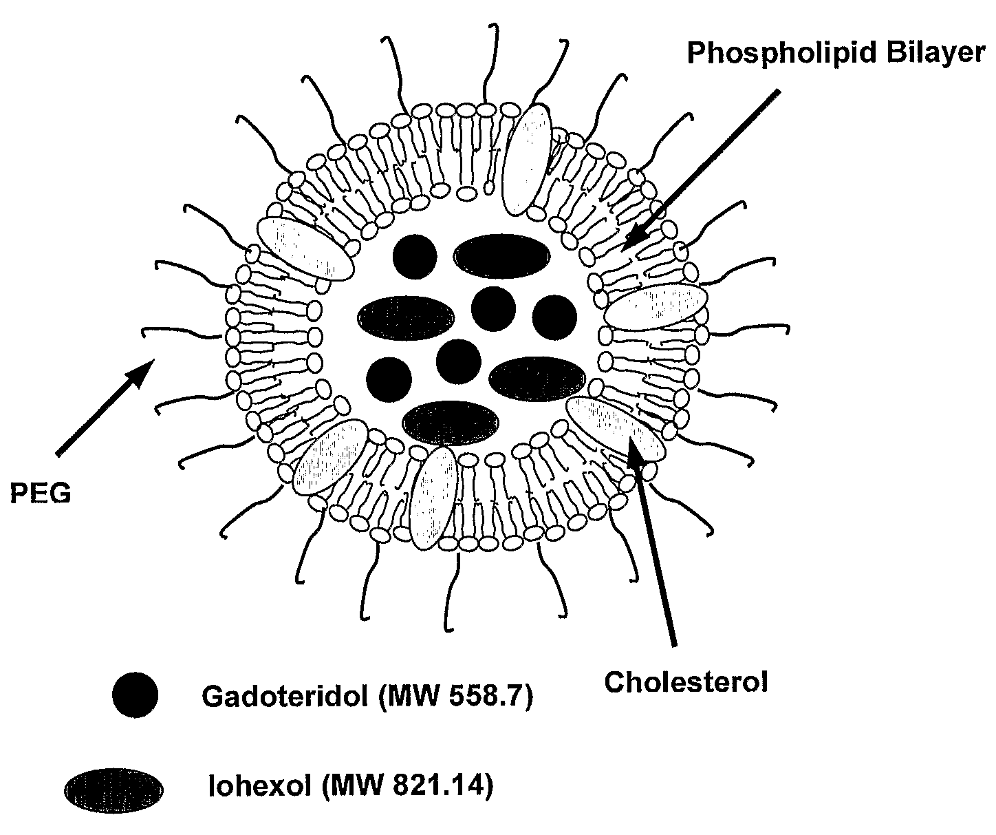
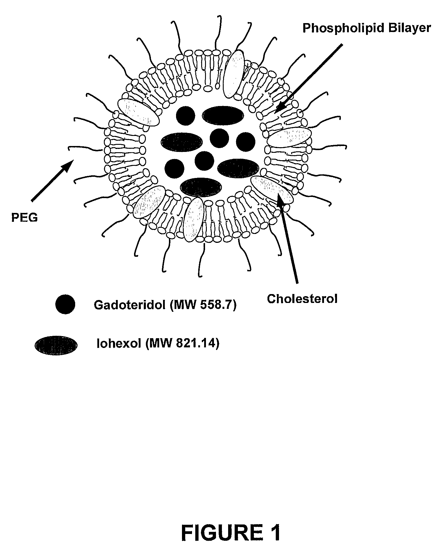
[0064]The components of liposomes: 1,2-Dipalmitoyl-sn-Glycero-3-Phosphocholine (DPPC, M.W. 734), Cholesterol (CH, M.W. 387) and 1,2-Distearoyl-sn-Glycero-3-Phosphoethanolamine-N-[Poly(ethylene glycol)2000] (PEG2000DSPE, M.W. 2774) were purchased from Northern Lipids Inc. (Vancouver, British Columbia, Canada). The CT signal modifying agent, Omnipaque® was obtained from Nycomed Imaging AS, Oslo, Norway. Omnipaque® (300 mg / mL of Iodine) contains iohexol (M.W. 821.14), an iodinated, water-soluble, non-ionic monomeric contrast medium. The MR signal modifying agent used was ProHance® from Bracco Diagnostics Inc. (Princeton, N.J., USA). ProHance® (78.6 mg / mL of gadolinium) contains gadoteridol (M.W. 558.7), a non-ionic gadolinium complex of 10-(2-hydroxy-propyl)-1,4,7,10-tetraazacyclododecane-1,4,7-triacetic acid.

Preparation of Liposome Formulations

[0065]Lipid mixtures (200 mmol / L) of DPPC, cholesterol and PEG2000DSPE in 55:40:5 percent mole ratios were dissol...

example 3

Materials

[0086]The following lipids: 1,2-Dipalmitoyl-sn-Glycero-3-Phosphocholine (DPPC, M.W. 734), Cholesterol (CH, M.W. 387) and 1,2- Distearoyl-sn-Glycero-3-Phosphoethanolamine-N-[Poly(ethylene glycol)2000] (PEG2000DSPE, M.W. 2774) were purchased from Northern Lipids Inc. (Vancouver, British Columbia, Canada). Omnipaque® was obtained from Nycomed Imaging AS, Oslo, Norway. Omnipaque® (300 mg / mL of iodine) contains iohexol (M.W. 821.14), an iodinated, water-soluble, non-ionic monomeric contrast medium. ProHance® from Bracco Diagnostics Inc. (Princeton, N.J., USA). ProHance® (78.6 mg / mL of gadolinium) contains gadoteridol (M.W. 558.7), a non-ionic gadolinium complex of 10-(2-hydroxy-propyl)-1,4,7,10-tetraazacyclododecane-1,4,7-triacetic acid.

Liposome Preparation

[0087]200 mmol / L of the DPPC, cholesterol and PEG2000DSPE (55:40:5 mole ratio) mixture was dissolved in ethanol at 70° C. and then hydrated with Omnipaque® and Prohance®. The total ethanol content was 10%vol. The resulting mul...


PUM


Abstract

Provided are signal modifying compositions for medical imaging comprising a carrier and two or more signal modifying agents specific for two or more imaging modalities. The compositions are characterized by retention efficiency, with respect to the signal modifying agents, which enables prolonged contrast imaging without significant depletion of the signal modifying agents from the carrier. The carriers of the present invention are lipid based or polymer based, the physico-chemical properties of which can be modified to entrap or chelate different signal modifying agents and mixtures thereof and to target specific organs or tumors or tissues within a mammal.

Description

FIELD OF THE INVENTION[0001]This invention relates to the field of medical imaging and more specifically to the use of signal modifying agents in medical imaging.BACKGROUND OF THE INVENTION[0002]In recent years significant effort has been devoted to the development of multimodality imaging. Since each medical imaging modality has unique strengths and limitations, it is often through the compound use of multiple modalities that the complete assessment of a patient is achieved. Interest in the area of multimodality imaging has also been prompted by the realization that such techniques offer much more sophisticated characterization of the morphology and physiology of tissues and organs, and that confidence gained in the accurate correspondence or registration of different modalities greatly enhances their value (Barillot C, Lemoine D, Le Briquer L, et al. Eur J Radiol 1993; 17:22-27.). Consequently, this improved value of imaging will ultimately allow for advances in diagnosis and eval...

Claims


Application Information

Patent Timeline
Patent Type & Authority Applications(United States)
IPC IPC(8): A61K49/00A61K51/00
CPCA61B6/00A61K49/04A61B5/055A61K9/127A61B8/00A61B6/5247A61B8/5238A61K9/1271A61K49/0002A61B5/415A61K49/1812A61K51/1234A61B5/418A61K49/0466
Inventor JAFFRAY, DAVIDALLEN, CHRISTINEZHENG, JINZIREILLY, RAYMOND MATTHEWPERKINS, GREGORY JASON
Owner UNIV HEALTH NETWORK
Why Eureka
Try Eureka