
Method for Drug Screening and Characterization by Calcium Flux

a technology of calcium flux and drug discovery, applied in the field of drug discovery, can solve the problems of low throughput of the patch clamp technique as a general method of pharmacological screening, inability to easily automate the technique, and extensive analysis by skilled electrophysiologists

Inactive Publication Date: 2008-10-16
CEDARS SINAI MEDICAL CENT
View PDF9 Cites 4 Cited by
  • Summary
  • Abstract
  • Description
  • Claims
  • Application Information

AI Technical Summary

Problems solved by technology

However, a major limitation of the patch clamp technique as a general method in pharmacological screening is its low throughput.
Furthermore, the technique is not easily amenable to automation, and produces complex results that require extensive analysis by skilled electrophysiologists.
However, cell populations are biologically heterogeneous, and the high spatial and temporal frequency of chemical and molecular information present within cells makes it impossible to extract high-content information from populations of cells using conventional techniques.
Indeed, conventional techniques for monitoring and analyzing, for example, ion oscillations using fluorescence has substantial drawbacks; for example, photobleaching and lack of specific information concerning individual cells.
Conventional techniques also are not fast or cost-efficient; for example, dose-dependent experiments using the patch-clamp technique typically require two days to complete.
However, these imaging methods are limited by the efficiency, photostability and toxicity of the fluorescence in the chosen system.
Other limitations of the related art will become apparent to those of skill in the art upon a reading of the specification and a study of the drawings.

Method used


Image

  • Method for Drug Screening and Characterization by Calcium Flux
  • Method for Drug Screening and Characterization by Calcium Flux
  • Method for Drug Screening and Characterization by Calcium Flux

Examples


example 1

Cell Culture

[0071]AtT-20 / D16-F2 mouse adrenocorticotropic hormone (ACTH)-secreting pituitary cells (CRL-1795; American Type Culture Collection; 2003) were grown in serum-containing low glucose Dulbecco's Modified Eagles Medium (DMEM) (obtained from Invitrogen; Carlsbad, Calif.) supplemented with 10% fetal bovine serum (obtained from Omega Scientifics; Tarzana, Calif.), with addition of 2 mM glutamine and 1% antibiotic / antimycotic (100 U / mL penicillin G sodium, 100 μg / mL streptomycin sulfate, 250 ng / mL amphotericin B (Fungisone® in 0.85% saline); obtained from Invitrogen), in 6% CO2, 37° C. humidified incubator.

[0072]For confocal imaging, cells were grown in 6 well plates on 25 mm cover slips coated with 10 μg / ml poly L-lysine (obtained from Sigma Aldrich; St. Louis, Mo.) to 70% confluence in low glucose DMEM with 0.3% Bovine Serum Albumin (BSA) and antibiotics 12 hours prior to analysis.

[0073]Ion-imaging cells were stained with Calcium Green-1 AM 488 dye (obtained from Molecular Pro...

example 2

Confocal Cytosolic Calcium Imaging

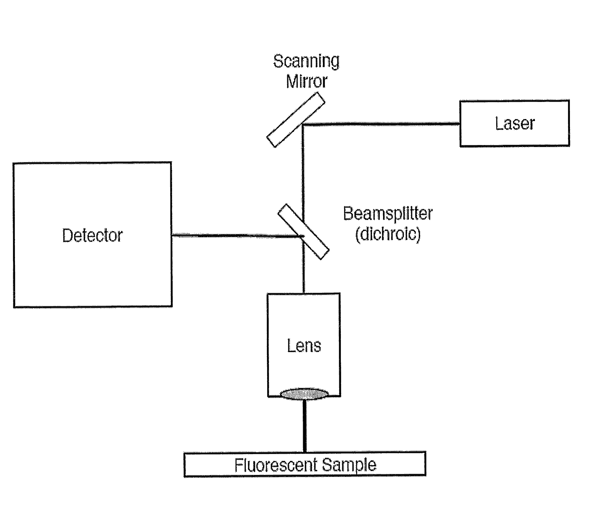
[0074]The Attofluor cell culture chamber was placed inside a PenCon temperature controlled incubator at 37° C. mounted on a DMIRB / E inverted microscope (obtained from Leica Microsystems; Wetzlar, Germany). Cells were imaged with a TCS SP confocal scanner (obtained from Leica Microsystems) using a temperature controlled 63× / 1.2 N.A. w / PlanApo water immersion objective. The digital temperature readout of the incubator was checked with a calibrated mercury thermometer to confirm the values within 0.2° C.

[0075]For excitation, the 488 nm Argon laser line was used with laser power set to minimum and 488 nm acousto-optic tunable filter (AOTF) line to 9% transmission.

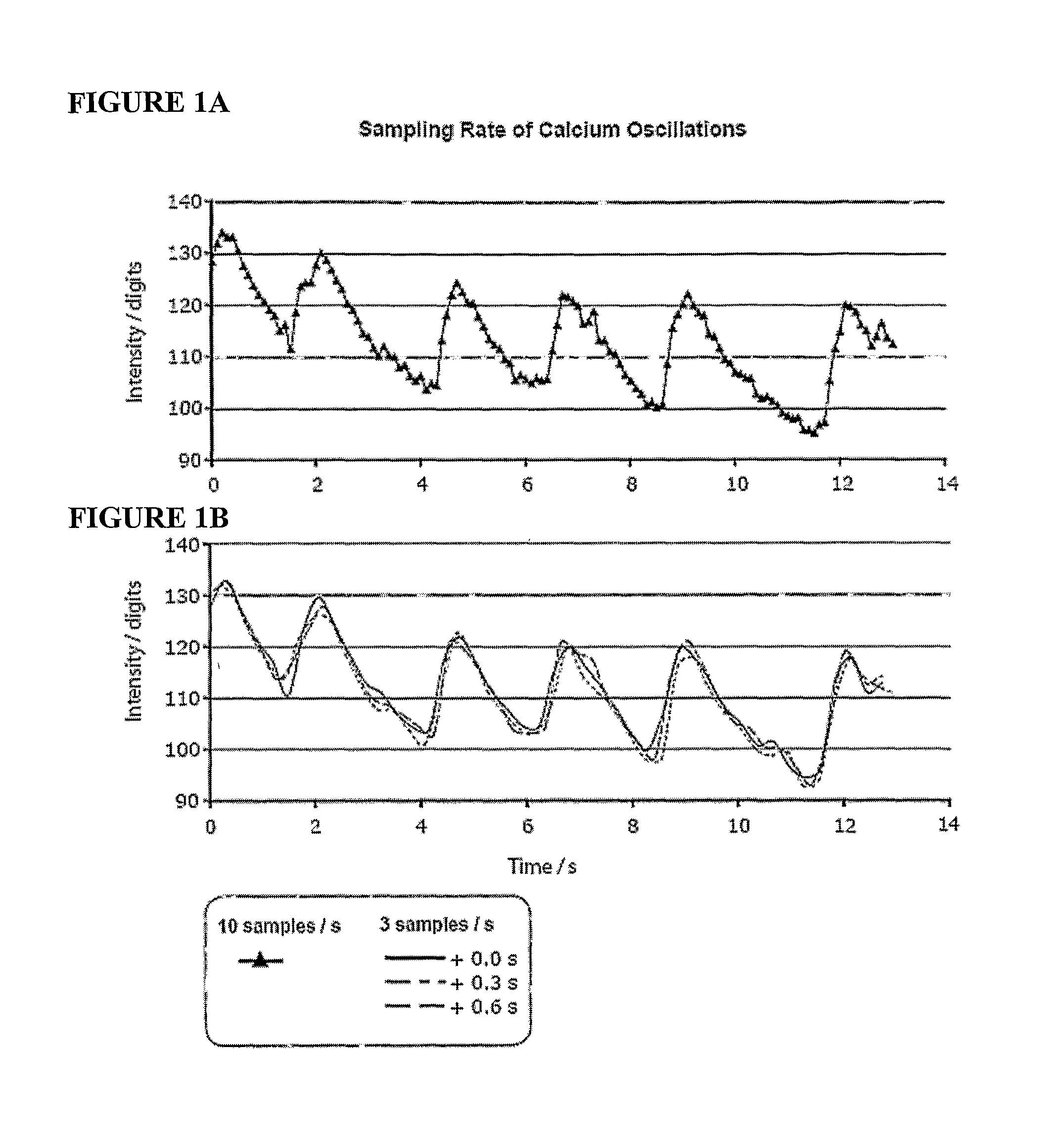
[0076]Time lapse sequences were acquired by setting the scanner to medium scan speed, bidirectional scan mode at 128 by 128 pixel resolution, resulting in a scan rate of about 3 fps (350 ms / frame). For the time resolution test, the scan rate was increased to 10 fps by changing to fast bidirecti...

example 3

Dose Response Acquisition

[0077]Cells were imaged over 5 minute time intervals for each dose. Baseline values were acquired with five 5 minute pre-treatment intervals. Consecutively, the drug of choice was added at increasing doses at one order of magnitude increments for each 5 minute interval. Total recording time was 45 minutes. Each 5 minute interval was stored as a separate time series of Tag Image File Format (TIFF) files within the duration of the 45 minute experiment.


PUM


Abstract

The instrumentation and methods described herein are based on imaging and measuring single cell dose response by fluorescent ion imaging that records live cell responses to drug doses with no apparent damage to the cells or toxic side effects. Dose response curves and other pharmacological parameters can be computed by imaging and measuring oscillation changes for each dose and each cell. Large variability of cellular responses under the same experimental conditions indicates that pharmacological benchmarks are determined by statistical distribution of cells and cellular physiology. The instrumentation and methods described herein provide a means of measuring statistical variability of cell populations for improved screening and development methods for drugs.

Description

FIELD OF THE INVENTION[0001]The present invention relates generally to the field of drug discovery; more particularly, to instrumentation and methods of analyzing ion oscillations to screen compounds.BACKGROUND OF THE INVENTION[0002]Ions move across plasma membranes and organelle membranes through channels created by proteins, which allows for the formation of concentration gradients between exterior and interior compartments. Ion channels participate in, and regulate, cellular processes as diverse as the generation and timing of action potentials, energy production, synaptic transmission, secretion of hormones and the contraction of muscles. In fact, many drugs exert their specific effects via modulation of ion channels. Examples include antiepileptic compounds like phenyloin and lamotrigine, which block voltage-dependent sodium channels in the brain, antihypertensive drugs like nifedipine and diltiazem, which block voltage-dependent calcium channels in smooth muscle cells, and sti...

Claims


Application Information

Patent Timeline
IPC IPC(8): G06K9/00C12N5/07C12N5/071
CPCG01N21/6428G01N21/6458G01N21/6486
Inventor BEN-SHLOMO, ANATMELMED, SHLOMOWAWROWSKY, KOLJA
Owner CEDARS SINAI MEDICAL CENT
Learn More