Photosensitizers and MRI Enhancers

a technology of enhancers and photosensitive materials, applied in the field of photosensitive materials and enhancers, can solve the problems of difficult preparation and purification, low selectivity of tumour tissues, and accumulation of slowly, and achieve the effect of accurately identifying malignant tissues

Inactive Publication Date: 2008-11-13
PHOTO DIAGNOSTIC DEVICES PDD
View PDF1 Cites 1 Cited by
  • Summary
  • Abstract
  • Description
  • Claims
  • Application Information

AI Technical Summary

Benefits of technology

[0052]The compound used in the first or second aspect of the present invention may be linked to a photosensitive material such as a nano-dot to enhance the luminescence and diagnostic capabilities and sensitivity for deep seated turnouts or pathology. Nano-dots, also called quantum-dots, are nanometer scale particles that are neither small molecules nor bulk solids. Their composition and small size (a few hundred to a few thousand atoms) give these dots extraordinary optical properties that can be readily customized by changing the size and composition of the dots. Quantum dots fluoresce intensely with illumination.
[0097]With the compound of the fourth aspect of the present invention, comprising the magnetic element, selectively attaching to for example tumours, the MRI more accurately identifies the malignant tissue. The compound of the fourth aspect of the present invention need not be photoactivated by illumination or sound for the MRI scan. The compound of the fourth aspect of the present invention acts as a carrier to concentrate the magnetic element in tumour or other diseased tissue, thereby making the tissue highly visible on the MRI scan in a way that is not possible with present technology.

Problems solved by technology

Most known photosensitizers have mayor drawbacks, for example, they may be difficult to prepare and purify, or they may only accumulate slowly in tumours.
However, the photosensitizers disclosed in RU-2183956 have a low selectivity for tumour tissues, a high toxicity to normal organs and tissues, and a low therapeutic photoactivity in tumour cells.
Moreover, they are chemically and photochemically unstable, but are only slowly metabolised and cleared from normal tissues.

Method used


Image

  • Photosensitizers and MRI Enhancers
  • Photosensitizers and MRI Enhancers
  • Photosensitizers and MRI Enhancers

Examples


example 1

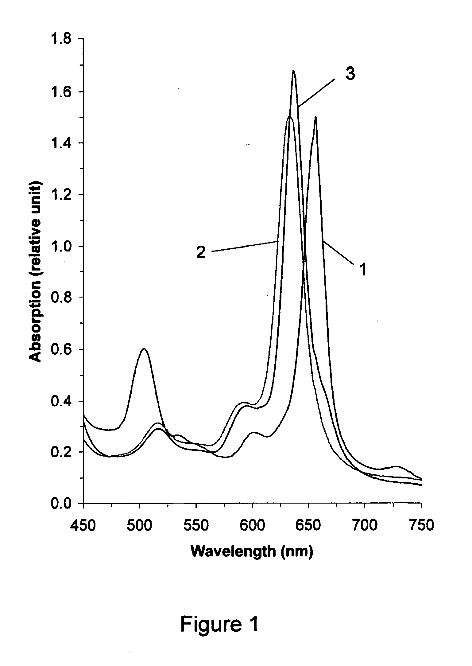
[0154]Ammonia was added to water until the pH of the solution was not less than 9. Then chlorine-e6 (1.0 g) was dissolved in the aqueous solution. An equimolar quantity of zinc acetate (0.22 g) was added and the reaction mixture was stirred for 15 minutes at about 20° C. to achieve the complex-formation reaction. The progress and completion of the reaction was monitored with the help of a spectrophotometer. On completion of the complex-formation reaction, serum humane albumin (SHA) (71 g) was added to the reaction mixture as an immobilizer. On completion of the immobilization reaction, which was monitored with a spectrophotometer, the product of the reaction, Zn-chlorine-e6 complex immobilized on SHA, was purified by dialysis.

[0155]FIG. 1 shows the long-wave region of the visible absorption spectra of (1) the starting material chlorine-e6 (λmax=656 nm), (2) Zn-chlorine-e6 complex (λmax=632 nm), and (3) Zn-chlorine-e6 complex immobilized on SHA (λmax=636 nm), all in water.

[0156]As ca...

example 2

[0158]The synthesis of immobilized Zn-chlorine-e6 was carried out as described in Example 1, except that as immobilizer polyvinylpyrrolidone (PVP) (62 g) was used instead of SHA.

[0159]As can be seen in FIG. 4, the spectral picture of the visible absorption spectra of (1) the starting material chlorine-e6 (λmax=656 nm), (2) Zn-chlorine-e6 complex (λmax=632 nm), and (3) Zn-chlorine-e6 complex immobilized on PVP (λmax=638 nm) are practically identical to the ones depicted in FIG. 1. One observes a significant 24 nm short-wave shift of the long-wave peak upon metal complex formation and a small 6 nm long-wave shift upon immobilization on polymer PVP. The medium intensity peak of chlorine-e6 at λmax=502 nm practically disappears, when forming the Zn-chlorine-e6 complex. All of these changes prove the completeness of the reactions and the purity and homogeneity of the products obtained.

example 3

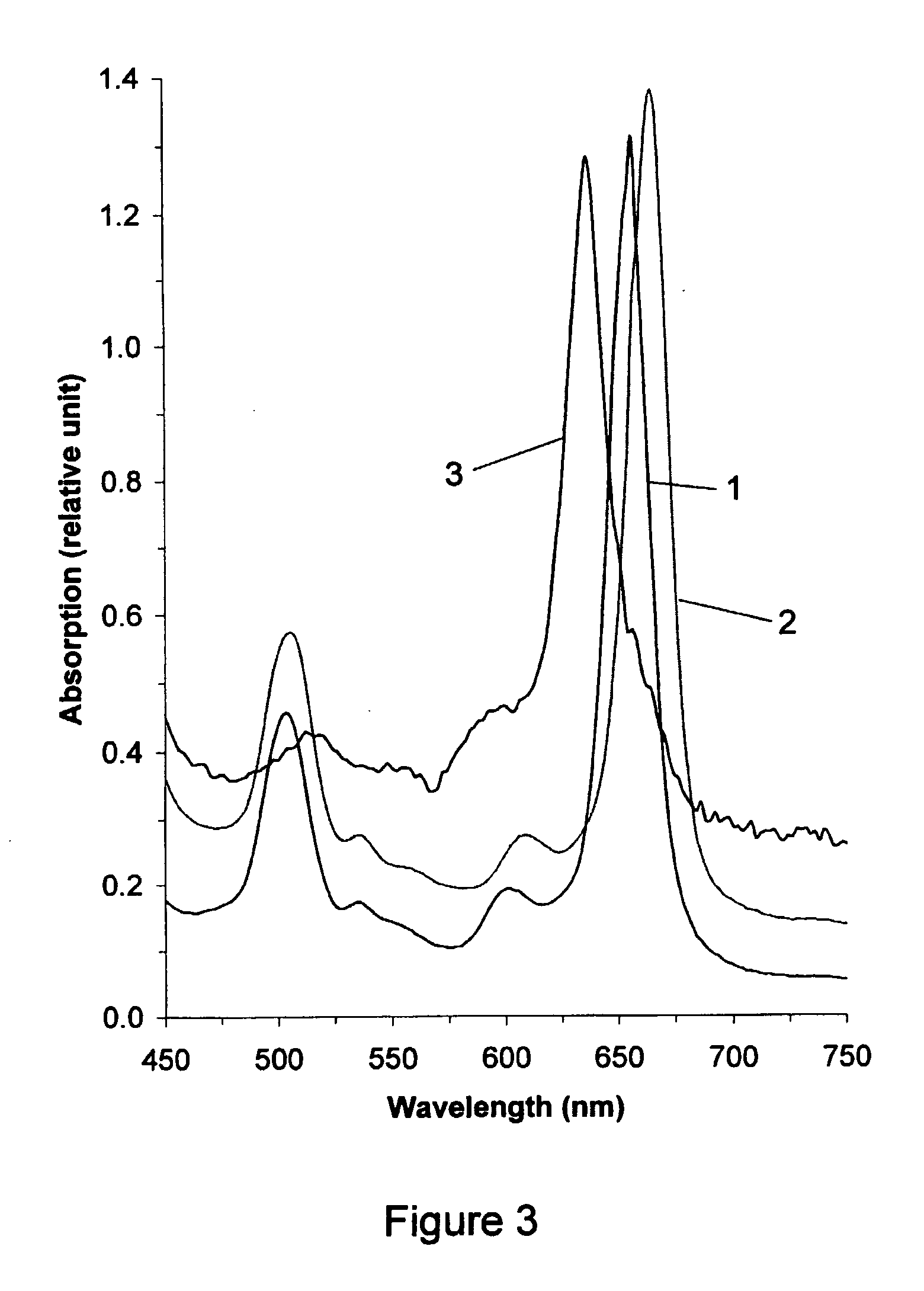
[0160]Ammonia was added to water until the pH of the solution was not less than 9. Then chlorine-e6 (1.0 g) was dissolved in the aqueous solution. An equimolar quantity of SHA (71 g) was added and the reaction mixture was stirred for 17 minutes at about 20° C. to immobilize chlorine-e6 on SHA. Then an equimolar quantity of zinc acetate (0.22 g) was added and the reaction mixture was stirred at room temperature to complex Zn into the chlorine-e6, which was monitored with a spectrophotometer. The product of the reaction, Zn-chlorine-e6 complex immobilized on SHA, was purified by dialysis.

[0161]FIG. 3 shows the long-wave region of the visible absorption spectra of (1) the starting material chlorine-e6 (λmax=656 nm), (2) chlorine-e6 immobilized on SHA (λmax=662 nm), and (3) Zn-chlorine-e6 complex immobilized on SHA (λmax=636 nm). Unlike the first method of synthesis (see Example 1), when forming chlorine-e6 immobilized on protein, first a 6 nm long-wave shift of the absorption peak occu...


PUM

PropertyMeasurementUnit
wavelengthaaaaaaaaaa

Abstract

The present invention relates to the use of a compound of formula 3 or a salt thereof for the manufacture of a medicament or phototherapeutic agent for the treatment of acne, Aids, viral hepatitis, diabetic retinopathy, infection with sars virus, coronary artery stenosis, carotid artery stenosis, intermittent claudication, Asian (chicken) flu virus, cervical dysplasia or cancer of the blood, cervix, naso-pharynx, trachea, larynx, bronchi, bronchioles, bladder, esophagus, stomach, rectum, colon, prostate, hollow organs, bile duct, ureter, kidney, uterus, vaginal or other female adnexa. The invention also relates to methods of treating these diseases.The present invention further relates to the use of a compound of formula 3 or a salt thereof for the manufacture of a photodiagnostic agent for the detection of the above diseases, as well as atherosclerosis, multiple sclerosis, diabetes, arthritis, rheumatoid arthritis, a fungal, viral, chlamydial, bacterial, nanobacterial or parasitic infectious disease, HIV, hepatitis, herpes simplex, herpes zoster, psoriasis, a cardiovascular disease, or a dermatological condition. The invention also relates to methods of detecting these diseases by photodiagnosis.The present invention further relates to a method of cold sterilising a surgical or other device, comprising the steps of: providing a compound of formula 3 or a salt thereof on the device and subjecting the device to irradiation or sound.The present invention further relates to a compound of formula 3 or a salt thereof, linked or attached to a magnetic element. Such a compound may be used as an MRI enhancer. The present invention also relates to a method of carrying out an MRI scan using such an MRI enhancer.

Description

TECHNICAL FIELD[0001]The present invention relates to the use of a compound of formula 3 or a salt thereof for the manufacture of a medicament or phototherapeutic agent for the treatment of acne, Aids, viral hepatitis, diabetic retinopathy, infection with sars virus, coronary artery stenosis, carotid artery stenosis, intermittent claudication, Asian (chicken) flu virus, cervical dysplasia or cancer of the blood, cervix, naso-pharynx, trachea, larynx, bronchi, bronchioles, bladder, esophagus, stomach, rectum, colon, prostate, hollow organs, bile duct, ureter, kidney, uterus, vaginal or other female adnexa. The invention also relates to methods of treating these diseases.[0002]The present invention further relates to the use of a compound of formula 3 or a salt thereof for the manufacture of a photodiagnostic agent for the detection of the above diseases, as well as atherosclerosis, multiple sclerosis, diabetes, arthritis, rheumatoid arthritis, a fungal, viral, chlamydial, bacterial, ...

Claims


Application Information

Patent Timeline
Patent Type & Authority Applications(United States)
IPC IPC(8): A61K49/06A61K31/555A61K49/00A61B5/055C07D209/56A61L2/18
CPCA61K49/0036A61K41/0071A61P1/16A61P15/00A61P17/00A61P17/06A61P17/10A61P19/02A61P25/00A61P27/02A61P29/00A61P31/04A61P31/10A61P31/12A61P31/16A61P31/18A61P31/22A61P33/00A61P35/00A61P35/02A61P43/00A61P7/02A61P9/00A61P9/10A61P3/10
Inventor AYER PORTER, WILLIAM H.AYER PORTER, MARGARETOVCHINNIKOV, ALEXANDER
Owner PHOTO DIAGNOSTIC DEVICES PDD
Why Eureka
Try Eureka