

Proteomic analysis of biological fluids
a biological fluid and proteome technology, applied in the field of proteome identification of biological fluids, can solve the problems of not providing information on the regulation of protein function through post-translational modifications, proteolysis or compartmentalization, and compromising the well-being of the mother and/or the fetus or newborn,
- Summary
- Abstract
- Description
- Claims
- Application Information
AI Technical Summary
Benefits of technology
Problems solved by technology
Method used
Image

Examples
example 1
General Protocols
[0127]Primate Model of Intra-Amniotic Infection
[0128]This protocol was approved by the Institutional Animal Care Utilization Committee of the Oregon National Primate Research Center, and guidelines for humane care were followed. Three pregnant rhesus monkeys (Macaca mulatia) with timed gestations were chronically catheterized as previously described (Haluska G J, et al., Temporal changes in uterine activity and prostaglandin response to RU 486 in rhesus macaques in late gestation., Am J Obstet Gynecol 157: 1487-95 (1987); and Gravett M G, et al., An experimental model for intramniotic infection and preterm labor in rhesus monkeys. Am J Obstet Gynecol 171: 1660-7 (1994)). Briefly, at approximately day 110 of gestation (term is 167 days) pregnant animals were conditioned to a jacket and tether system (Ducssay C A, et al., Simplified vest and tether system for maintenance of chronically catheterized pregnant rhesus monkeys. Lab. Anim Sci 38:343-4 (1988)). After conditi...
example 2
Identification of Proteins and Polypeptides Expressed in the Amniotic Fluid
[0164]Using the materials and methods described in Example 1, proteins and polypeptides expressed in normal and infected amniotic fluid were identified. Human and primate amniotic fluid samples (pooled and individual) were subjected to protein separation techniques (1-D, 2-D and HPLC fractionation) as described in Example 1. The separated proteins (gel bands, spots and fractions) were digested with trypsin to generate peptide pools. The peptide pools were analyzed using tandem MS to decipher their amino acid sequence and composition.
[0165]Five thousand MS spectra were selected using spectral verification programs. These spectral files were analyzed using de novo sequencing programs (LUTEFISK™, PEAKS™) to generate the amino acid sequence corresponding to each peptide. The de novo sequences generated from the peptide pool were used to search protein and DNA databases as described in Example 1.
[0166]Using homolo...
example 3
Protein Expression Profiles of Primate Amniotic Fluid Following Intrauterine Infection
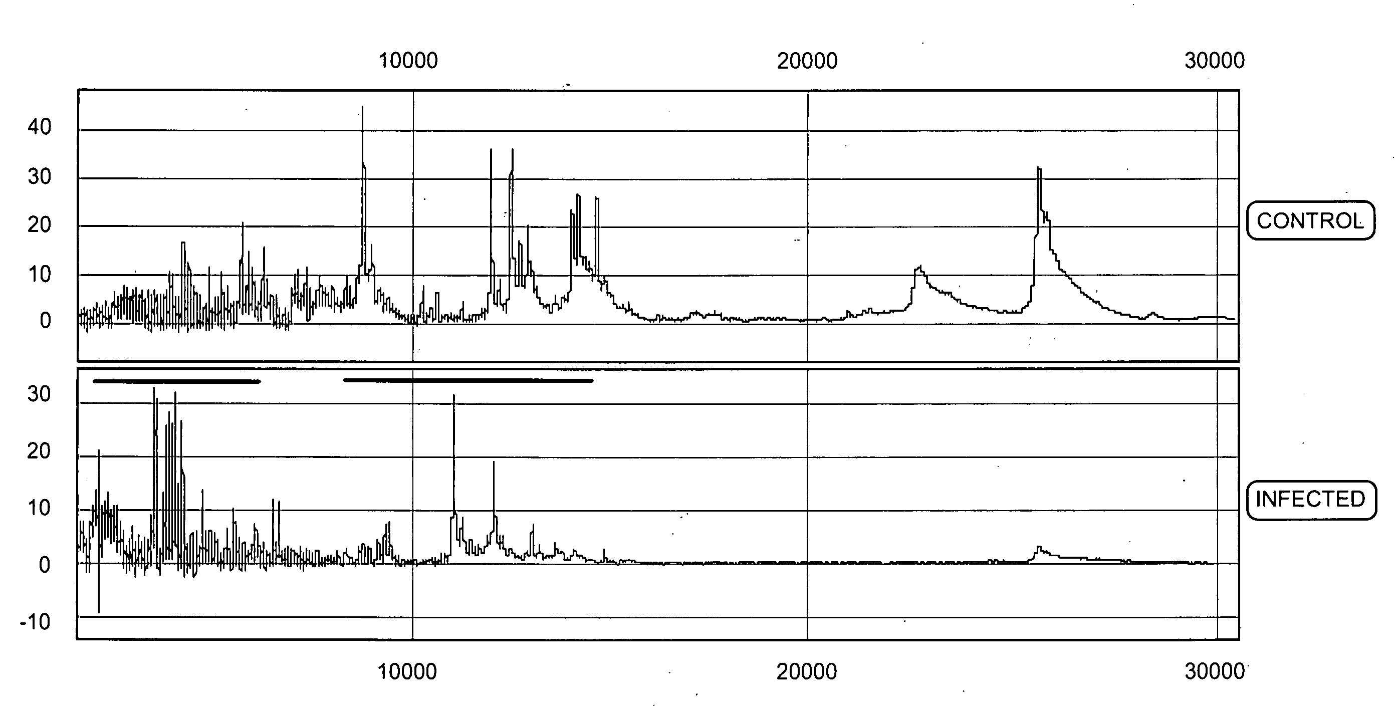
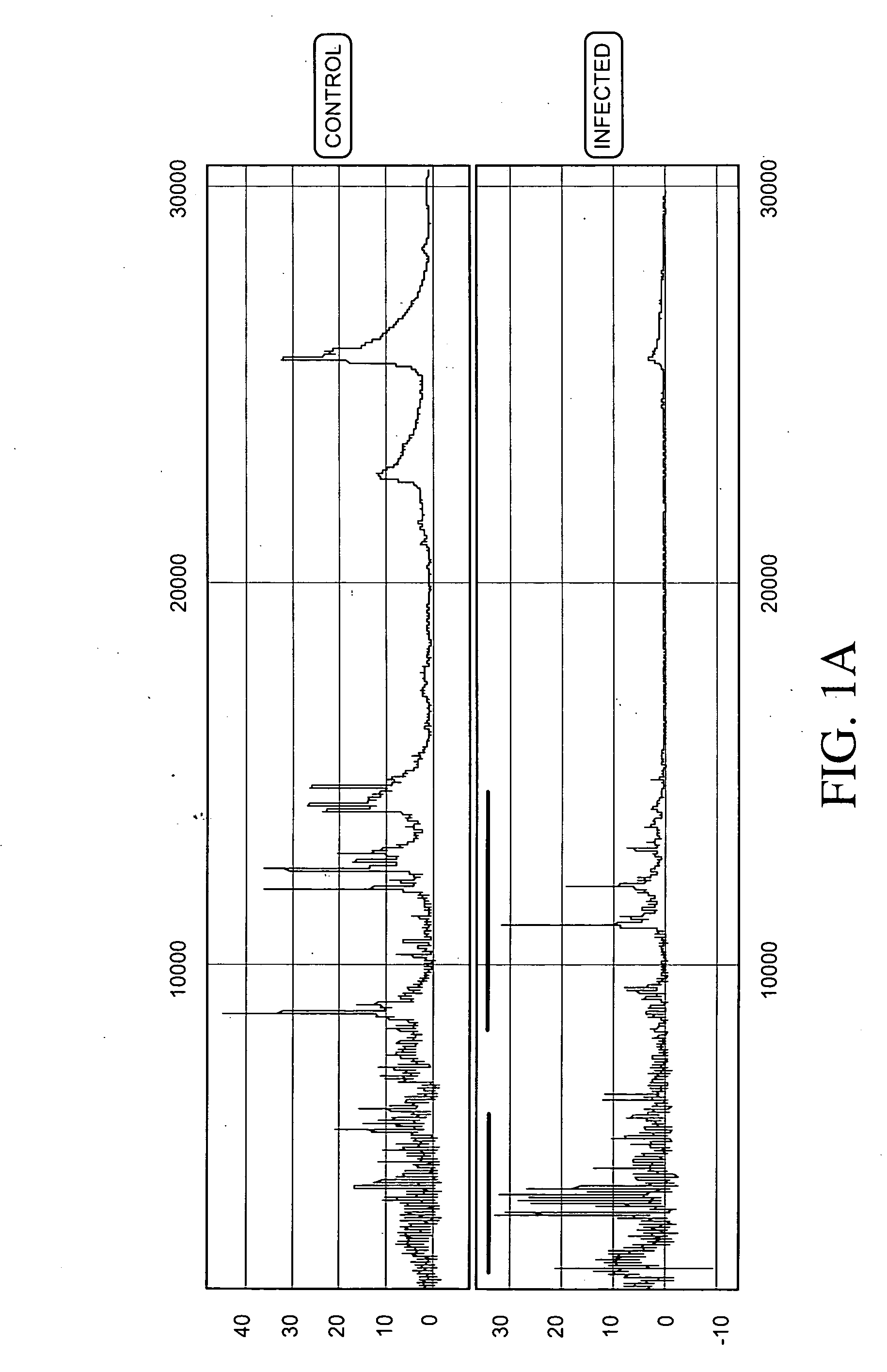
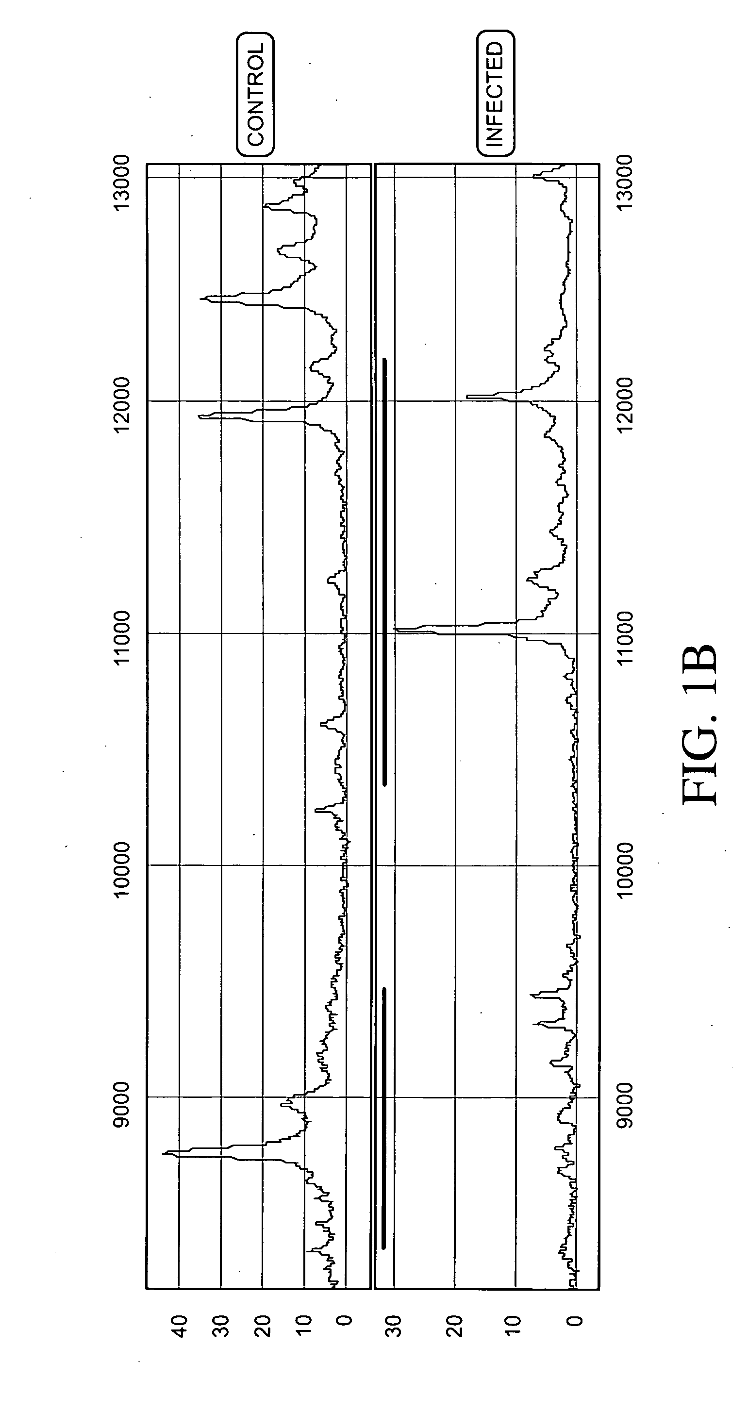
[0187]Protein expression profiles of primate amniotic fluid following intrauterine infection, compared with the corresponding normal expression profiles, are shown in FIGS. 1A-C.
[0188]As illustrated in FIGS. 1A-C, the global protein expression profiles of control and infected amniotic fluid are distinct. A detailed spectra of amniotic fluid profiles in a smaller mass range (FIGS. 1B and 1C), shows distinct and characteristic differences between the protein expression profiles of control and infected samples approximately in the 3-5 KDa and 10-12 KDa range. This illustrates global regulation of protein expression in response to intrauterine infection and the ability to detect a unique expression signature diagnostic of intrauterine infection.
PUM
| Property | Measurement | Unit |
|---|---|---|
| molecular weight | aaaaa | aaaaa |
| concentration | aaaaa | aaaaa |
| concentration | aaaaa | aaaaa |
Abstract
Description
Claims
Application Information