
Portable Materials and Methods for Ultrasensitive Detection of Pathogen and Bioparticles

a bioparticle and ultrasensitive technology, applied in the field of portable materials and methods for ultrasensitive detection of pathogens and bioparticles, can solve the problems of significant death, laborious and time-consuming methods, and time constraints and ease of on-site analysis are major limitations, and achieve rapid and highly sensitive and specific detection.

Inactive Publication Date: 2008-12-18
UNIV OF FLORIDA RES FOUNDATION INC
View PDF4 Cites 3 Cited by
  • Summary
  • Abstract
  • Description
  • Claims
  • Application Information

AI Technical Summary

Benefits of technology

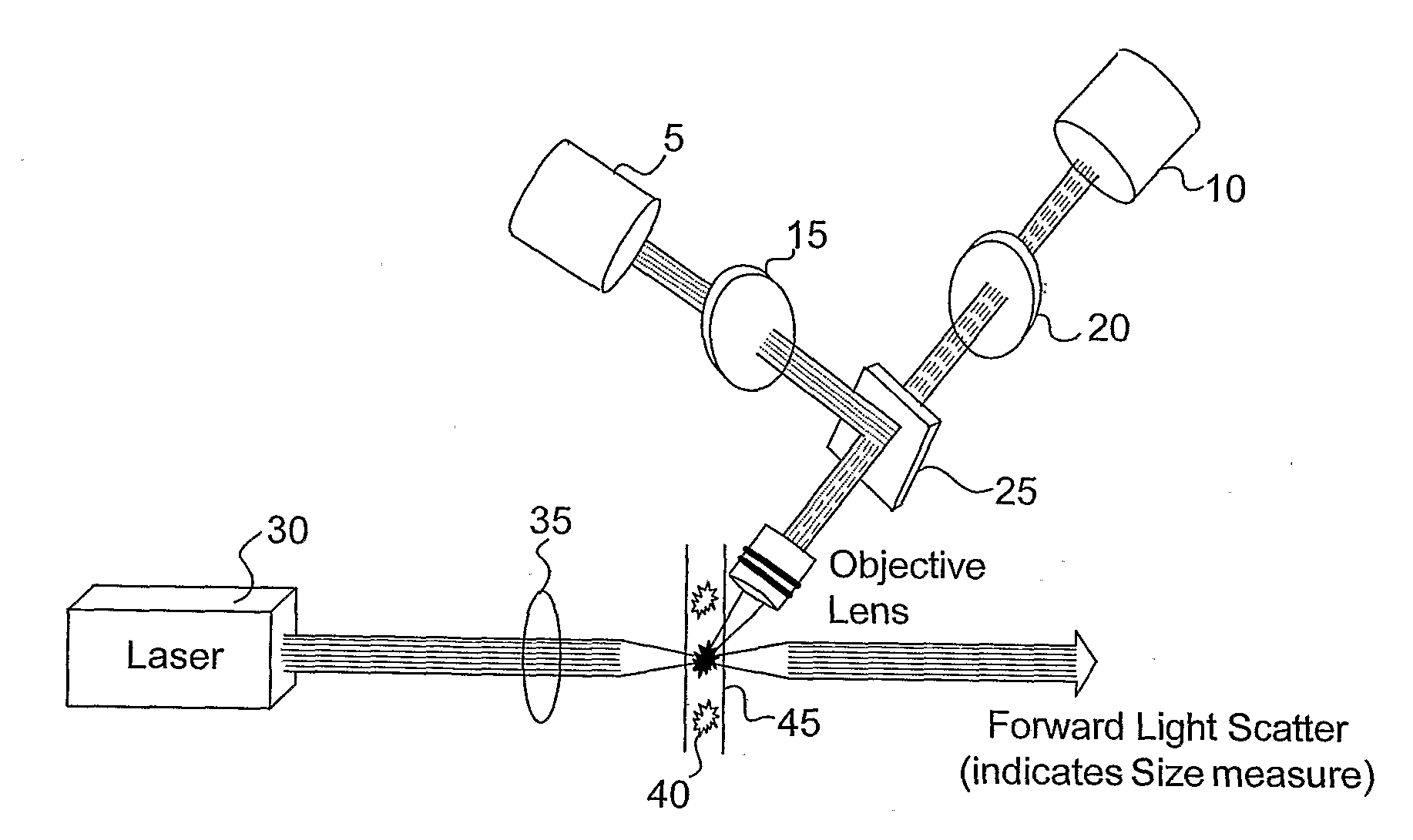
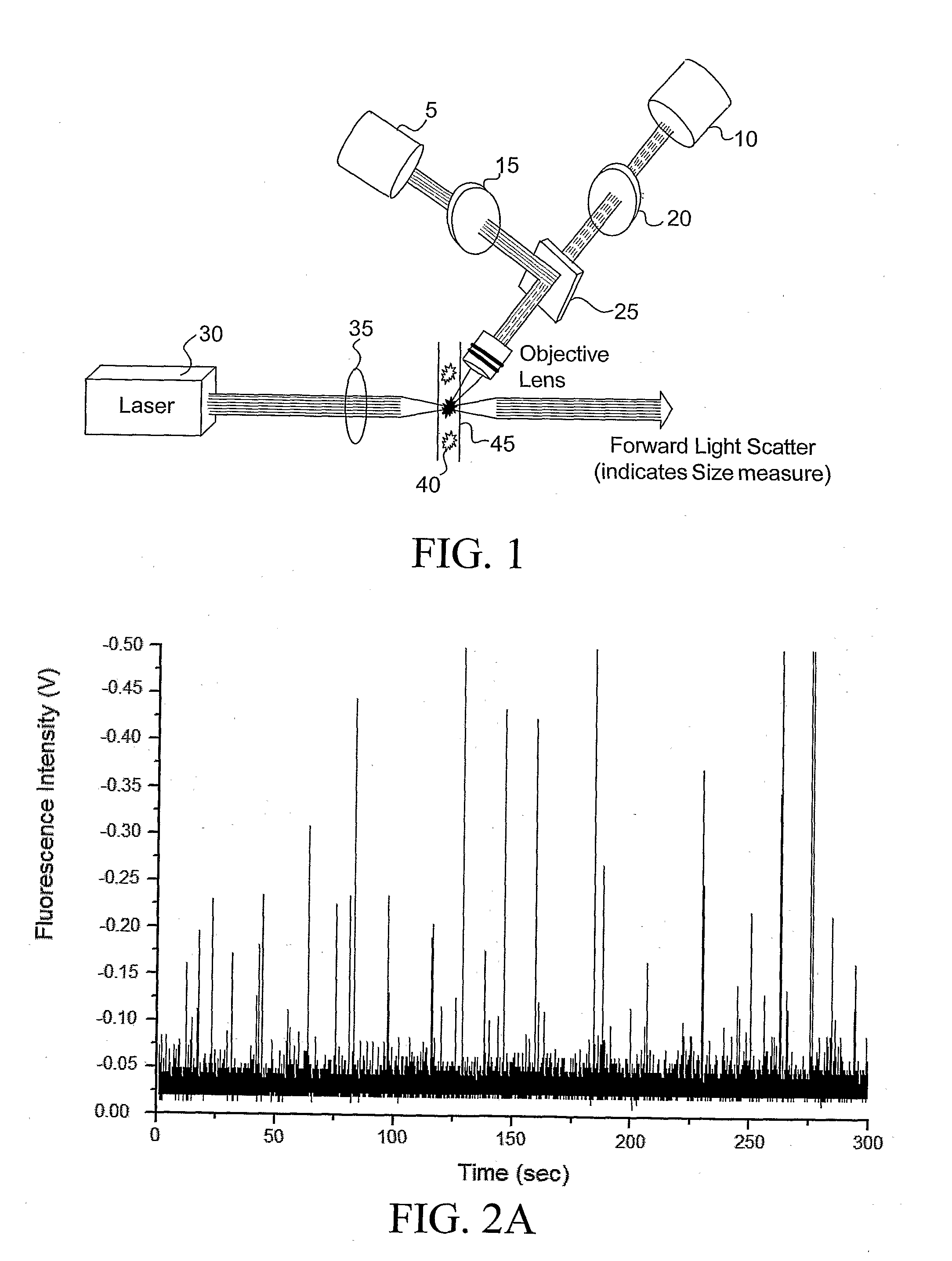
[0006]The present invention provides systems for ultrasensitive detection of pathogens and bioparticles. Advantageously, the systems of the subject invention are simple and can be made portable.
[0007]In one embodiment, the present invention provides a simple flow channel detection system for rapid and sensitive analysis of bacterial cells. In a preferred embodiment, the system utilizes dye doped silica nanoparticles (NP) that provide highly luminescent signals and that can be easily used for bioconjugation with molecular probes for bioanalysis. The use of luminescent silica NPs not only provides significant signal amplification in bacterial antibody-antigen recognition, but also presents highly photostable luminescent signals for reproducible measurements.
[0008]The system of the subject invention is rapid, technically simple, highly sensitive and efficient. Using antibodies specific for various bacterial pathogens, this assay can be adapted for the detection of a wide variety of bacterial pathogens with high sensitivity, accuracy and fast speed.
[0011]The system allows for the rapid and highly sensitive and specific detection of bacteria or other bioparticles without the need for amplification or enrichment of the sample.

Problems solved by technology

Outbreaks of E. coli O157:H7 infections have caused serious illnesses and led to a significant number of deaths.
However, time constraints and ease of-on-site analysis are major limitations because many of these methods rely on the ability of microorganisms to grow into visible colonies over time in special growth media, which may take about 1-5 days.
Moreover, detection of trace amounts of bacteria typically requires amplification or enrichment of the target bacteria in the sample.
These methods tend to be laborious and time consuming because of the complicated assay procedures.
Though the detection time is dramatically reduced, an improved sensitivity is still a challenge.

Method used


Image

  • Portable Materials and Methods for Ultrasensitive Detection of Pathogen and Bioparticles
  • Portable Materials and Methods for Ultrasensitive Detection of Pathogen and Bioparticles
  • Portable Materials and Methods for Ultrasensitive Detection of Pathogen and Bioparticles

Examples


example 1

Synthesis of Dye-Doped Silica Nanoparticles

[0047]FIG. 6 is a diagram showing a reverse microemulsion procedure for nanoparticle synthesis. In one embodiment, using a reverse microemulsion method (also known as water-in-oil microemulsion), generally uniformly sized 60±4 nm spherical RuBpy-doped silica NPs are synthesized and characterized with respect to uniformity and luminescence properties. With a water-to-surfactant molar ratio (W0) of 10, a reverse microemulsion is prepared by mixing about 7.5 mL cyclohexane, about 1.8 mL n-hexanol, about 1.77 mL triton x-100, about 80 μl of 0.01 M RuBpy, and about 400 μl water, followed by continuous stirring for about 20 minutes at room temperature. The size of the nanoparticles can be manipulated, as needed, by changing the water-to-surfactant molar ratio.

[0048]After adding about 100 μL of TEOS and about 60 μl of NH4OH solution, which initiates the polymerization of Si(OH)4 generated from the hydrolysis of TEOS, the reaction proceeds with con...

example 2

Immobilization of the Monoclonal Antibodies onto the Silica Nanoparticle Surface

[0049]The surface of the nanoparticle serves as a universal biocompatible and versatile substrate for the immobilization of biomolecules. In one embodiment of the present invention, after a thorough water wash, the silica surfaces of the RuBpy-doped carboxylated nanoparticles are activated, using about 100 mg / ml of 1-ethyl-3-3(3-dimethylaminopropyl) carbodiimide hydrochloride (EDC) and about 5 ml of 100 mg / ml N-hydroxy-succinimide (NHS) in a Z-morpholinoethanesulfonic acid (Mes) buffer (pH 6.8), for about 25 minutes at room temperature with continuous stirring. Water-washed particles are dispersed in about 10 ml of 0.1M PBS (pH 7.3) and reacted with monoclonal antibodies (mAbs) against E. coli O157: H7 for about 3 hours at room temperature with continuous stirring. To covalently immobilize the monoclonal antibodies onto the NP surface, about 5 ml of 0.1 mg / ml nanoparticles is reacted with about 2 ml of 5...

example 3

Detection of Bacteria

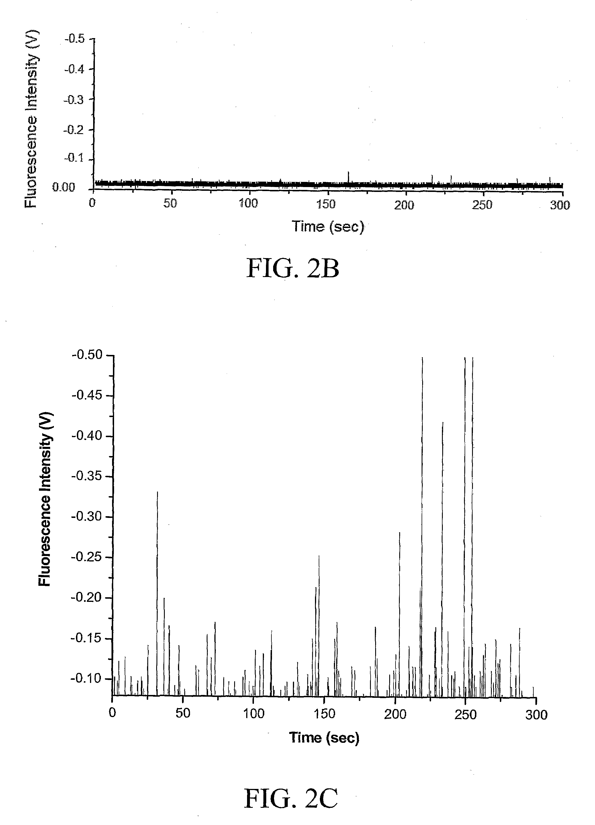
[0051]A 500 μL bacterial sample, which contains 25 bacteria based on plate-counting results, is dispersed into about 500 μL of 0.1 mg / ml of antibody conjugated NPs in a 0.1 M PBS buffer (pH 7.3) for about ten minutes. To remove the free antibody conjugated NPs that did not bind to the bacteria, the samples are centrifuged at about 14,000 rpm for about 30 seconds, and then the supernatant is removed. The samples are washed again to remove all unbound antibody conjugated NPs, and about 1.0 ml of PBS buffer is added to the samples. Samples are pumped through the capillary using a 1 ml syringe and a mechanical microliter syringe pump. This allows for a steady flow of sample through the channel at controllable various flow rates, including, for example, sample flow rates ranging from about 1 μL / hr to about 2 mL / hr. In another embodiment, control samples are obtained using the same experimental procedures but without the addition of bacteria.


PUM


Abstract

The present invention provides systems for ultrasensitive detection of pathogens and bioparticles. One embodiment of the system comprises an optical detection scheme that allows for the detection of the fluorescence signal of bacteria or other bioparticles in less than about 20 minutes. A microflow channel allows for an assay probing volume of as little as a few picoliters. In one embodiment, the system uses RuBpy dye-doped silica nanoparticles bioconjugated with specific monoclonal antibodies of the target bioparticles. The system allows for the rapid and highly sensitive and specific detection of bacteria or other bioparticles without the need for amplification or enrichment of the sample.

Description

GOVERNMENT SUPPORT[0001]The subject invention was made with government support tinder NIH Grant No. GM-66137, NIH Grant No. NS-045174 and NSF Grant No. EF-0304569. The government has certain rights in the invention.BACKGROUND OF THE INVENTION[0002]The rapid and accurate detection of trace amounts of organisms such as pathogenic bacteria is important in food safety, clinical diagnosis, and military / civilian warfare. Recently, there has been much interest in the identification of various microorganisms due to the increased risks of terrorism via biological warfare agents. Escherichia coli O157:H7 (E. coli O157:H7) is one of the most dangerous food borne bacterial pathogens. It is commonly found in raw beef, fruits, vegetables, salad bar items, salami, and other food products. Outbreaks of E. coli O157:H7 infections have caused serious illnesses and led to a significant number of deaths. Therefore, in order to prevent accidental outbreaks or intentional terrorist acts, early detection ...

Claims


Application Information

Patent Timeline
IPC IPC(8): G01N33/53C12M1/34B01J19/00G01N21/00G01N21/76
CPCG01N15/1459G01N21/6428G01N2015/1486
Inventor TAN, WEIHONGMECHERY, SHELLY JOHN
Owner UNIV OF FLORIDA RES FOUNDATION INC
Learn More