

Microscope system and screening method for drugs, physical therapies and biohazards
a microscope and screening method technology, applied in the field of tunneling nanotube screening, can solve the problems of pathological effects of multicellular organisms, easy destruction, extreme sensitivity, etc., and achieve the effects of high throughput, low cost, and minimal cell clustering
- Summary
- Abstract
- Description
- Claims
- Application Information
AI Technical Summary
Benefits of technology
Problems solved by technology
Method used
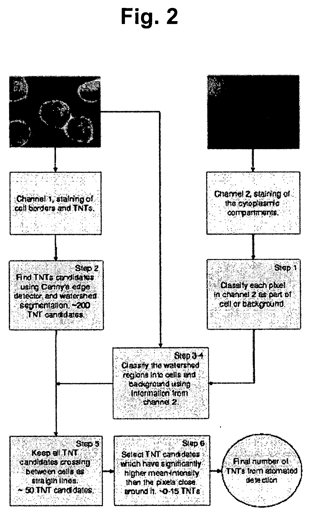
Image

Examples
examples
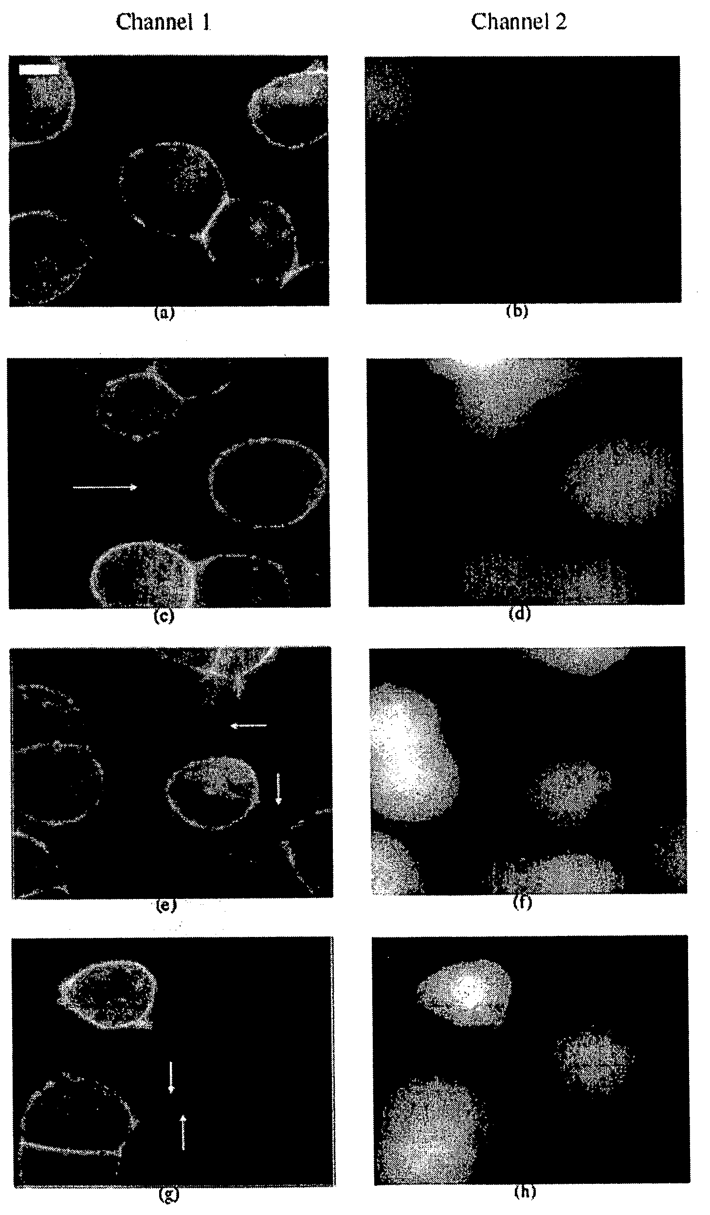
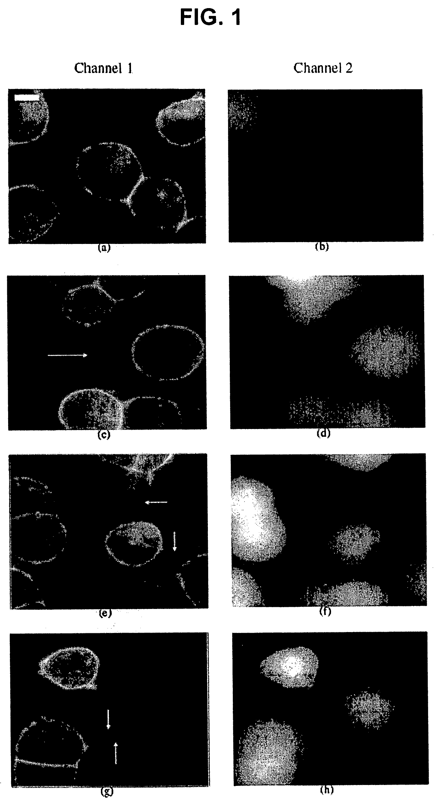
A. Preparation of the Microscopic Images
[0063]All image analyses were applied to mono-layers of cells from the living rat neuroendocrine cell line PC12 (rat pheochromocytoma cells, clone 251, gift of R. Heumann). This cell line was first generated in 1976 by Greene and Tischler [PNAS USA 1976; 73:2424-2428] from a transplantable rat adrenal pheochromocytoma. It is a single cell clonal line which grows monolayer forming small clusters. The PC12 cells also represent a common convenient model system for the study of secretory, neuron-like cells in cell culture. For comparative studies, NRK cells (normal rat kidney, Mrs. M. Freshney, Glasgow, UK) were used.
[0064]PC12 and NRK cells were cultured in DMEM supplemented with 10% fetal calf serum and 5% horse serum. For high-resolution fluorescence microscopy and light microscope analysis, PC 12 cells were plated in LabTek™ chambered swell cover glasses (Nalge Nunc Int., Wiesbaden, Germany). Two hours after plating, the cells were stained wit...
PUM
| Property | Measurement | Unit |
|---|---|---|
| diameter | aaaaa | aaaaa |
| length | aaaaa | aaaaa |
| length | aaaaa | aaaaa |
Abstract
Description
Claims
Application Information