
System for detecting protein-protein interactions

a protein interaction and protein technology, applied in the field of complementation assays for the study of protein interactions, can solve the problems of only working protein interaction screens that depend on placing complimenting fragments at the ends of fusion partners, protein interactions in which the ends of two proteins are too far apart, and protein interactions that are los

Inactive Publication Date: 2009-06-11
MONTANA STATE UNIVERSITY +1
View PDF22 Cites 2 Cited by
  • Summary
  • Abstract
  • Description
  • Claims
  • Application Information

AI Technical Summary

Benefits of technology

[0017]The present invention involves the placement of complementing fragments of a reporter protein within the structure of two interacting proteins or two interacting protein surfaces or protein domains. When the reporter protein fragments are placed at interacting protein surfaces, the two fragments are brought close enough to one another to fold together to form a functional reporter protein. This approach does not rely upon attaching the complementing fragments to the ends of the interacting fragments. This has several advantages. First, there are protein interactions that will be blocked, due to steric hindrance, by the addition of reporter fragments to the ends of the interacting proteins. Second, protein interaction screens that depend upon placing the complimenting fragments at the ends of the fusion partners will only work when the ends of the interacting proteins are physically close enough to one another for the complementation to take place. Protein interactions in which the ends of the two proteins are too far apart will be lost. Third, appending the reporter fragments to the interacting fragments often involves fragmenting or truncating the interacting proteins. The present invention works with full length proteins, where at least one complementing fragment is fused at an internal position within the interacting protein.
[0018]Thus, the present invention concerns methods of detecting interaction of at least a first and second protein or protein domain by complementation of at least two fragments of a reporter protein, wherein at least two fragments of said reporter protein are genetically fused to the first and second proteins or protein domains, respectively, and at least one complementing fragment is fused at an internal position within one of the first or second interacting proteins or protein domains. The present invention also encompasses methods wherein each of the complementing fragments is fused at an internal position within one of the interacting proteins or protein domains. While there are many ways to construct the interacting fusion proteins of the invention, as described herein, a convenient means of construction is to use a transposon to randomly insert each complimenting fragment into each member of a pair of interacting proteins. This enables one to quickly and easily generate a library of fusions containing the complimenting fragments fused at different locations, and screen this library for interacting members by observing those that bring the complimenting fragments of the reporter together to form a functional reporter protein.
[0022]The present invention is useful for detecting or measuring the binding of two or more interacting proteins, for instance by measuring functional reporter protein activity following complementation of the complementing fragments fused to each binding member. The methods of the invention are also useful for measuring or detecting protein folding, for instance, where the two or more protein domains are located in a single protein. The present invention also includes methods for measuring dimerization or aggregation of protein subunits, for instance, where the complementing fragments are located in two or more subunits of a dimeric or multimeric protein complex, including receptor complexes. The present invention also includes methods for detecting or measuring the interaction of a ligand with its receptor, for instance where the complementing fragments are located in interacting ligand and receptor proteins, respectively. The methods of the present invention may also be used to detect protein interactions within a cell, for instance where the complementing fragments are located in different proteins that participate in a common signal transduction pathway. The constructs of the present invention may also be used in methods of identifying compounds that modulate binding of a first protein to a second protein, for instance by observing changes in reporter protein activity via complementation of fragments in the first and second proteins in response to exposure to different compounds. Such assays may be readily performed at the high throughput level.

Problems solved by technology

First, there are protein interactions that will be blocked, due to steric hindrance, by the addition of reporter fragments to the ends of the interacting proteins.
Second, protein interaction screens that depend upon placing the complimenting fragments at the ends of the fusion partners will only work when the ends of the interacting proteins are physically close enough to one another for the complementation to take place.
Protein interactions in which the ends of the two proteins are too far apart will be lost.
Third, appending the reporter fragments to the interacting fragments often involves fragmenting or truncating the interacting proteins.

Method used


Image

  • System for detecting protein-protein interactions
  • System for detecting protein-protein interactions
  • System for detecting protein-protein interactions

Examples


example 1

Construction of a Synthetic Tn5 Transposon

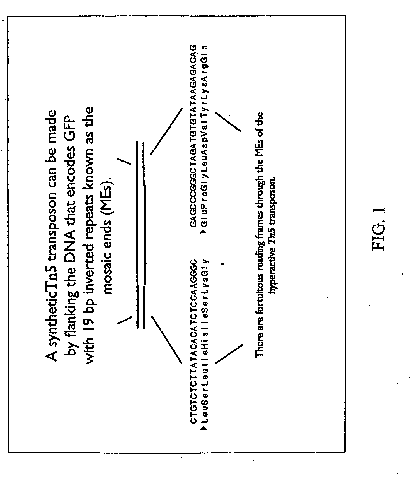
[0079]To create a transposon for generating random fusions to complementing fragments of a GFP reporter, we used the mosaic ends from a hyperactive Tn5 transposon. The mosaic ends are simply 19 base pair inverted repeats (SEQ ID Nos. 1 and 2) that can be placed on either side of any stretch of DNA to create a Tn5 transposon (see FIG. 1). These Tn5 ends are particularly suitable for creating fusion proteins in that there is an open reading frame that crosses the ends in either orientation.

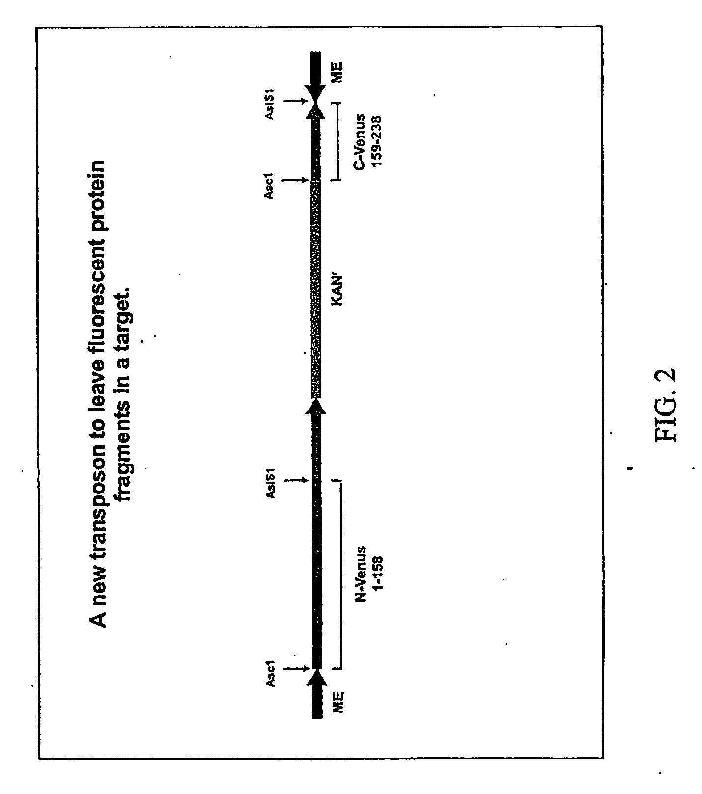
[0080]We created a Tn5 transposon (pBonjovi) with PCR and standard subcloning techniques that carried the mosaic ends on either end of a segment of DNA that would, in combination with recombinant Tn5 transposase become inserted into a target plasmid. This is an in vitro reaction in which the transposase recognizes the mosaic ends of the transposon and inserts the transposon in a reasonably random fashion into any other DNA present in the reaction. We positio...

example 2

Isolation of YFP Complementing Fragments Fused Internally to Shaker Subunits

[0082]To test the process, we targeted the Shaker potassium channel. The Shaker potassium channel is a voltage-gated ion channel that is composed of 4 identical subunits. Our rationale was that if we created many different versions of the subunit, containing the two different fragments of the fluorescent protein, we could use pairwise expression of the different subunits to determine whether any complementation could occur between adjacent subunits.

[0083]The sequence encoding the Shaker subunit was first moved as a restriction fragment into a small CMV expression plasmid. An in vitro reaction with the plasmid containing the Shaker subunit coding region, the transposon pBonjovi, and recombinant Tn5 transposase was used to insert the transposon sequence. Transformation of Top 10f′E. coli with 1 ul of the 15 ul in vitro reaction produced greater than 3,000 colonies that displayed both the ampicillin resistance ...

example 3

Measuring Voltage Dependent Changes Using Shaker Complementing Subunits

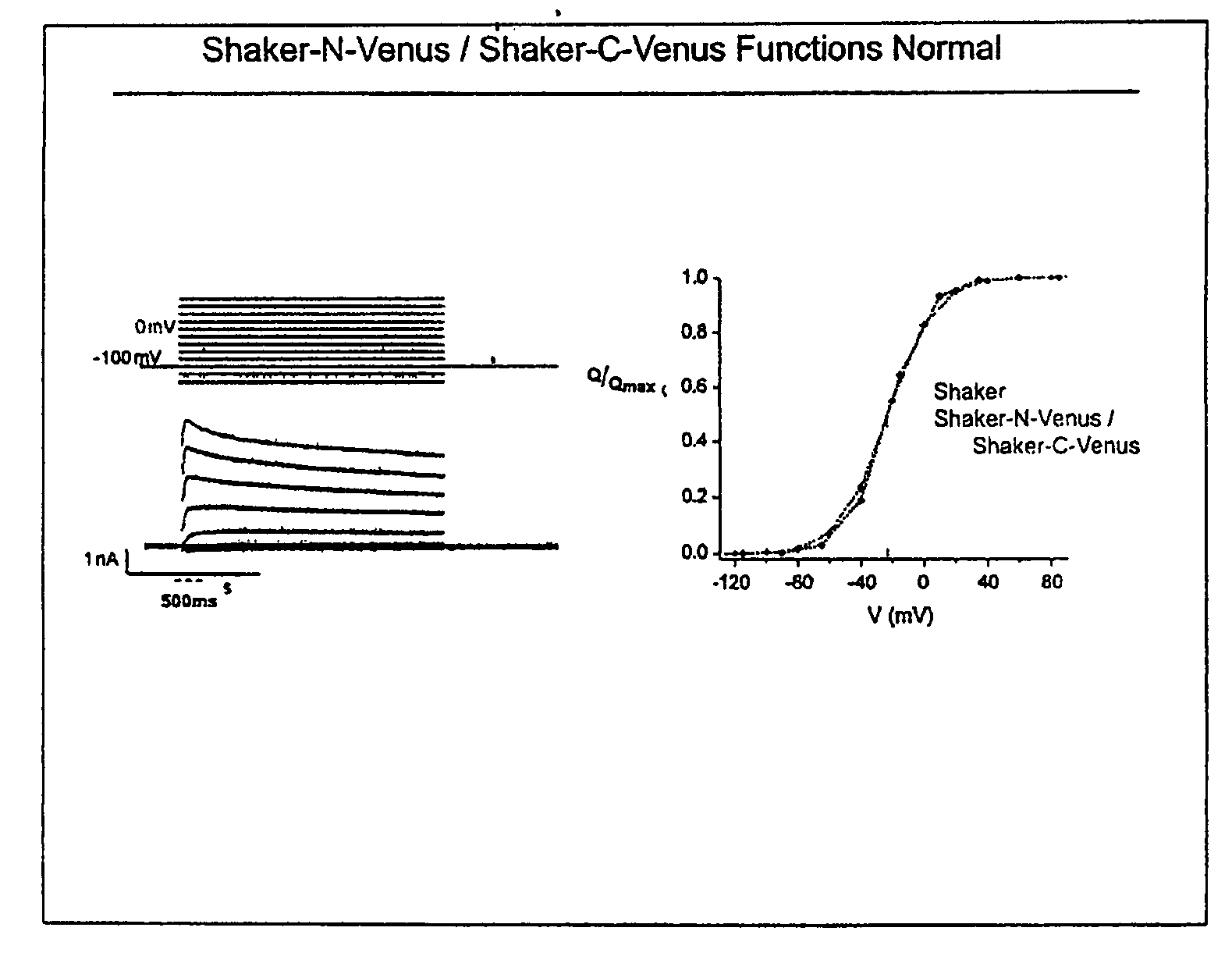
[0088]When the complementing pairs were screened for voltage dependent changes in fluorescence, two pairs of subunits were identified that produced changes in fluorescence of approximately 20% as a result of depolarization of the cells with high concentrations of extracellular potassium (see FIG. 11). Thus, interacting complementing subunits may also be used as new biosensors for detecting changes in extracellular potassium. Voltage versus gating measurements indicate that some pairs of the complementing subunits produce a normally functioning channel when expressed in HEK 293 cells, showing that complementing pairs may be isolated that retain the function of the interacting subunits (see FIG. 12).

[0089]The results of our work show that GFP fragments can be inserted deep in the structure of two interacting proteins and still complement one another to form a fluorophore. This means that the approach can be used wi...


PUM

PropertyMeasurementUnit
Concentrationaaaaaaaaaa
Electrical resistanceaaaaaaaaaa
Protein activityaaaaaaaaaa

Abstract

A method of detecting protein interactions is described wherein reporter protein fragments are genetically fused at internal positions of suspected interacting proteins. When proteins interact, the fluorescent fragments are brought close enough together to form a functional reporter protein providing visible confirmation of interaction.

Description

CROSS-REFERENCE TO RELATED APPLICATIONS[0001]This application claims benefit of priority to U.S. provisional application 60 / 722,764, filed Sep. 30, 2005, which is herein incorporated by reference in its entirety.FIELD OF INVENTION[0002]This invention relates to the use of complementation assays for the study of protein interactions.BACKGROUND OF THE INVENTION[0003]Every process within a cell involves highly orchestrated interactions between different proteins. Determining which proteins interact, where they interact, and when they interact is critical to understanding most cellular processes. To date, the best strategy for identifying and characterizing protein interactions is complementation. There are now several complementation systems and most involve bringing two different protein domains together to produce a functional signal. The best example is the two hybrid approach in yeast where a DNA binding domain and a transcription activator have to be brought together by fusion pro...

Claims


Application Information

Patent Timeline
IPC IPC(8): C40B30/04C12Q1/68C12N15/11C07K14/00
CPCC07K2319/60C12N15/1055G01N2500/00G01N33/54386C12N15/1082
Inventor HUGHES, THOMAS E.MEALER, ROBERT G.BERLOT, CATHERINE H.
Owner MONTANA STATE UNIVERSITY
Learn More