Method for detecting intracellular interaction between biomolecules

Active Publication Date: 2009-08-27
CIS BIO INT
View PDF17 Cites 14 Cited by
  • Summary
  • Abstract
  • Description
  • Claims
  • Application Information

AI Technical Summary

Problems solved by technology

Very few kinetic observations are currently possible and rare events easily go undetected.
However, the use of such technologies is limited by numerous disadvantages:Microscopic studies are tricky and difficult to automate and require complex image analysis tools, specific softwares and very experienced users; they also require a great deal of time.Image analysis is complicated and often a source of errors, and these technologies are difficult to reproduce and rather unreliable (low value of the parameter Z′).They make it possible to follow biological events in living cells with a moderate throughput of molecule screening that limits their use in the primary screening steps.The visualized effects are generally downstream of the signaling cascade.
It is for these main reasons that these techniques are not compatible with high-throughput molecule screening.
Furthermore, these techniques do not provide direct information on the interactions between biomolecules or on the conformational changes within one and the same molecule, both of which are direct indicators of the biological event which it is desired to detect and quantify.
However, the use of these fluorescent proteins has certain major disadvantages:The overlap spectra of the two fluorescent molecules (CFP and YFP) are broad and cause a strong parasitic excitation of the acceptor (YFP) at the wavelengths used to excite the donor (CFP).
Such analyses then exhibit the same disadvantages as those described above for the first detection approach.The limited number of variants of GFP restricts the use of such proteins to applications where the proximate changes are consistent with the Förster radius (Ro) of the CFP / YFP pair.These proteins are fused with the target proteins.
The fluorescence of these proteins is detected as soon as the cell expresses it, and it cannot be triggered or controlled by the experimenter without providing much more complex expression systems.
In the same way as for the CFP / YFP pair, numerous disadvantages limit the use of the luciferase / GFP pair in high-throughput screening (HTS):The signal-to-noise ratio of the BRET tests is rather low.The fluorescence signal can easily be masked by an autofluorescence of the test products.
BRET technology is not sufficiently robust for screening molecules because of the low stability of the signal over time, which leaves a very short reading window that is not very compatible with high-throughput molecule screening.
The main disadvantage of these approaches using functional complementation lies in the fact that the measurement of interaction between biomolecules is dependent on a perfect reconstitution of the 3rd protein in its active form, such as β-galactosidase, and sometimes requires displacement of the interaction complex into a cellular compartment other than that in which the biomolecules normally interact.
The use of functional complementation is more complex in the case of a single biosensor type biomolecule.
Depending on the intrinsic properties of the proteins used in these energy transfer processes, the technologies using fluorescent proteins such as GFP (or its other variants) or bioluminescent proteins such as luciferase do not allow a great flexibility in the choice of the donor / acceptor pair involved in the energy transfer process.
However, it is difficult to label the biomolecules specifically with these compounds in the cell.
Furthermore, it does nothing to qualify an interaction between two biomolecules of interest and serves only to reveal the presence of a messenger RNA of interest.

Method used


Image

  • Method for detecting intracellular interaction between biomolecules
  • Method for detecting intracellular interaction between biomolecules
  • Method for detecting intracellular interaction between biomolecules

Examples


example 1

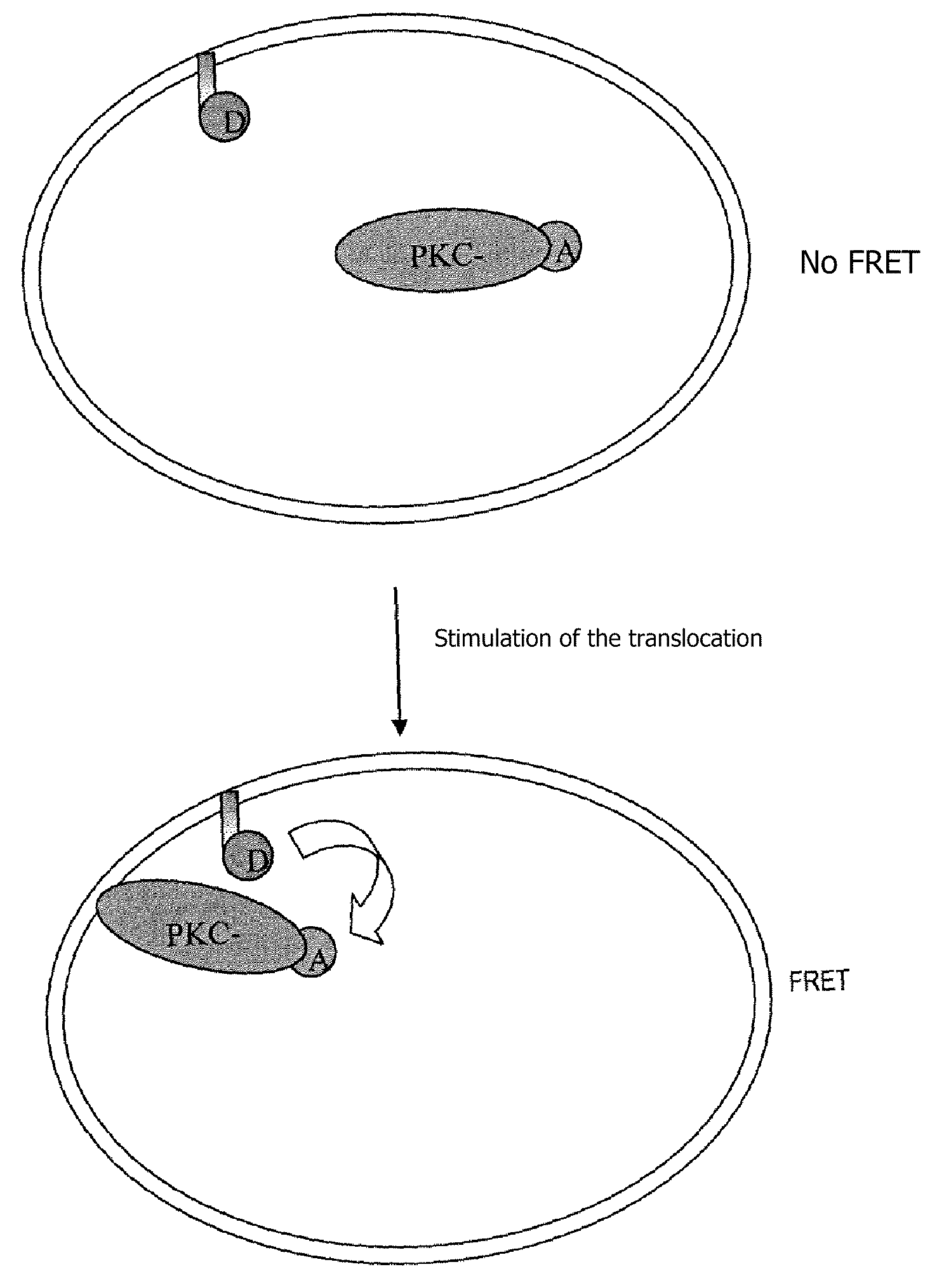
Measurement of the Translocation of Protein Kinase C-gamma (PKC-γ) (cf. Flow Chart, FIG. 1)

[0109]Protein kinases C are proteins belonging to a group of phospholipid-dependent serine / threonine kinases. These proteins play a major role in numerous cell signaling pathways. Physiologically PKC-γ is activated in a calcium-dependent manner by phosphatidylserines (PS) and it binds diacylglycerol (DAG), but PKC-γ can be activated independently of the presence of DAG by tumor-inducing phorbol esters (PMA).

[0110]After stimulation, PKC-γ undergoes a translation of the cytoplasm into the plasma membrane, it being possible for this translocation to be measured by means of a fusion protein with GFP after stimulation with PMA (Sakai N. J., Cell Bio. (1997) 139, 1465-1476).

[0111]A fusion protein was created between PKC-γ and a suicide enzyme, called HaloTag, in order to label the PKC-γ with an acceptor fluorescent organic compound, and a tool was developed to label the plasma membrane specifically ...

example 2

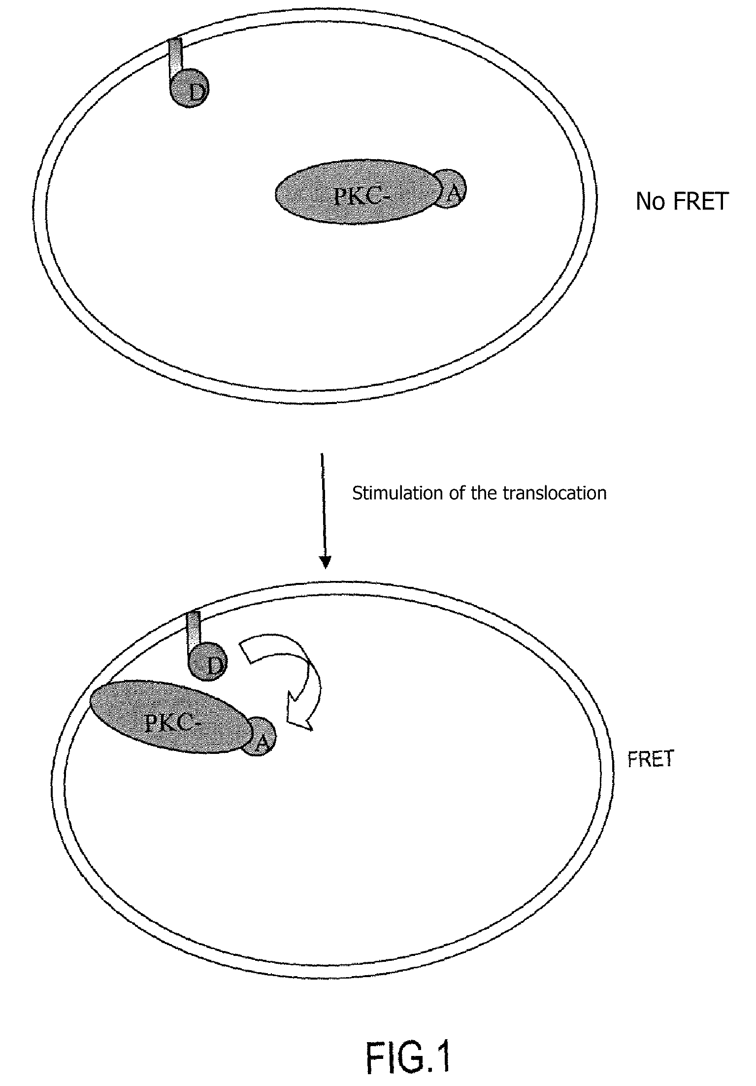
Revealing Androgen Receptor / Coactivator or Androgen Receptor / Corepressor Interactions (cf. Flow Chart, FIG. 2)

[0116]Androgens and their functional receptor (AR) are responsible for normal differentiation of the external male phenotype. Cytosolic androgen receptors, in the inactive state, are associated with heat shock proteins. After binding with the ligand, the receptors dimerize and are subjected to a translocation into the nucleus. AR coregulators (coactivators or corepressors) will or will not then interact with the receptor and the result of these interactions will optionally cause the complex to bind to the DNA at a specific sequence (called ARE=androgen receptor responsive element) and thus to regulate the transcription.

[0117]The method according to the invention is used to reveal the interactions between a corepressor, a coactivator and an AR. This is done by introducing expression vectors into the cell in order to express the following in the intracellular medium:[0118]an A...

example 3

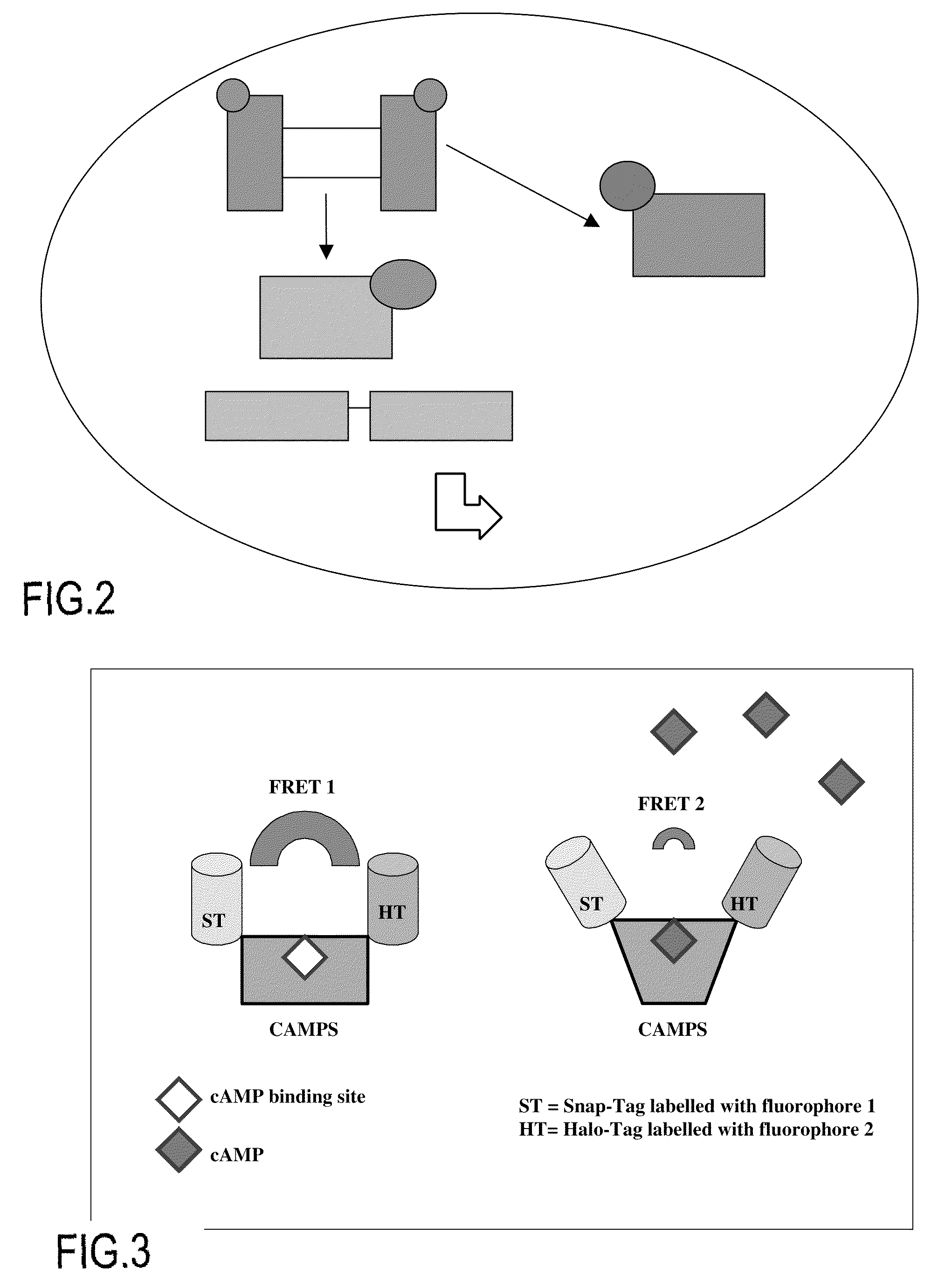
cAMP Biosensor: Detection of Conformational Modifications of a Fusion Protein Comprising a cAMP Binding Domain (cf. Flow Chart, FIG. 3)

[0123]In this Example the variations in the TR-FRET signal of a pair of donor / acceptor fluorescent compounds is measured by coupling these fluorophores with a cAMP binding domain (e.g. that of the βII regulatory subunit of PKA, or that of cAMP-activated exchange proteins, which are known by the term “CAMPS”). These molecular constructs can be used to quantify, in living cells, the levels of cAMP after pharmacological stimulation of a G protein-coupled receptor (GPCR) (cf. Nikolaev et al., JBC, vol. 279, no. 36, pp 37215-37218, 2004).

[0124]The plasmid coding for a fusion protein of the SnapTag / CAMPS / HaloTag type is transfected into HEK cells. 24 hours or 48 hours after transfection, the cells are incubated (1 hour at 37° C.) with the SnapTag and HaloTag substrates, each of which is coupled with a member of a TR-FRET donor / acceptor pair.

[0125]When the ...


PUM

PropertyMeasurementUnit
emission wavelengthsaaaaaaaaaa
fluorescence lifetimeaaaaaaaaaa

Abstract

The present invention relates to a quantitative non-microscopic method of detecting intracellular interactions between biomolecules, on living cells, in response to a biological or pharmacological stimulation, by a time-resolved proximate energy transfer effect between two members of a fluorescence donor / acceptor pair.

Description

[0001]The present invention relates to a quantitative non-microscopic method of detecting intracellular interactions between biomolecules, on living cells, in response to a biological or pharmacological stimulation, by a time-resolved proximate energy transfer effect between two members of a fluorescence donor / acceptor pair, and to its applications such as, in particular, the high-throughput screening of molecules and the detection of intracellular signaling pathways.TECHNICAL FIELD AND PRIOR ART[0002]Molecules with pharmacological activity (e.g. drugs) act on molecular targets located either on the plasma membrane or inside the living cells themselves. To select and optimize candidate molecules in the search procedures within the pharmaceutical industry, it is necessary to determine their molecular actions, as far as possible in a native environment so as to integrate the modulations that may be introduced by interactions between different partners. From a more fundamental point of...

Claims


Application Information

Patent Timeline
Patent Type & Authority Applications(United States)
IPC IPC(8): C12Q1/02
CPCG01N33/542
Inventor PRIBILLA, IRISBAZIN, HERVEGHOSE, SRABONITINEL, NORBERTFINK, MICHELTRINQUET, ERICMATHIS, GERARD
Owner CIS BIO INT
Why Eureka
Try Eureka