

METHOD FOR DETECTING IGF1R/Chr 15 in CIRCULATING TUMOR CELLS USING FISH
a technology of circulating tumor cells and igf1r/chr 15 is applied in the field of oncology and diagnostic imaging, which can solve the problems of difficult detection and elimination, inability to treat all patients successfully, and inability to improve the survival rate of cancer patients over the past two decades
- Summary
- Abstract
- Description
- Claims
- Application Information
AI Technical Summary
Benefits of technology
Problems solved by technology
Method used
Image

Examples
example 1
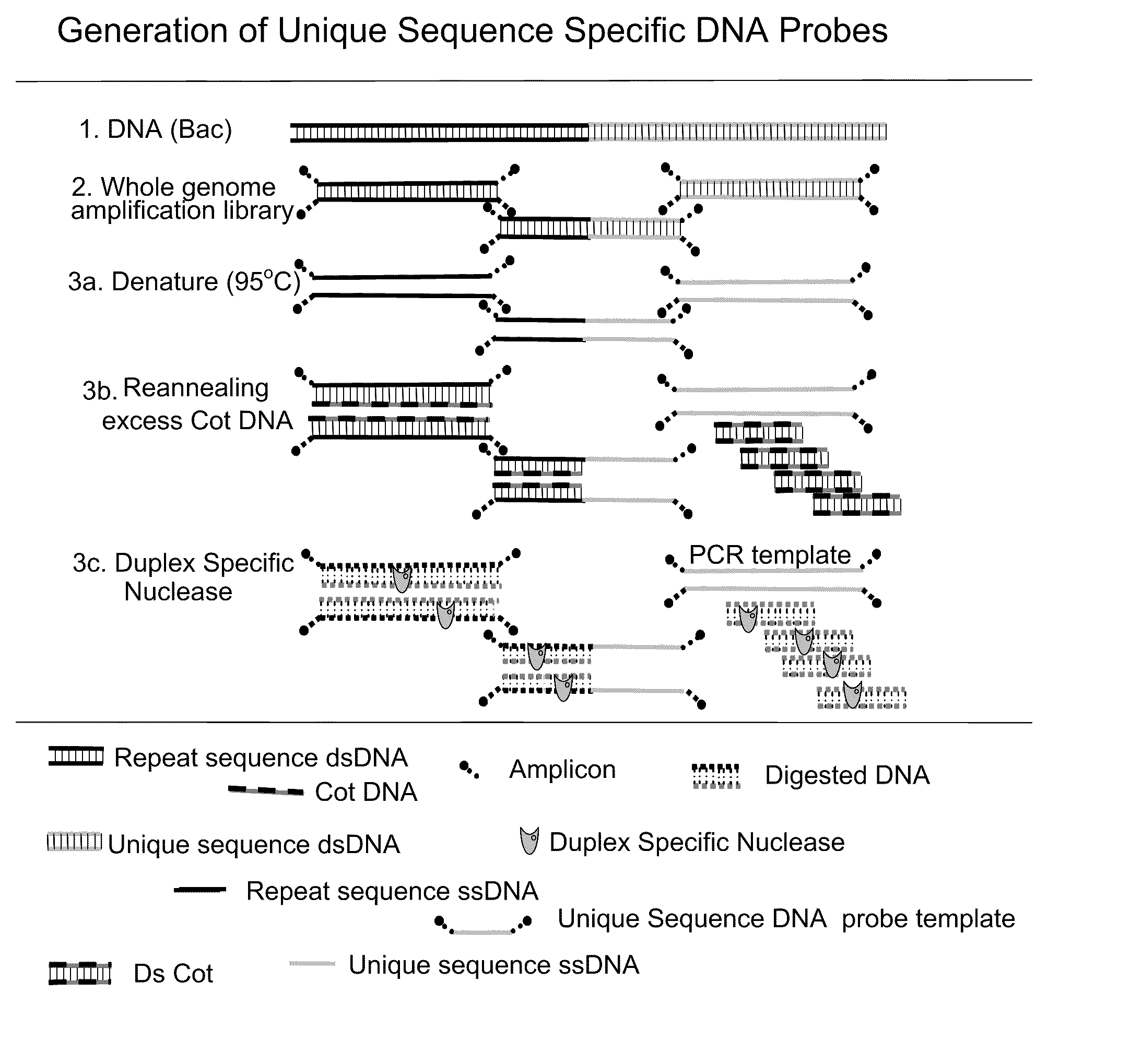
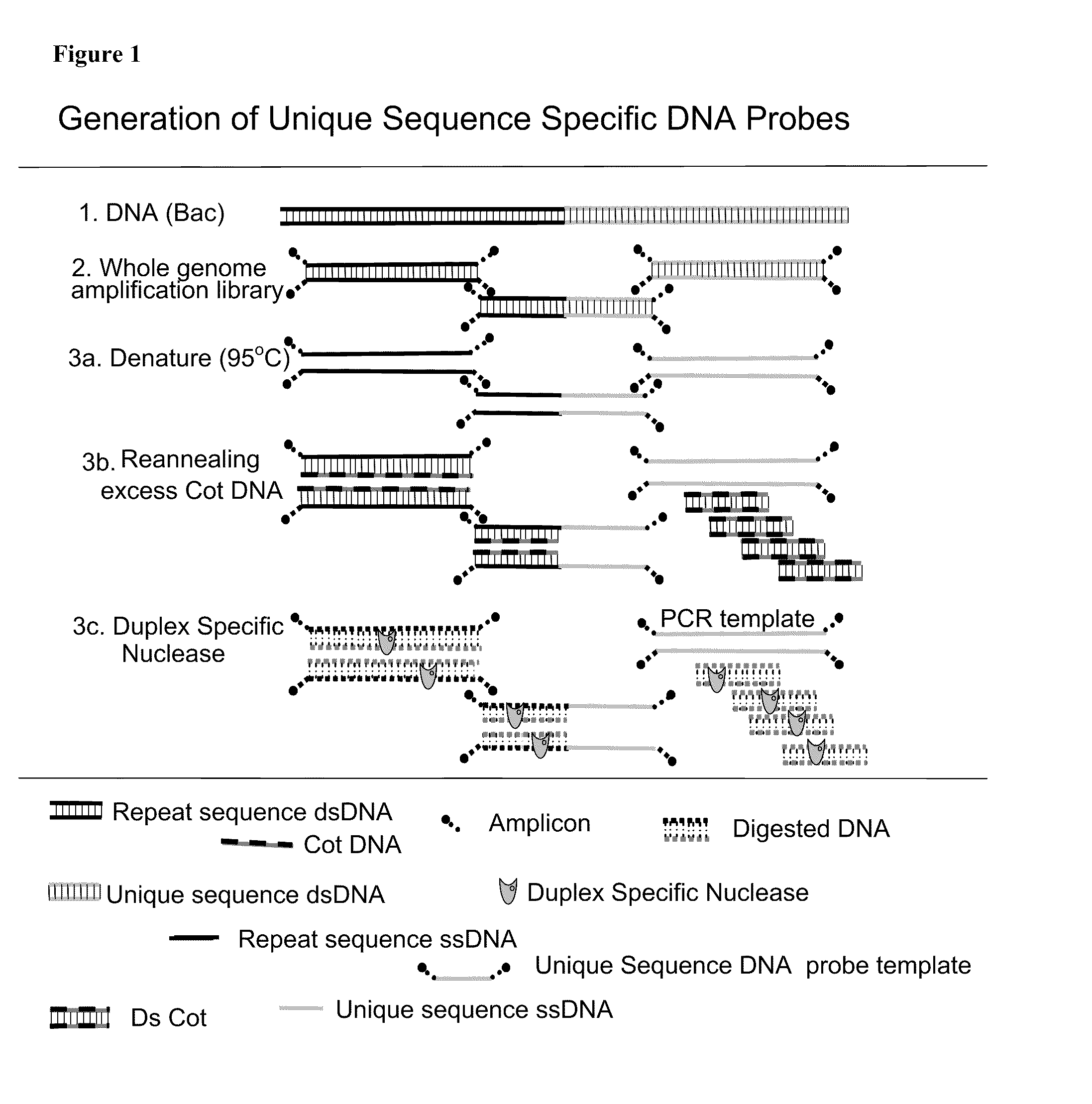
Development of a Probe Set for an IGF-1R / Chr 15 FISH Assay
[0055]Two types of probes are needed for the assay. One type is a satellite enumeration (SE) probe. These probes bind the satellite (repetitive) sequence near the centromere of the chromsome and is used for chromosome enumeration and aneuploidy detection. The second type of probe is the unique sequence probe. As the name implies these probes bind unique sequences, like genes. Using bioinformatics, unique sequence probes can be designed for any location in the genome. Unique sequence probes usually contain repetitive elements like Alu or Kpn repeats which can cause non-specific binding of the probe. Suppression of non-specific binding is typically done by the incorporation of unlabeled blocking DNA in the hybridization. Blocking DNA has been shown to interfere with hybridization of unique sequence and satellite probes. The present method eliminates this step with the use of repeat-free probes which allow faster, brighter hybri...
example 2
Cell Line Evaluation of the IGF-1R / SE-15 Probe Configuration
[0059]The combination of the alpha satellite probe for chromosome 15 and the IGF-1R clones A & B (IGF-1R / SE-15) were tested for assessing genetic aberrations. The IGF1R / SE-15 probe configuration was used on several cell lines to see if the probe could detect aneupliody or gene amplification / deletion. A549 (lung), BT474 (Breast), PC3 (prostate), LNCAP (prostate), H1299 (lung), and MCF7 (Breast) cell lines were tested with this configuration. The images in FIG. 5 show that LNCAP have four copies of SE-15 and four copies of IGF1R, indicating aneupliody with no gene amplification. The remaining five cell lines tested showed cross-hybridization of the SE-15 probe to other chromosomes. BT 474 cells contain 2-3 copies of IGF 1R and numerous SE-15 signals indicating cross-hybridization. The other cell lines had varying amounts of cross hybridization of the SE-15 probe. Since the degree of cross hybridization varied among the cell l...
example 3
Evaluation of Probe Configuration from Donor Samples
[0063]After obtaining the optimum conditions for the assay which included the use of 4 ng / μl of each probe in the reaction, the probe configuration was evaluated on a total of 350 CD45+ / CK− cells (leukocytes) from three different donors and analyzed using the CellTracks-FISH System. The number of copies of each target scored and the results are listed in the tables below. FISH signals were able to be scored in greater than 95% of the cells relocated by the CellTracks-FISH System.
[0064]Table II shows the results from the three donors. The expected 2 copies of IGF1R occurred in 87%, 81%, and 93% of the WBC examined from three donors. It was expected that ≧75% of WBC should show the expected result of 2 dots per WBC for IGF1R and >85% of the WBC evaluated should be scoreable. The three donors showed the expected 2 copies of Chr 15 in 91%, 97%, and 93% of the WBC examined. For this reason we set a QC specification that ≧80% of WBC shou...
PUM
| Property | Measurement | Unit |
|---|---|---|
| concentrations | aaaaa | aaaaa |
| magnetic field | aaaaa | aaaaa |
| immunofluorescent microscopy | aaaaa | aaaaa |
Abstract
Description
Claims
Application Information