Surgical sutures incorporated with stem cells or other bioactive materials

a technology of stem cells and bioactive materials, applied in the direction of surgical staples, prostheses, transportation and packaging, etc., can solve the problems of fluids, affecting the treatment effect of endogenous tissues, and affecting the patient's recovery, so as to reduce the concentration of stem cells and reduce the migration of cells.

Inactive Publication Date: 2009-12-24
BIOACTIVE SURGICAL
View PDF42 Cites 59 Cited by
  • Summary
  • Abstract
  • Description
  • Claims
  • Application Information

AI Technical Summary

Benefits of technology

[0012]Central to the instant invention is the provision of a surgical suture or elongate, linear or thread-like tissue scaffold in which the radial cross section comprises at least two concentric zones, which are differentiated in part by structure and by functionality, and wherein at least one interior zone comprises, in part, stem cells or other bioactive or therapeutic agents, and that said construct can be assembled prior to introduction into the intended host tissue. The two zones can share common elements or be of completely separate compositions. In the simplest construct, a hollow longitudinal structure, or sheath, of either porous or biodegradable nature (or a combination thereof), surrounds a longitudinal interior zone, or core, which comprises, in part, the stem cells or therapeutic materials. Alternatively, the sheath and core can comprise common structural elements and the differentiating factor can be the nature and density of the bioactive material present in each, for example a woven suture which forms a porous structure and wherein the core comprises an inner concentric zone in which the pores contain a higher concentration of stem cells and the sheath comprises an outer concentric zone in which the pores contain a barrier material, which tends to reduce the concentration of stem cells in that zone and thus reduce the migration of cells or other therapeutic materials out from the core into surrounding tissue for a period of time.

Problems solved by technology

However, while selected growth factors introduced from synthetic production have proven to have benefit, they are costly to produce and may provide adverse reactions in the patient.
In addition, the selected mix of components may not have the requisite range of therapeutic activity associated with endogenous tissues and fluids.
However, such prefabricated synthetic coating systems often fail in the context of a biological environment, with the coating of interest either being substantially removed in the course of insertion (e.g., wiped or stripped through contact with neighboring tissue) or rapidly dispersed after introduction.
Though this effect may be countered by providing the suture surface with an overabundance of bioactive material, high local concentrations of bioactive material can result in deleterious, even toxic effects.
Moreover, given the high cost of manufacture for certain bioactive materials, particularly natural and synthetic growth factors, this is not a cost effective solution.
In addition to the above-noted disadvantages, the prefabricated coating systems also cannot readily be adapted for use with transplant and graft materials, whether extracted from an intended recipient (autografts) or a matched donor (allografts).
Presently available options for delivery of graft material generally involve either the direct injection of the material into an area of interest or the material is injected into to a bone graft or bone graft substitute prior to insertion; however, in either case, there is typically no system to immobilize the material in the area of interest.

Method used


Image

  • Surgical sutures incorporated with stem cells or other bioactive materials
  • Surgical sutures incorporated with stem cells or other bioactive materials
  • Surgical sutures incorporated with stem cells or other bioactive materials

Examples


example 1

A Surgical Suture Kit

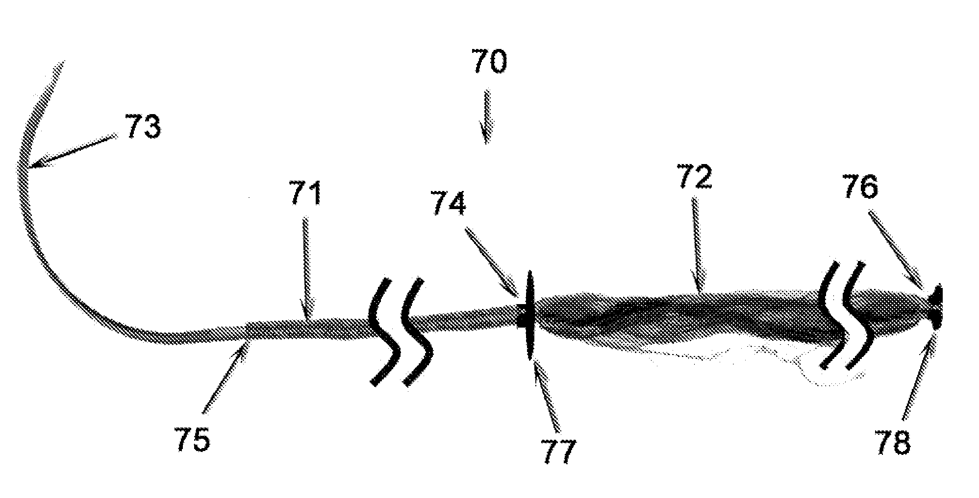
[0195]As noted above, the present invention contemplates the provision of a surgical suture thread and needle kit contained within a sterile package, illustrative examples of which are depicted in FIGS. 8-11. The suture and needle combination used in connection with the instant example is analogous to that depicted in FIGS. 7A and 7B, being composed of a woven exterior sheath (71) housing a filamentous interior core (72). However, alternate configurations, such as those discussed above and / or depicted in FIGS. 5 and 6, may be adapted as well. In either case, the proximal end (75) of the suture thread (70) is attached to the distal end of a suture needle (73), leaving the sharp needle tip (the proximal end) free for tissue penetration.

[0196]Referring to FIGS. 7 and 9, the exterior suture sheath (71) is a woven construction of a biodegradable polyhydroxyalkanoate material such as that manufactured by Tepha, Inc. (Lexington, Mass.) while the interior suture core (7...

example 2

Surgical Suture

[0200]The surgical suture of the instant example is analogous to that depicted in FIGS. 5 and 7, being composed of a woven exterior sheath (71) housing a filamentous interior core (72). In particular, the suture was formed with a woven polyester (PET) sheath using 40 denier bundles of 20 filaments each and a pick count of 80 picks / inch, woven over a core comprised of five 220 denier bundles of 100 filaments of UHMW polyethylene (PE). The finished construct was approximately 0.7 mm diameter. The material was cut into 45 cm sutures. The proximal end was connected to a surgical suture needle. The sheath was retracted from the distal end of the suture towards the proximal end of the suture, such that 35 cm of the interior core was exposed. The core fibers were induced to spread through shear motion applied perpendicularly to the linear axis of the core. The construct was then sterilized. The suture core was coated with fibronectin and then exposed to a stem cell bearing f...

example 3

Surgical Suture

[0201]The surgical suture of the instant example is analogous to that depicted in FIGS. 5 and 7, being composed of a woven exterior sheath (71) housing a filamentous interior core (72). In particular, the suture was formed with a woven polyester (PET) sheath using 40 denier bundles of 20 filaments each and a pick count of 80 picks / inch, woven over a core comprised of a 220 denier bundle of 100 filaments of UHMW polyethylene terephthalate (PE) and a 128 denier bundle of 48 filaments of polyglycolic acid (PGA). The finished construct was in the 0.35 to 0.40 mm diameter range. The material was cut into 40 cm sutures. The proximal end was connected to a surgical suture needle. The sheath was retracted from the distal end of the suture towards the proximal end of the suture, such that 31 cm of the core was exposed. The core fibers were induced to spread through shear motion.

[0202]The distal end of the suture sheath was secured to the inside of a package using a biodegradab...


PUM

PropertyMeasurementUnit
sizeaaaaaaaaaa
compressed lengthaaaaaaaaaa

Abstract

Materials and Methods for immobilizing bioactive molecules, stem and other precursor cells, and other agents of therapeutic value in surgical sutures and other tissue scaffold devices are described herein. Broadly drawn to the integration and incorporation of bioactive materials into suture constructs, tissue scaffolds and medical devices, the present invention has particular utility in the development of novel systems that enable medical personnel performing surgical and other medical procedures to utilize and subsequently reintroduce bioactive materials extracted from a patient (or their allogenic equivalents) to a wound or target surgical site.

Description

PRIORITY[0001]The instant application claims the benefit of U.S. Provisional Application Ser. Nos. 61 / 075,122 filed Jun. 24, 2008 (Attorney Docket No.: BST-002P), 61 / 086,879 filed Aug. 7, 2008 (Attorney Docket No.: BST-002P2), and 61 / 165,055 filed Mar. 31, 2009 (Attorney Docket No.: BST-002P3), the entire contents of which are incorporated by reference herein.FIELD OF THE INVENTION[0002]The present invention relates to the immobilization of bioactive materials, such as stem cells, other biological cells, bioactive molecules, particularly growth factors, and other materials of therapeutic value, in medical devices, particularly surgical sutures and tissue scaffolds, and the use of such sutures and scaffolds to afford controlled delivery of such materials to a patient, more particularly a target tissue site in need thereof. In addition to medical device constructs, precursors, kits and packaging systems therefore, the present invention provides methods of making and using such materia...

Claims


Application Information

Patent Timeline
Patent Type & Authority Applications(United States)
IPC IPC(8): A61B17/04A61L17/06
CPCA61B17/06114A61B17/06166A61B2017/00884A61B2017/00893A61L17/005A61L17/06D06B1/10A61L2300/42A61L2300/604A61L2300/64A61L2300/45A61L2300/414A61B2017/06185A61L2300/404A61B17/064A61F2/02A61L17/08A61L27/38
Inventor SPEDDEN, RICHARD H.PINGEL, LAURA J.SCHON, LEW C.
Owner BIOACTIVE SURGICAL
Why Eureka
Try Eureka