Method for representing interventional instruments in a 3D data set of an anatomy to be examined as well as a reproduction system for performing the method

a technology of interventional instruments and 3d data, which is applied in the direction of instruments, diagnostic recording/measuring, radiation beam directing means, etc., can solve the problems of total occlusion, coronary arteries, and blockage of blood flow, so as to achieve faster and safer minimally invasive therapy

Active Publication Date: 2010-01-28
SIEMENS HEALTHCARE GMBH
View PDF11 Cites 27 Cited by
  • Summary
  • Abstract
  • Description
  • Claims
  • Application Information

AI Technical Summary

Benefits of technology

[0022]The underlying object of the invention is to specify a method of the type mentioned at the start, which makes possible a three-dimensional reconstruction of an instrunent together with a 3D data set. A further object of the invention is to embody a display system of the type mentioned at the start such that 3D image presentations at the correct location, i.e. in the immediate vicinity and in the correct spatial orientation relative to the patient, are made possible for faster and safer minimally-invasive therapy.

Problems solved by technology

This can lead to a total occlusion (“blockage”) of coronary vessels and thereby to a blocking of the blood flow.
This was associated with high risks and long recuperation times of up to six weeks for the patient.
The disadvantage of the new minimally-invasive therapies is that they can only be performed with the aid of x-ray fluoroscopy.
This meant that a spatial assignment was barely possible.
However, this technology has not become established in medicine because of the additional eyeglasses required.
This solution has the disadvantage that only one observer in each case perceives the impression of the 3D image.
In addition the solution does not always operate reliably and has not established itself in the field of medical therapy.
In practice however this is not a simple matter since many medical imaging systems, especially angiography systems, cannot generate any 3D data at a sufficiently high repetition rate in real time in order to make guidance possible.
The known methods of 2D / 3D overlaying are also not able to be used for holographic representation, so that it is not at all clear how such holographic images can be used for guiding an intervention.

Method used


Image

  • Method for representing interventional instruments in a 3D data set of an anatomy to be examined as well as a reproduction system for performing the method
  • Method for representing interventional instruments in a 3D data set of an anatomy to be examined as well as a reproduction system for performing the method
  • Method for representing interventional instruments in a 3D data set of an anatomy to be examined as well as a reproduction system for performing the method

Examples


Embodiment Construction

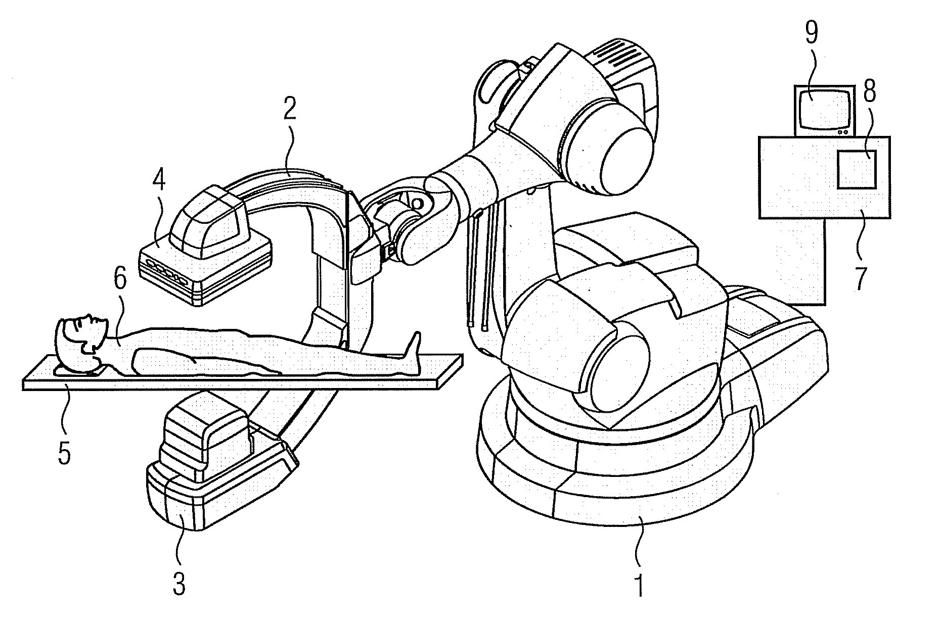
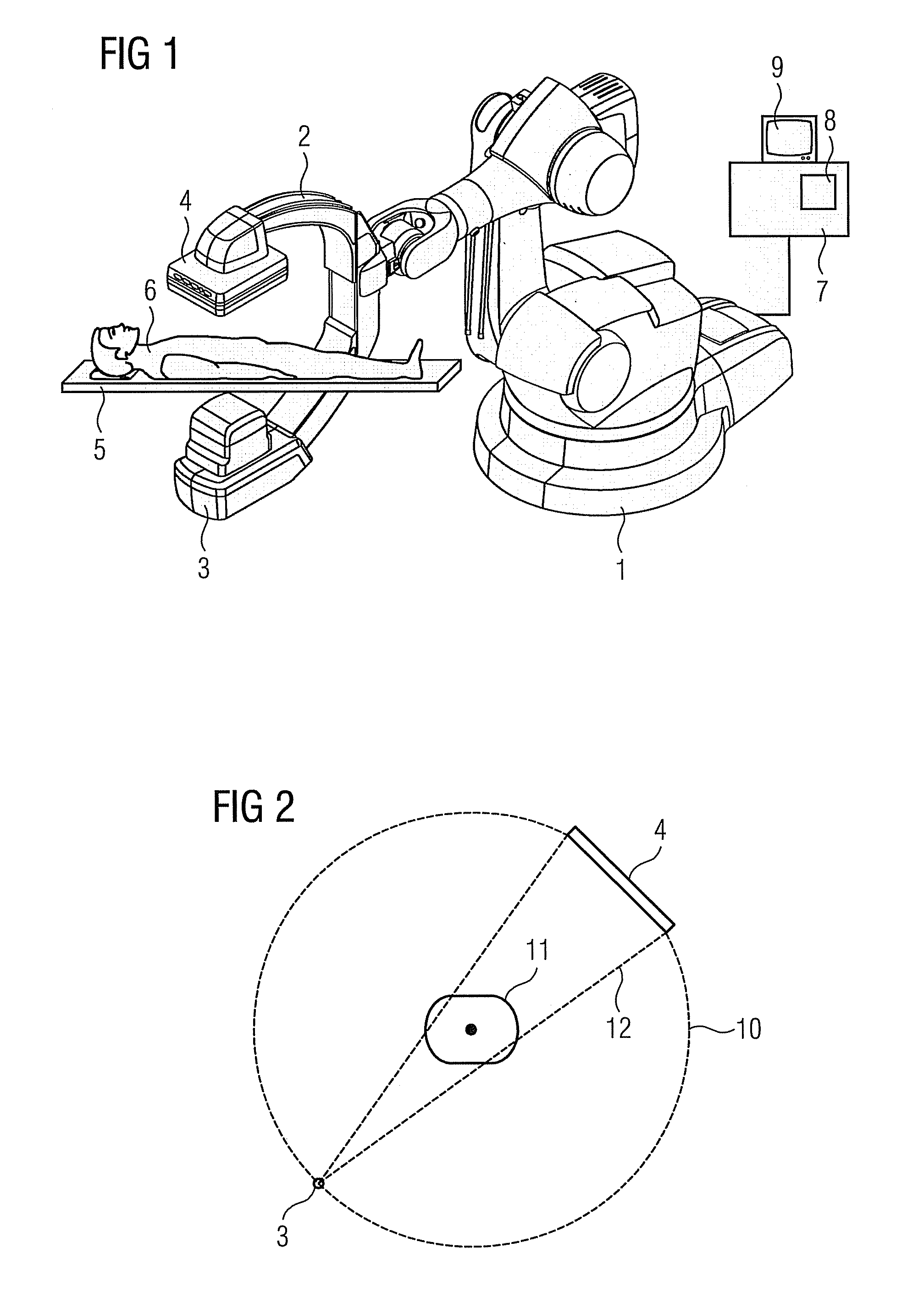
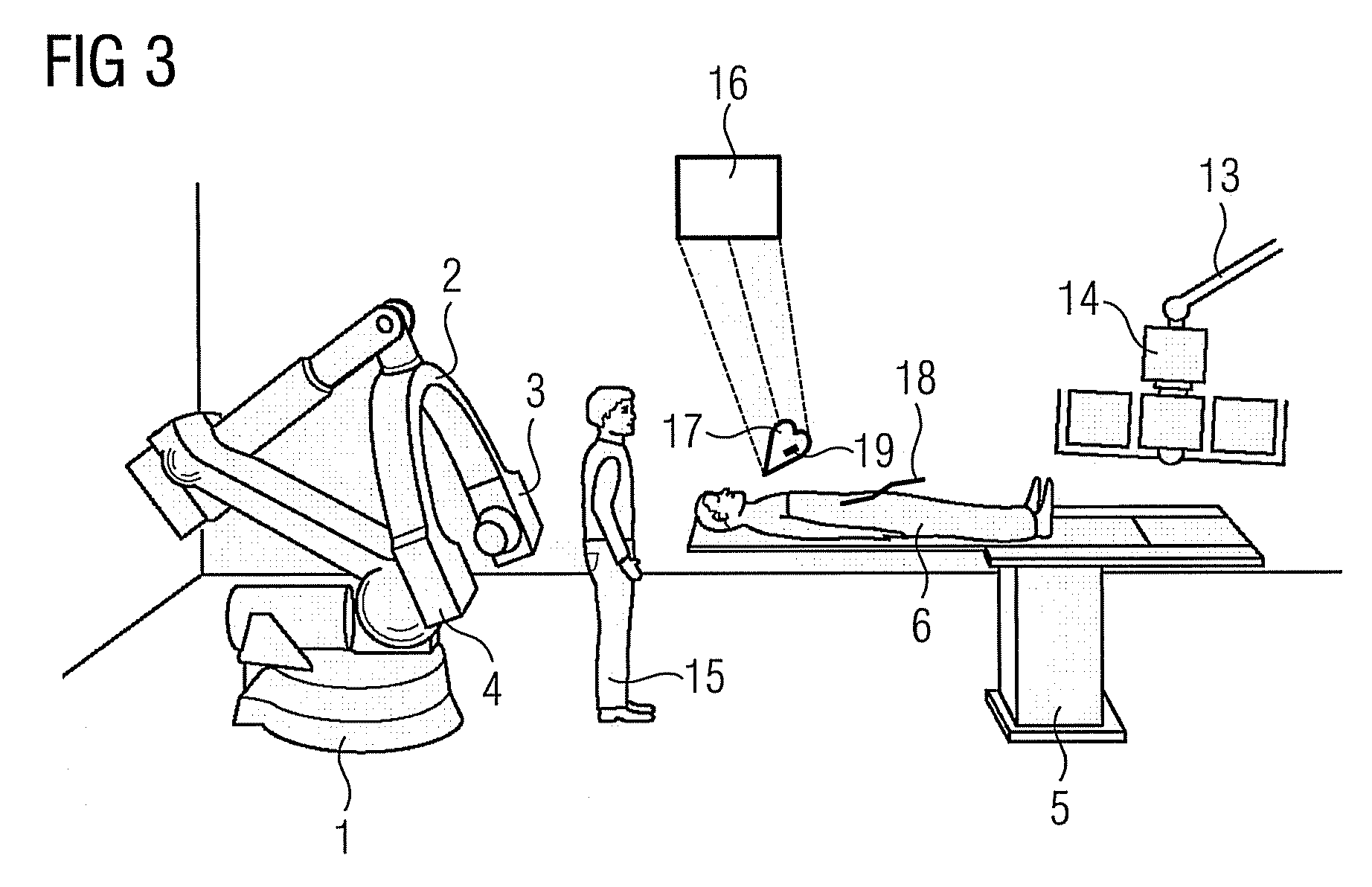
[0046]FIG. 1 shows an x-ray diagnostic device which has a C-arm able to be rotated on a stand in the form of an industrial robot 1, with an x-ray radiation source, for example an x-ray emitter 3, and an x-ray image detector 4 being attached to the ends of said C-arm.

[0047]The x-ray image detector 4 can be a rectangular or square, flat semiconductor detector which is preferably made of amorphous silicon (a-Si).

[0048]Located on a patient bed in the beam path of the x-ray emitter 3 is a patient to be examined so that an image of their heart can be recorded for example. Connected to the x-ray diagnostic device is a system control unit 7 with an image system 8, which receives and processes the image signals of the x-ray detector 4. The x-ray images can then be observed on a monitor 9.

[0049]By means of the industrial robot 1 known from DE 10 2005 012 700 A1 for example, which preferably has six axes of rotation and thus six degrees of freedom, the C-arm 2 can be repositioned as required, ...


PUM

PropertyMeasurementUnit
degrees of freedomaaaaaaaaaa
coloraaaaaaaaaa
Magnetic Resonance Imagingaaaaaaaaaa

Abstract

The invention relates to a method for presenting interventional instruments in a 3D data set of an anatomy to be treated. A 3D data set of the anatomy is recorded before introduction of an interventional instrument. Once the interventional instrument has been applied, the spatial position of the instrument is determined by x-ray fluoroscopy from images created at two different angulations. A 3D model of the instrument is formed from the x-ray images. The 3D model of the instrument is fused with the 3D data set of the anatomy. A 3D hologram is reproduced from the fused 3D data set. The 3D hologram is repeatedly reproduced in real time to follow the application of the instrument in the presentation.

Description

CROSS REFERENCE TO RELATED APPLICATIONS[0001]This application claims priority of German application No. 10 2008 034 686.1 filed Jul. 25, 2008, which is incorporated by reference herein in its entirety.FIELD OF THE INVENTION[0002]The invention relates to a method for representing interventional instruments in a 3D data set of the anatomy to be examined. The invention further relates to a reproduction system for diagnostic imaging systems for performing the method for representing interventional instruments in a 3D data set of the anatomy to be examined.BACKGROUND OF THE INVENTION[0003]In medicine the switch from surgical interventions to minimally-invasive therapies is becoming established for serious illnesses.[0004]Among the most frequent diseases with a fatal outcome are vascular angiopathies, with the resulting diseases such as heart attacks or strokes. The heart attack (myocardiac infarction=MI) is caused by diseases of the coronary vessels. In such cases arteriosclerotic plaque...

Claims


Application Information

Patent Timeline
Patent Type & Authority Applications(United States)
IPC IPC(8): G01N23/223G03H1/08
CPCA61B5/06A61B6/12A61B6/466A61B6/5229A61B6/5241A61B19/5225A61B6/4464A61B2019/5238A61B2019/5293A61B2019/5295A61B5/745A61B6/4458A61B19/5244A61B2090/366A61B2090/367A61B34/20A61B2090/376A61B90/37
Inventor BOESE, JANKLINGENBACK, KLAUS
Owner SIEMENS HEALTHCARE GMBH
Why Eureka
Try Eureka