

Addressable antibody arrays and methods of use
a technology of antibody arrays and antibodies, applied in the field of antibody arrays and methods of use, can solve the problems of inability to parallel, high-throughput analysis of multiple auto antibodies, and lack of universal standards of current methods used in clinical laboratories, so as to enhance sensitivity and specificity, and minimize the amount of biological samples
- Summary
- Abstract
- Description
- Claims
- Application Information
AI Technical Summary
Benefits of technology
Problems solved by technology
Method used
Image

Examples
example 1
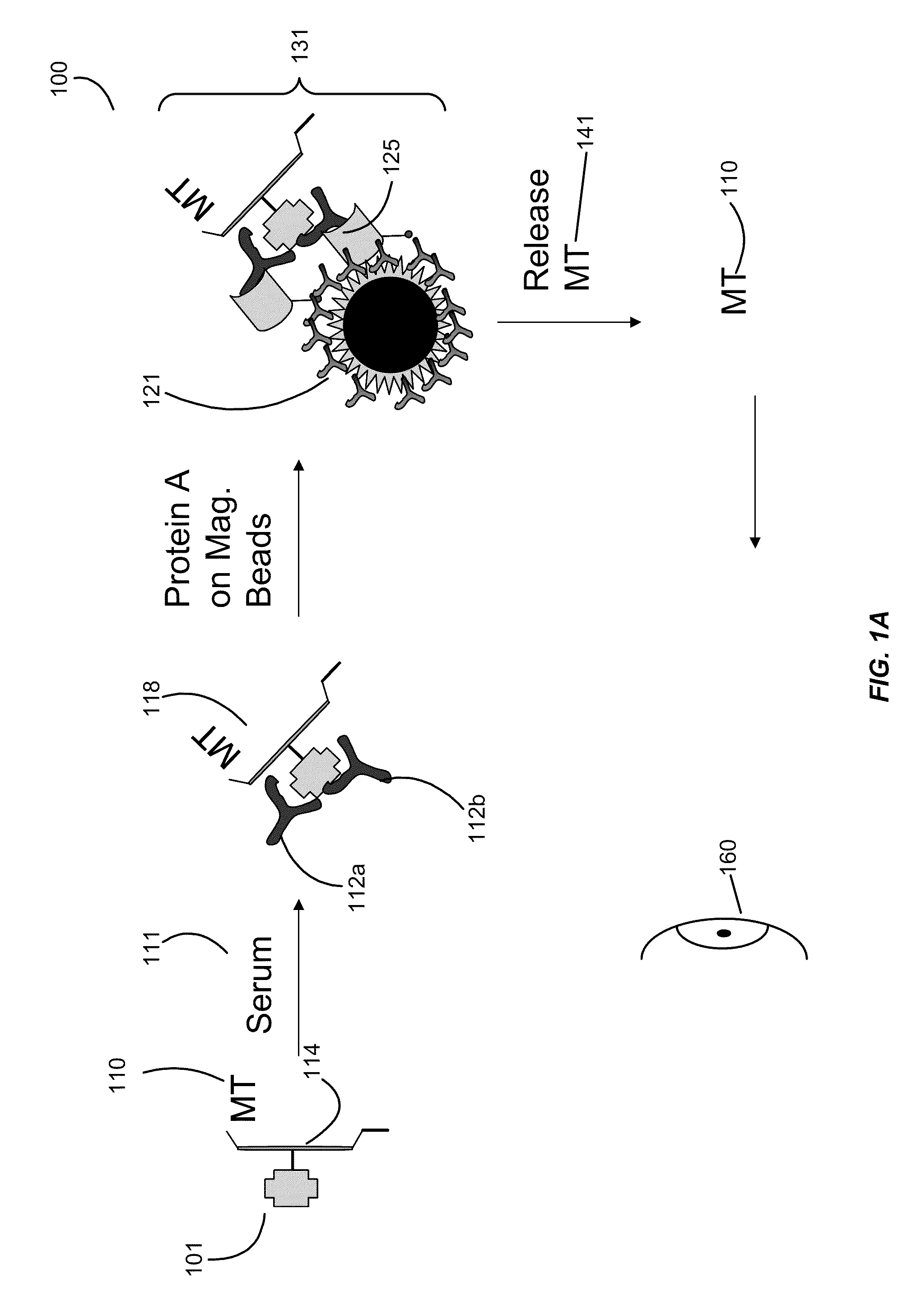
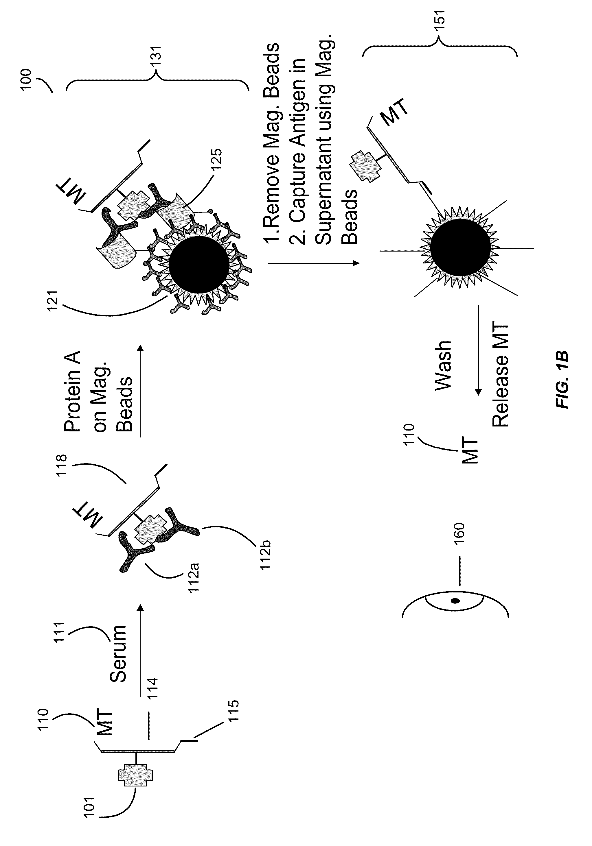
[0109]Example 1 illustrates a mass tag being attached to an antigen.
[0110]Purified GAD-65 is allowed to react with a 50-fold molar excess of benzyl oxy-propionic acid 3-sulfo-N-hydroxysuccinimide ester (1) in 50 mmol / L HEPES, 9 g / L NaCl, pH 7.4, for 4 h at room temperature. Unconjugated reagent is removed by gel filtration on a NAP-5 column (Amersham Biosciences) with 50 mmol / L HEPES, 9 g / L NaCl, 0.5 g / L sodium azide, pH 7.4, as elution buffer. The conjugated protein is stored at 4° C., either in solution or freeze-dried.
[0111]Insulin: Purified Insulin is allowed to react with a 50-fold molar excess of 2-fluoro benzyl oxy-propionic acid 3-sulfo-N-hydroxysuccinimide ester (2) in 50 mmol / L HEPES, 9 g / L NaCl, pH 7.4, for 4 h at room temperature. Unconjugated reagent is removed by gel filtration on a NAP-5 column (Amersham Biosciences) with 50 mmol / L HEPES, 9 g / L NaCl, 0.5 g / L sodium azide, pH 7.4, as elution buffer. The conjugated protein is stored at 4° C., either in solution or freez...
example 2
[0113]Example 2 illustrates a plurality of antigens being bound to an antigen support such as a polymer.
[0114]20 molar excess of purified GAD-65 is allowed to react with benzyl oxy-propionic acid amide conjugated (˜1:1 conjugate), 3-sulfo-N-hydroxysuccinimide ester activated carboxy dextran (500 KD) in 50 mmol / L HEPES, 9 g / L NaCl, pH 7.4, for 4 h at room temperature. Unconjugated reagent is removed by gel filtration (Amersham Biosciences) with 50 mmol / L HEPES, 9 g / L NaCl, 0.5 g / L sodium azide, pH 7.4, as elution buffer. The conjugated dextran protein is stored at 4° C., either in solution or freeze-dried. The insulin and IA-2 conjugates were prepared in identical fashion (the mass tags were different).
[0115]20 molar excess of purified GAD-65 and an addressable oligo with a 5′ thiol (50 molar excess) is allowed to react with biotin and bromoacetyl conjugated (˜1:1 conjugate), 3-sulfo-N-hydroxysuccinimide ester activated carboxy dextran (500 KD) in 50 mmol / L HEPES, 9 g / L NaCl, pH 7.4,...
example 3
[0119]Example 3 illustrates an assay of the present invention.
[0120]This example illustrates the assay with a specific example and detects the tag with mass spectrometry.
[0121]Serum samples (2 μL) were incubated with a mixture of mass-tag labeled IA-2, insulin and GAD-65 aliquots (40 ng) in 50 μL of 50 mmol / L Tris-HCl, 150 mmol / L NaCl, pH 7.4, containing 1 mL / L Tween 20 (TBST) overnight at 4° C. The formed immune complexes were captured by adding 5 μL of Protein A magnetic or Sepharose beads (Amersham Biosciences). Protein A magnetic beads are also available from New England Biolabs, Invitrogen Dynal AS (Dynabeads® Protein A), GenScript Corporation (Protein A MagBeads), Polysciences, Inc. (BioMag® Protein A), and Thermo Scientific Pierce Protein Research Products (MagnaBind™ Protein A Beads). After a 1 h incubation on a shaker at 4° C., the samples were transferred to a 96-well opaque filtration plate with a 0.45 μm Durapore filter at the bottom of each well (Millipore). The samples...
PUM
 Abstract
Description
Claims
Application Information