Forward-Looking Optical Coherence Tomography Endoscope
an endoscope and optical coherence technology, applied in the field of endoscopy, can solve the problems of not being satisfactory, unable to readily determine the depth of tumor invasion by either method, and the relative survival rate of 5 years is less than 20%, and achieves the effects of low external vibration of the probe, low cost, and high resolution
- Summary
- Abstract
- Description
- Claims
- Application Information
AI Technical Summary
Benefits of technology
Problems solved by technology
Method used
Image
Examples
example 1
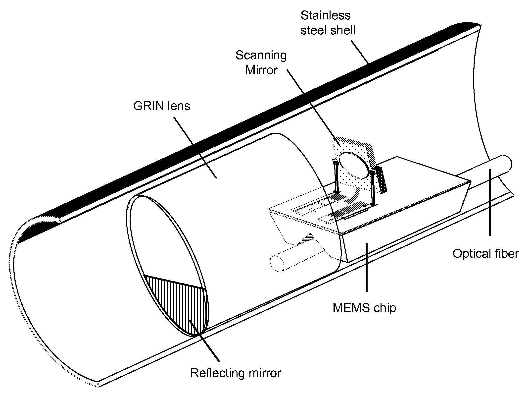
[0038]A simulation of the optical system was first conducted with large scale components. The distributed focusing of a π / 4 GRIN lens was approximated with a series of three equally-spaced conventional lenses. A HeNe laser beam at a wavelength of 0.633 μm was focused by a microscope objective to a numerical aperture of 0.12 at the entrance to the lens, approximately the same numerical aperture as that for light entering the GRIN lens in the system depicted in FIG. 3. The laser light then made three passes through the lenses and was reflected from the mirrors as shown, before coming to a sharp focus downstream from the lens. This experiment demonstrated in principle the successful use of the novel optical system.
example 2
[0039]FIG. 4 depicts a side-viewing endoscope OCT system for comparison. In many respects, except for those features of the new system that are novel, the new system may be operated in a generally similar manner to that used for a side-viewing endoscope OCT system. There is a superluminescent light source having a center wavelength of 1300 nm and a 100 nm bandwidth (Superlum D1300-HP, Moscow, Russia), for a coherence length on the order of 10 μm. A center wavelength of 1300 nm was chosen because of the ready availability of photonics components, and because tissue attenuation at that wavelength is low, allowing relatively deep imaging. Light from the source is split by a fiber-based Michelson interferometer into two paths: one path travels to the sample arm, where light is focused onto tissue or other sample; and one path travels to a reference arm, which includes a Fourier-domain, rapid-scanning optical delay line to modulate the optical path length. A relatively low coherence leng...
example 3
[0041]To demonstrate the feasibility in principle of imaging the interior of a rat stomach through OCT, a side-firing OCT system was used to image excised specimens. The stomachs of three rats were excised, opened, rinsed with saline, and pinned flat to a wax block. The light source used was an 890 nm superluminescent diode. The OCT system had a resolution of approximately 8 μm. Images 1 mm deep by 6 mm lateral were successfully taken. The OCT system easily imaged through the thin (250-300 μm) layers of the stomach, and all major layers could be identified. Blood vessels were also visible. This preliminary study demonstrated that OCT can perform imaging that would be useful, for example, in diagnosing gastric cancer. To enhance in vivo image quality, it is preferred to place the animals (or patients) on a liquid diet (e.g., Pedialyte™) for ˜12 hours before imaging, to reduce solids content in the stomach.
PUM
 Abstract
Description
Claims
Application Information
 - R&D
- Intellectual Property
- Life Sciences
- Materials
- Tech Scout
- Unparalleled Data Quality
- Higher Quality Content
- 60% Fewer Hallucinations
Browse by: Latest US Patents, China's latest patents, Technical Efficacy Thesaurus, Application Domain, Technology Topic, Popular Technical Reports.
© 2025 PatSnap. All rights reserved.Legal|Privacy policy|Modern Slavery Act Transparency Statement|Sitemap|About US| Contact US: help@patsnap.com


