Method and Apparatus for the Ablation of Gastrointestinal Tissue

a technology for gastrointestinal tissue and ablation, which is applied in the field of medical equipment and procedures, can solve the problems of high risk of complications, bleeding and perforation, and difficult removal of flat sessile polyps

Active Publication Date: 2010-05-06
SANTA ANNA TECH LLC
View PDF106 Cites 104 Cited by
  • Summary
  • Abstract
  • Description
  • Claims
  • Application Information

AI Technical Summary

Problems solved by technology

While most polyps are detected on colonoscopy and easily removed using a snare, flat sessile polyps are hard to remove using the snare technique and carry a high risk of complications, such as bleeding and perforation.
The presence of submucosal uterine fibroids and a large uterus size have been shown to decrease the efficacy of standard endometrial ablation.
Conventional techniques have inherent limitations, however, and have not found widespread clinical applications.
This ablation mechanism is operator dependent, cumbersome and time consuming.
Second, because the target tissue is moving due to patient movement, respiration movement, normal peristalsis and vascular pulsations, the depth of ablation of the target tissue is inconsistent and results in a non-uniform ablation.
Superficial ablation results in incomplete ablation with residual neoplastic tissue left behind.
Deeper ablation results in complications such as bleeding, stricture formation and perforation.
All of these limitations and complications have been reported with conventional devices.
However, the rigid electrode does not accommodate for variations in esophageal size and is ineffective in ablating tortuous esophagus, proximal esophageal lesions as an esophagus narrows towards the top, and esophagus at the gastroesophagal junction due to changes in the esophagus diameter.
Nodular disease in Barrett esophagus also cannot be treated using the rigid bipolar RF electrode.
Due to its size and rigidity, the electrode cannot be passed through the scope.
In addition sticking of sloughed tissue to the electrode impedes with delivery of radiofrequency energy resulting in incomplete ablation.
The tissue is then exposed to a selected wavelength of light which activates the photo-sensitizer and results in tissue destruction.
PDT is associated with complications such as stricture formation and photo-sensitivity which has limited its use to the most-advanced stages of the disease.
In addition, patchy uptake of the photosensitizer results in incomplete ablation and residual neoplastic tissue.
These techniques suffer the shortcoming of the traditional hand-held devices.
Treatment using this probe is cumbersome and requires operator control under direct endoscopic visualization.
Continuous movement in the esophagus due to respiration or cardiac or aortic pulsations or movement causes an uneven distribution of the ablative agent and results in non-uniform and / or incomplete ablation.
Close or direct contact of the catheter to the surface epithelium may cause deeper tissue injury, resulting in perforation, bleeding or stricture formation.
Too distant a placement of the catheter due to esophageal movement will result in incomplete Barrett ablation, requiring multiple treatment sessions or buried lesions with a continued risk of esophageal cancer.
Expansion of cryogenic gas in the esophagus results in uncontrolled retching which may result in esophageal tear or perforation requiring continued suctioning of cryogen.
Both these treatments, have the previously mentioned limitations.
Hence, most large flat polyps undergo surgical resection due to high risk of bleeding, perforation and residual disease using traditional endoscopic resection or ablation techniques.
When the balloon catheter is deflated, the epithelium sticks to the catheter and sloughs off, thereby causing bleeding.
Blood can interfere with delivery of energy i.e. energy sink.
In addition reapplication of energy will result in deeper burn in the area where superficial lining has sloughed.
Further, balloon catheters cannot be employed for treatment in non cylindrical organs, like the uterus or sinuses, and also do not provide non-circumferential or focal ablation in a hollow organ.
Additionally, if used with cryogens as ablative agents, which expand exponentially upon being heated, balloon catheters may result in a closed cavity and trap the escape of cryogen, resulting in complications such as perforations and tears.

Method used


Image

  • Method and Apparatus for the Ablation of Gastrointestinal Tissue
  • Method and Apparatus for the Ablation of Gastrointestinal Tissue
  • Method and Apparatus for the Ablation of Gastrointestinal Tissue

Examples


Embodiment Construction

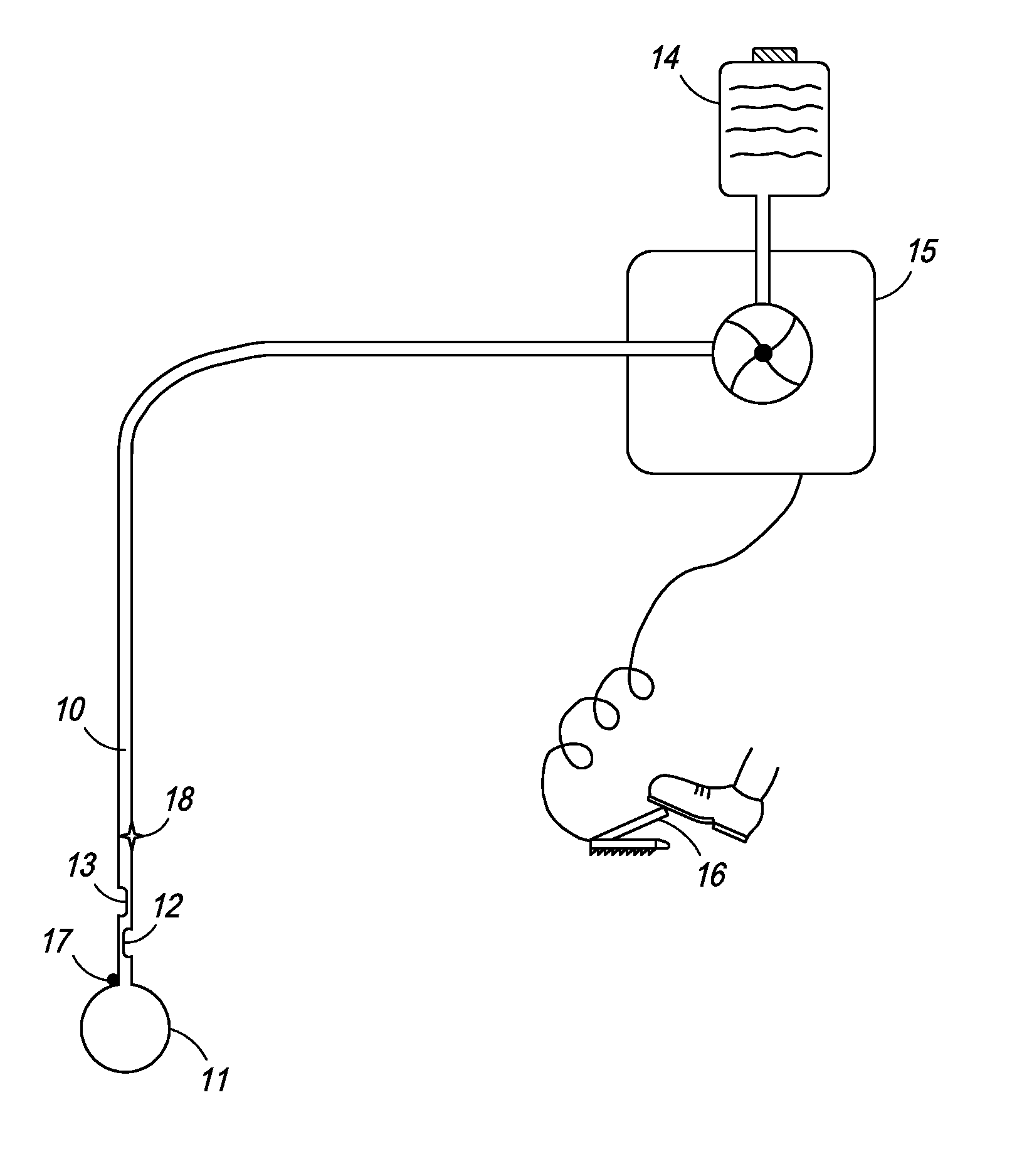
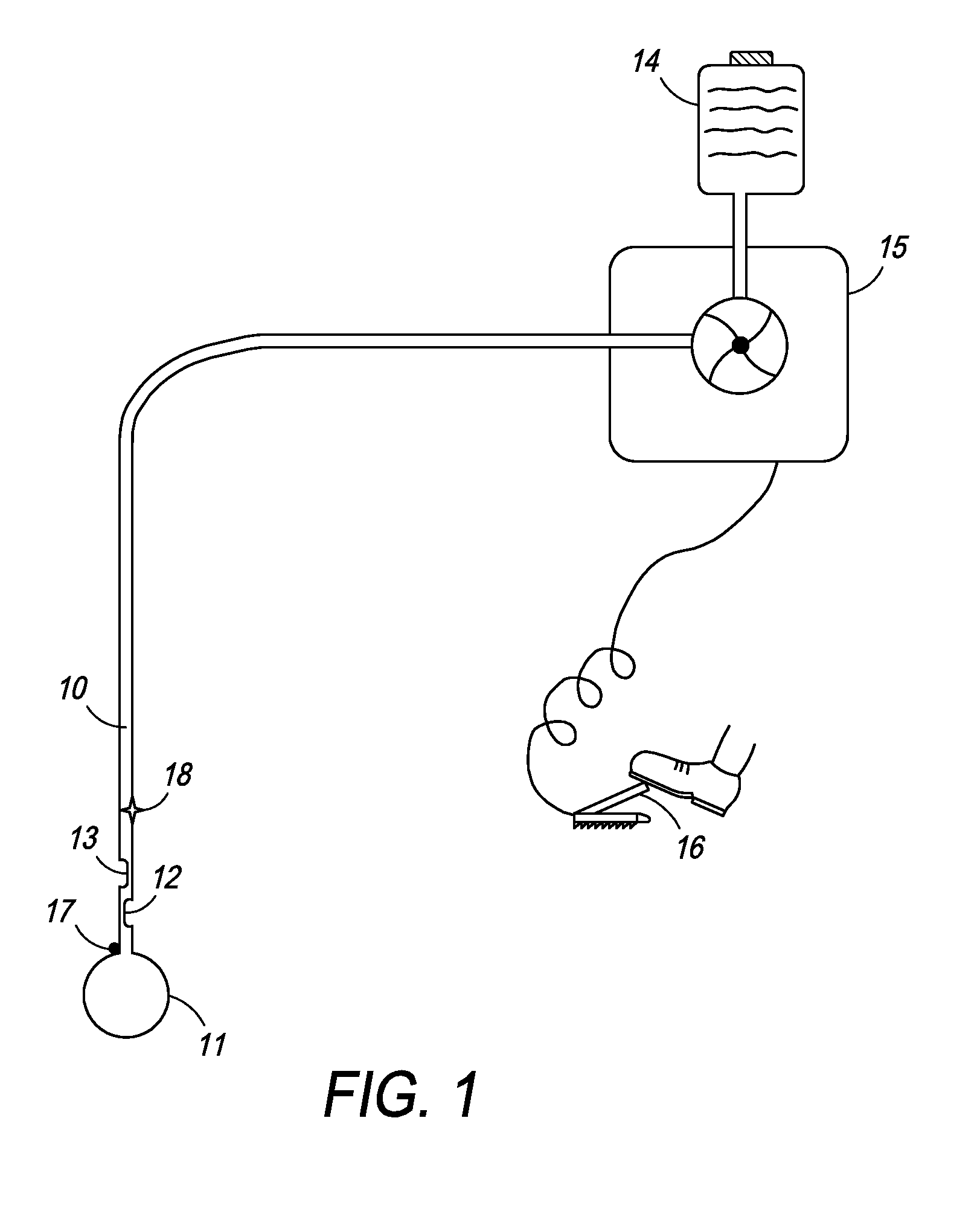
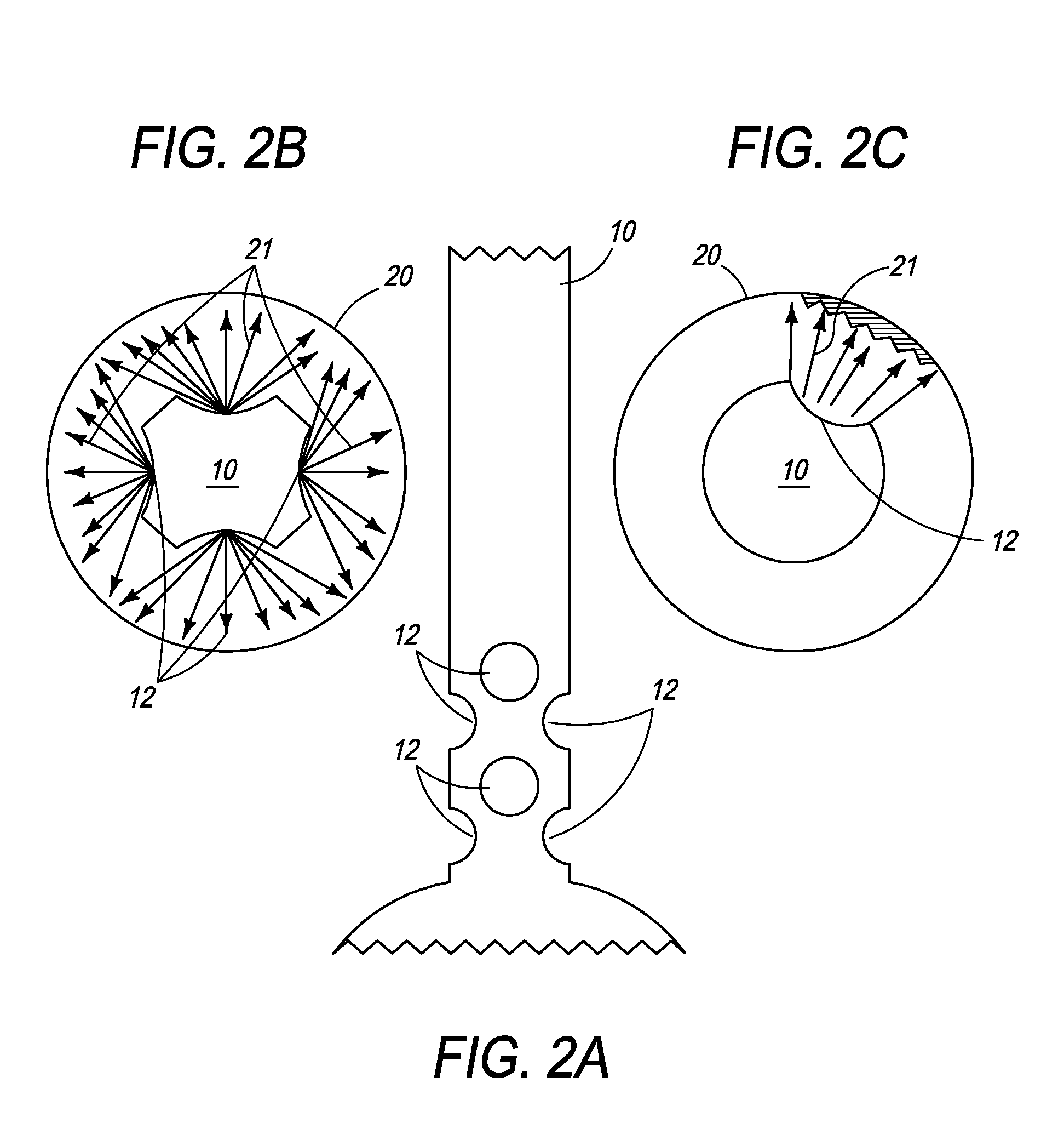
[0057]The present invention provides an ablation device comprising a catheter with one or more centering or positioning attachments at one or more ends of the catheter to affix the catheter and its infusion port at a fixed distance from the ablative tissue which is not affected by the movements of the organ. The arrangement of one or more spray ports allows for uniform spray of the ablative agent producing a uniform ablation of large area such as Barrett esophagus. The flow of ablative agent is controlled by the microprocessor and depends upon one or more of the length or area of tissue to be ablated, type and depth of tissue to be ablated and distance of the infusion port from the tissue to be ablated.

[0058]“Treat,”“treatment,” and variations thereof refer to any reduction in the extent, frequency, or severity of one or more symptoms or signs associated with a condition.

[0059]“Duration” and variations thereof refer to the time course of a prescribed treatment, from initiation to co...


PUM


Abstract

The present invention is directed toward a device that performs ablation of tissue. The device has a catheter with a shaft through which an ablative agent can travel, a first positioning element attached to the catheter shaft at a first position and a second positioning element attached to the catheter shaft at a second position. The shaft also has ports through which the ablative agent can be released.

Description

CROSS-REFERENCE[0001]The present invention relies on U.S. Provisional Application No. 61 / 102,885, filed on Oct. 6, 2008, for priority and is hereby incorporated herein by reference.FIELD OF THE INVENTION[0002]The present invention relates to medical apparatus and procedures. More particularly, the present invention relates to a device for ablation of tissue in a hollow organ comprising a centering or positioning attachment in order to position the device at a consistent distance from the tissue to be ablated.BACKGROUND OF THE INVENTION[0003]Colon polyps affect almost 25% of the population over the age of 50. While most polyps are detected on colonoscopy and easily removed using a snare, flat sessile polyps are hard to remove using the snare technique and carry a high risk of complications, such as bleeding and perforation. Recently, with improvement in imaging techniques, more flat polyps are being detected. Endoscopically unresectable polyps require surgical removal. Most colon can...

Claims


Application Information

Patent Timeline
Patent Type & Authority Applications(United States)
IPC IPC(8): A61B18/04
CPCA61B5/1076A61B2018/00488A61B18/04A61B2017/00084A61B2018/048A61M25/1011A61M2205/3368A61B2018/00577A61B2018/00642A61B2018/00791A61B18/1492A61M2202/0468A61M25/10A61B2560/04A61M2205/3331A61B2018/00773A61B17/24A61B2018/00279A61B2018/00029
Inventor SHARMA, VIRENDER K.
Owner SANTA ANNA TECH LLC
Why Eureka
Try Eureka