
Ultrafast laser system for biological mass spectrometry

a laser system and mass spectrometry technology, applied in the field of mass spectrometry, can solve the problems of incomplete backbone fragmentation, limited application of mass spectrometry and associated methodologies for comprehensive proteome analysis, and significant limitations of mass spectrometry and associated methodologies, so as to prevent the redistribution of energy and fast fragmentation of ions

Active Publication Date: 2010-05-20
BOARD OF TRUSTEES OPERATING MICHIGAN STATE UNIV
View PDF49 Cites 30 Cited by
  • Summary
  • Abstract
  • Description
  • Claims
  • Application Information

AI Technical Summary

Benefits of technology

"The patent text describes a new system for mass spectrometry that uses a laser to analyze biological samples. The laser emits a short laser beam pulse that causes the ultrafast loss of an electron from charged ions, resulting in the ultrafast loss of an election from the charged ions for optional further fragmentation and more detailed mass spectrometry analysis. The system also uses a femtosecond laser beam pulse to cause the ultrafast loss of an election from the charged ions for optional further fragmentation and more detailed mass spectrometry analysis. The use of a laser system for biological mass spectrometry provides greater control and selectivity over the fragmentation reactions of peptide ions, allowing for more comprehensive proteome analysis. The patent text also describes a method for using a shaped laser pulse to isolate and further ionize a specimen for further analysis. Overall, the patent text describes a new system and method for mass spectrometry that improves the accuracy and efficiency of identifying and characterizing peptides and post-translational modifications in biological samples."

Problems solved by technology

However, the generally limited ability to selectively control or direct the fragmentation reactions of peptide ions during CID-MS / MS towards the formation of structurally informative ‘sequence’ ions (i.e., those resulting from amide peptide bond cleavages) or ‘non-sequence’ ions (i.e., those resulting from cleavage of amino acid side chains that are characteristic of the presence of post translational modifications), placed significant limitations on the application of mass spectrometry and associated methodologies for comprehensive proteome analysis.
However, these approaches typically did not have bond-selective control over the site of energy absorption from the laser pulse, due to rapid intramolecular vibrational relaxation that occurred prior to bond cleavage, and typically required the presence of a chromophore that was able to absorb energy at the wavelength of the laser to induce fragmentation.
In practice, collision induced dissociation, whereby energy deposition occurs through ion-molecule collisions followed by internal vibrational energy redistribution prior to dissociation, often resulted in incomplete backbone fragmentation, or the dominant loss of labile groups from side chains containing post-translational modifications such as phosphorylation, particularly for peptides observed at low charge states.
However, each of these methods suffers from certain limitations.
The drawback to this approach is that short-wavelength laser wavelengths are difficult to generate especially with high energy per pulse.
The newly created ion can also acquire additional energy which leads to fragmentation.

Method used


Image

  • Ultrafast laser system for biological mass spectrometry
  • Ultrafast laser system for biological mass spectrometry
  • Ultrafast laser system for biological mass spectrometry

Examples


Embodiment Construction

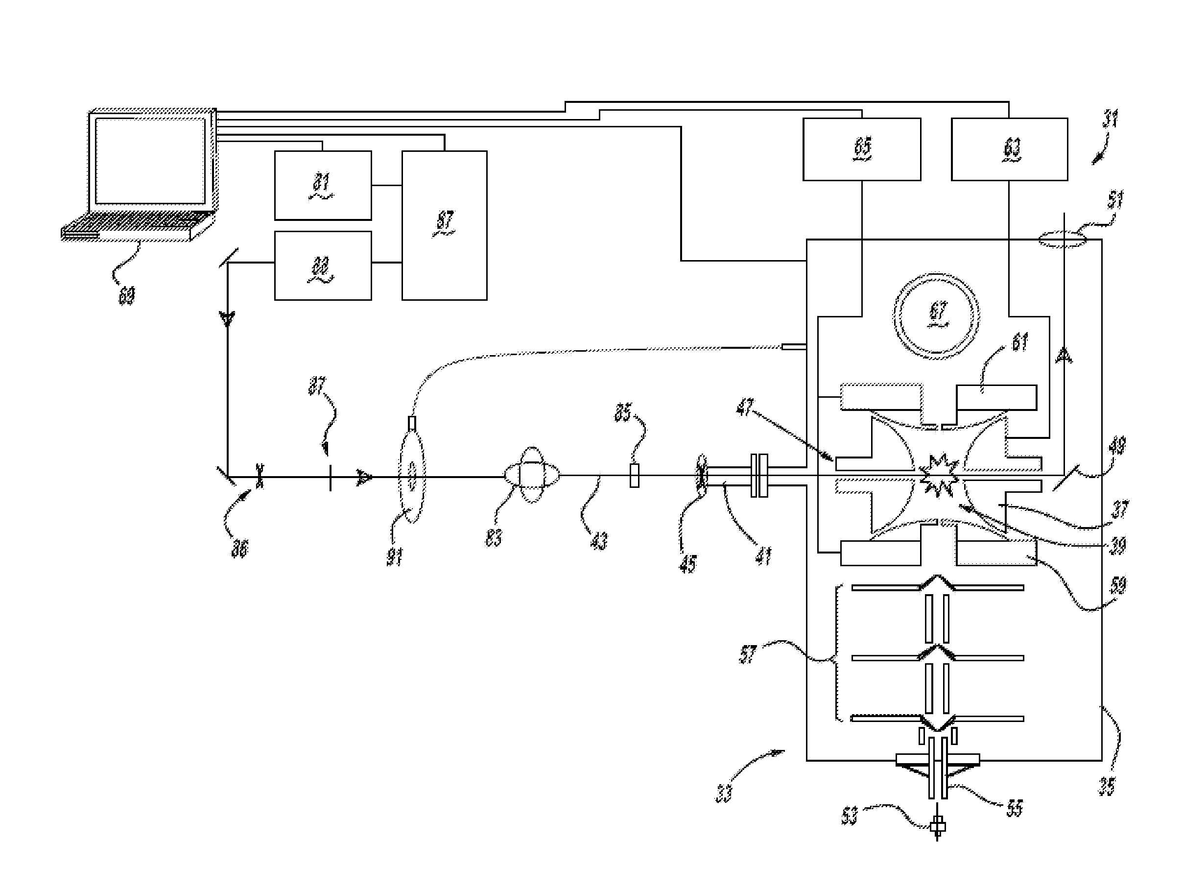
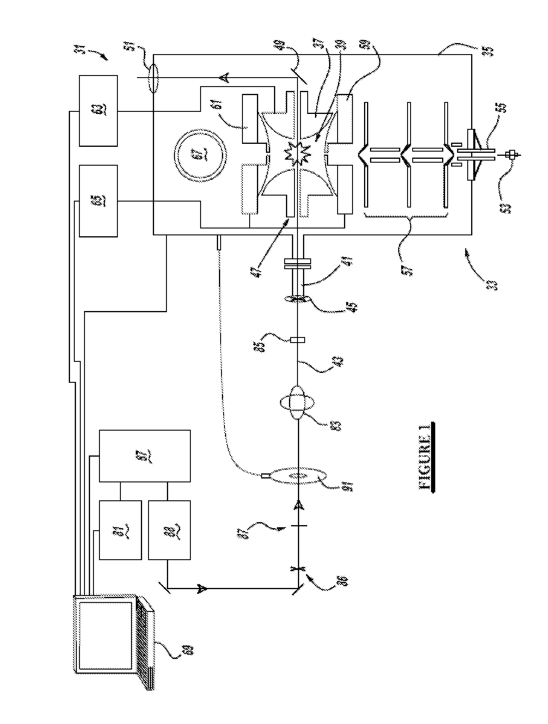
[0036]Referring to FIG. 1, a preferred embodiment of the ultrafast laser system 31 of the present invention includes a 3D ion-trap mass spectrometer 33 (model LCQ Deca XP Plus, Thermo Scientific, San Jose, Calif.) modified using the following specific conditions: a ½″ hole is drilled through the left hand side of a vacuum manifold 35 at a 60° downward angle, in line with the center of a ring electrode 37 of an ion-trap 39, and an aluminum conflat nipple is welded to the manifold. A series of ½″ KwikFlange components are used to construct a vacuum-sealed entrance port 41 for a laser pulse 43, capped with a 1″ diameter fused silica window 45. A 5 mm hole 47 is drilled through ring electrode 37 of the ion-trap, and quartz spacers on either side of the ring electrode are notched to allow clear passage of the laser pulse through the ion-trap. A silver mirror 49, mounted on a custom-cut aluminum block, is then fixed to vacuum manifold 35 on the far side of the trap to direct laser beam pu...


PUM


Abstract

One aspect of the system provides the use of a laser with a mass spectrometer. Another aspect of the present application employs a laser emitting a pulse of less than one picosecond duration into an ion-trap mass spectrometer. In yet another aspect of the present application, a femtosecond laser beam pulse is emitted upon an ionized specimen to remove at least one electron therefrom.

Description

CROSS-REFERENCE TO RELATED APPLICATIONS[0001]This application Claims the benefit of U.S. Provisional Application No. 61 / 114,809, filed on Nov. 14, 2008, which is incorporated by reference herein.STATEMENT OF GOVERNMENT INTEREST[0002]A portion of this invention was made with U.S. government support under Grant Nos. CHE-0547940 and CHE-0647901 from the National Science Foundation. The U.S. government may have certain rights in this invention.BACKGROUND AND SUMMARY[0003]This application relates generally to mass spectrometry and more particularly to an ultrafast laser system for biological mass spectrometry.[0004]Over the past decade, mass spectrometry (“MS”) has become a key analytical tool for analyzing proteins and metabolites. MS has been used to identify post-translational modifications (“PTMs”) of proteins, which are in some cases the signature of aging processes and malignant disease, making them valuable markers for medical diagnosis. Typically, complex protein mixtures or indi...

Claims


Application Information

Patent Timeline
Patent Type & Authority Applications(United States)
IPC IPC(8): H01J49/26B01D59/44
CPCH01J49/0059H01J49/4205H01J49/162
Inventor DANTUS, MARCOSREID, GAVIN
Owner BOARD OF TRUSTEES OPERATING MICHIGAN STATE UNIV
Learn More