Method for detection, characterization and/or identification of microorganisms in a sealed container
- Summary
- Abstract
- Description
- Claims
- Application Information
AI Technical Summary
Benefits of technology
Problems solved by technology
Method used
Image
Examples
example 1
Blood Culture (Culture Medium and Blood Sample) in Quartz Cuvette in Waterbath Adapter
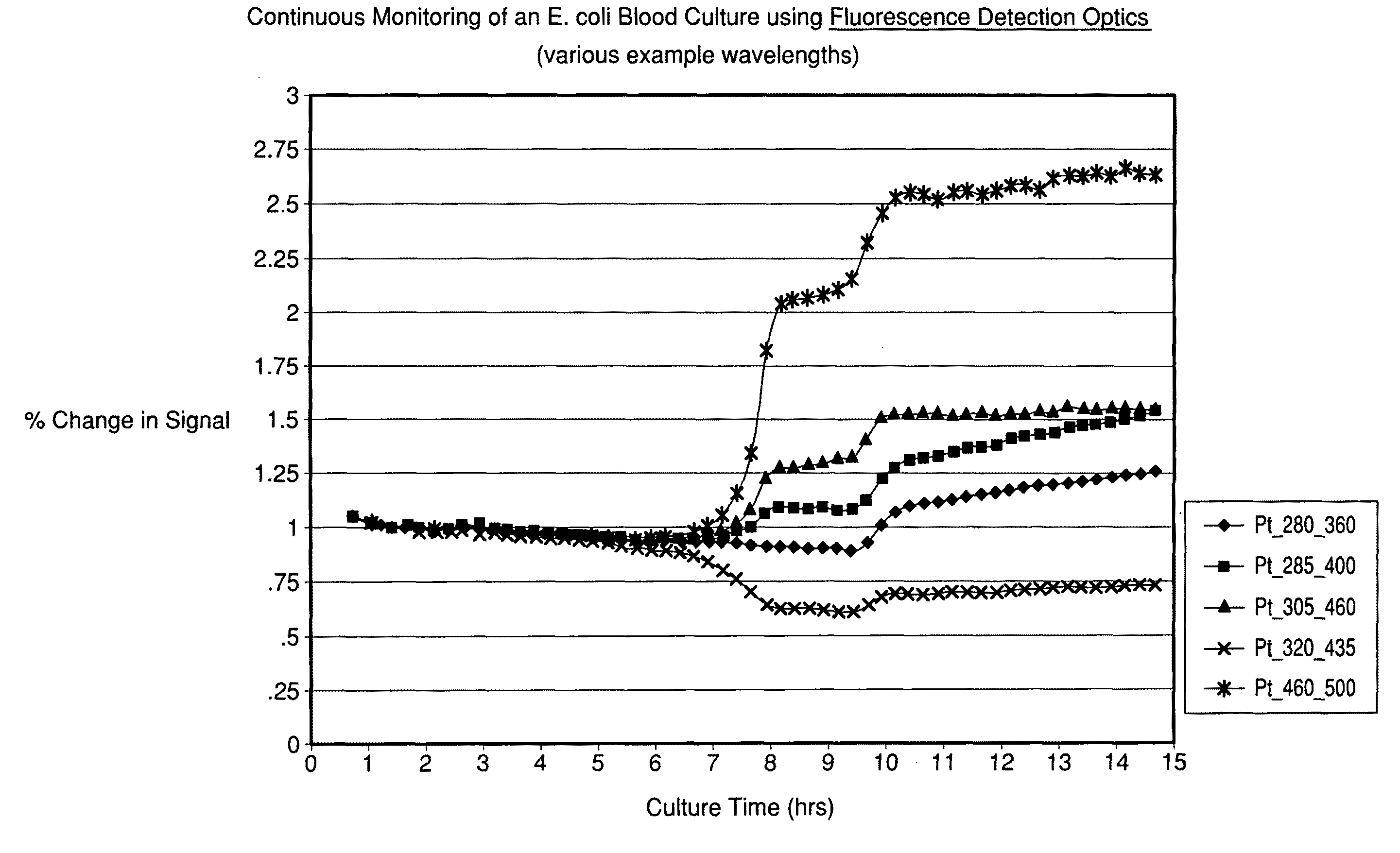
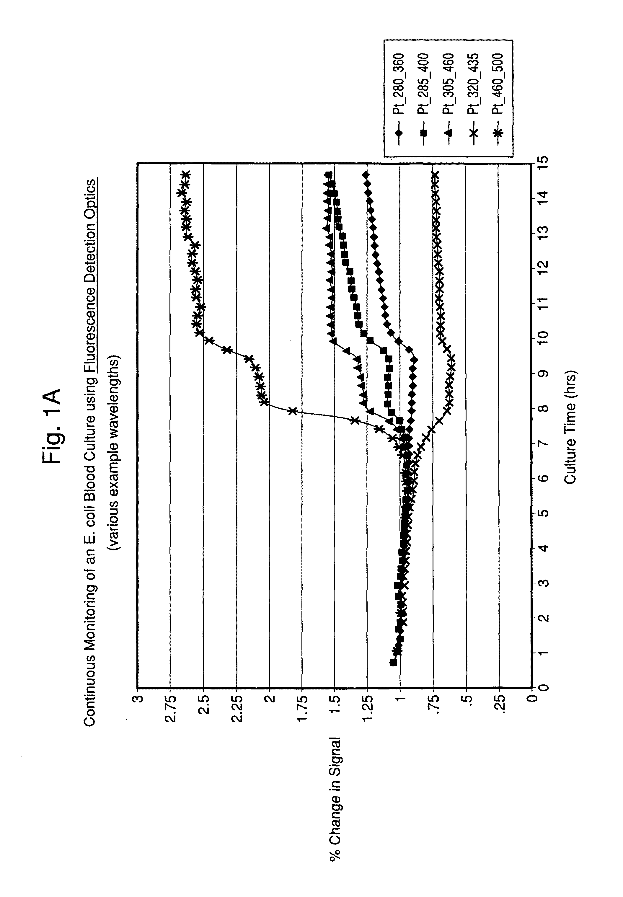
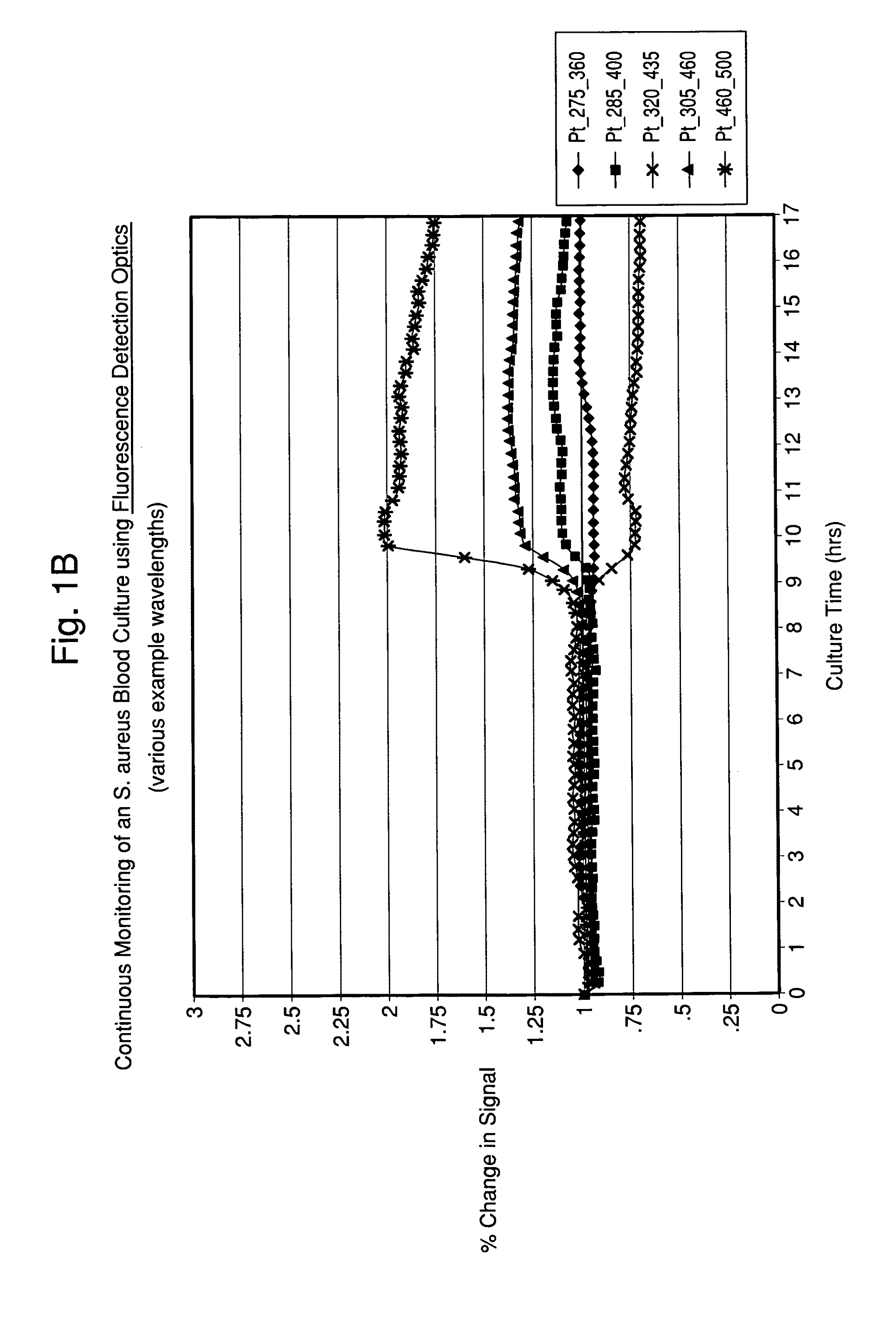
[0103]A Fluorolog® 3 fluorescence spectrophotometer system (Horiba Jobin-Yvon Inc., New Jersey) was modified with a temperature-controlled front-face cuvette holder to enable the incubation and continuous monitoring of seeded blood cultures contained in sterile cuvettes. The fluorescence was recorded using a photomultiplier tube. Fluorescence signal strength was taken at several different wavelengths and saved. The readings were compared to experimentally determined strengths for different types of microorganism. In addition, further analysis of the spectra was conducted so that microbial identification to a given level of confidence was achieved.
[0104]Seeded blood cultures were set up in autoclaved 1.0 cm screw-capped quartz cuvettes (Starna, Inc.) containing a stir bar for agitation. To the cuvette was added 2.4 mL of standard blood culture medium, 0.6 mL of fresh normal human blood and 0.05 mL o...
example 2
Identification of Microorganisms Using Measurements of Microbial Pellets and Comparison to Microbial Suspensions
[0107]Several investigators have previously described using right-angle fluorescence measurements of dilute suspensions of pure microorganisms in order to identify them. We compared the effectiveness of this traditional method with our novel approach of front face measurement of a sedimented microbial pellet within the base of a proprietary UV-transparent container. Further, we compared the effectiveness of two detectors for the front face measurements; a PMT detector connected to a double grating spectrometer, and a CCD detector connected to a single grating spectrometer. These experiments were conducted using microbial colonies grown on agar plates. A panel of 42 strains representing 7 species (S. aureus, S. epidermidis, E. coli, K. oxytoca, C. albicans, C. tropicalis and E. faecalis) were tested in each of the following three optical configurations:[0108]1. A 0.40 OD at...
example 3
Method for the Rapid Identification of Blood Culture Isolates By Intrinsic Fluorescence
[0114]To establish the utility of this method, we built a database of 373 strains of microorganisms representing the most prevalent 29 species known to cause sepsis. These organisms were “seeded” at a low inoculum into BacT / ALERT® SA bottles containing 10 mLs of human blood. Positive blood culture broth was treated as follows:[0115]1. A 2.0 mL sample of positive broth was mixed with 1.0 mL of selective lysis buffer (0.45% w / v Brij® 97+0.3M CAPS, pH 11.7), then placed in a 37° C. water bath 1 minute.[0116]2. A 1.0 mL sample of lysate was overlayed onto 0.5 mL of density cushion (24% w / v cesium chloride in 10 mM Hepes ph 7.4+0.005% Pluronic F-108) contained in a custom-built optical separation tube. A polypropylene ball was present on the surface of the density cushion to facilitate loading without disturbing the two aqueous phases.[0117]3. The optical separation tube was sealed with a screw-cap and...
PUM
 Abstract
Description
Claims
Application Information
 - R&D
- Intellectual Property
- Life Sciences
- Materials
- Tech Scout
- Unparalleled Data Quality
- Higher Quality Content
- 60% Fewer Hallucinations
Browse by: Latest US Patents, China's latest patents, Technical Efficacy Thesaurus, Application Domain, Technology Topic, Popular Technical Reports.
© 2025 PatSnap. All rights reserved.Legal|Privacy policy|Modern Slavery Act Transparency Statement|Sitemap|About US| Contact US: help@patsnap.com


