Integrated Bioanalyzer

a bioanalyzer and integrated technology, applied in the field of integrated bioanalyzers, can solve the problems of increasing the potential for contamination, unable to differentiate viable from non-viable organisms, and unable to differentiate viable from non-viable organisms, and achieve the effect of adequate clarity

Active Publication Date: 2010-05-27
PHOTONIC BIOSYST
View PDF18 Cites 24 Cited by
  • Summary
  • Abstract
  • Description
  • Claims
  • Application Information

AI Technical Summary

Benefits of technology

[0029]The invention is preferably used for identification and quantification of microorganisms, in particular with analyses that require or are based on growth and propagation of the organisms. The method and filter apparatus for the collection of the organisms provides additionally for the culture of said organisms with concurrent optical analyses of the contents of the filter apparatus. The invention enables a simple means of preparing a fluid sample and the culturing of cells and their analysis, in the protection of the enclosed environment of the filter apparatus, which is vital to the assay as exposure to contaminant organisms external to the assay can invalidate the analysis. Said protection of the filtered sample for the analytical procedure is likewise importantly applicable to other fluid sample assays where contamination of the filtered entity is to be avoided, as is exposure of the user to the sample.
[0034]Flushing and filling of the Integrated Filter Assay Cartridge (IFAC) with selective media containing multiple fluorescent and / or chromogenic substrates can be accomplished by one of two methods. First, preparation of a “full-fill” device is achieved by exposing both sides of the filter to media, which allows for planktonic growth of the captured organisms. Indications of the presence or absence of the organism and kinetics of the reporter conversion are then measured. Second, preparation of a “half-fill” device is achieved by exposing only one side of the filter to media which allows growth of individual colonies of captured organisms that can be visually examined. This enables a visual evaluation of individual colonies and their enumeration in addition to the optical instrument analysis of the presence or absence of the organism and kinetics of the reporter conversion.
[0035]Preferably the use of fluorogenic β-D-glucuronide substrate such as resorufin-β-d-glucuronide or 4-MU-glucuronide allows the detection of the glucruronidase enzyme activity. Fluorgenic β-d-galactopyranoside substrate such as EHC-galactopyranoside or 4-MU-galactopyranoside is similarly used for the detection of the galactopyranosidase enzyme activity. Additionally, in cells growing on the filter membrane, chromogenic glucuronide substrate such as X-glucuronide was used for the detection of glucuronidase activity. Chromogenic galactopyranoside substrate such as Magenta-galactopyranoside was used for the detection of galactopyranosidase enzyme activity in cells growing on the filter membrane. Combinations of chromogenic and / or fluorogenic substrates for simultaneous visual and instrument detection of enzyme activity is also possible, and can be aided by increasing the buffer concentration in the enclosed filter apparatus.
[0038]The filtration cartridge contains a housing with inlet and outlet ports and a porous membrane interposed between these ports such that a fluid sample introduced into the housing via the inlet port must pass through the filter prior to exiting through the outlet port. The housing is preferably non-opaque to luminescence emissions and has adequate clarity for viewing purposes and visual interrogation. A vent is also included on the inlet chamber to allow gas exchange without risk of contamination.
[0039]The filtration apparatus enables the introduction as well as removal of reagents and culture media, incubation of the apparatus, and optical measurement of the apparatus contents, while protecting the contents from external contamination. Preferably the filtration apparatus provides for all of the above cited procedures that are to be performed after filtration without requiring the filter element and its contents within the filter apparatus be removed, transferred to another apparatus, or exposed to external contamination in order to conduct the subsequent procedures. The instrument providing for optical interrogation of the filter apparatus enables selectively the interrogation of different aspects of the filter apparatus, from different perspectives, such as an outlet chamber side or inlet chamber side, or preferably the interrogation of all of the contents of the filter apparatus simultaneously.

Problems solved by technology

However, large numbers of organisms are typically required, and the methods are unable to differentiate viable from non-viable organisms.
However, these techniques require highly trained personnel manipulating samples in a laboratory to achieve reproducible results, and it is still not possible to differentiate viable from non-viable organisms.
Obvious disadvantages of this method are the need to handle the membrane, which increases the potential for contamination, and the long incubation time.
The obvious advantage of this method is its simplicity; however, the method does not provide for quantification of the contamination level and still requires 18 hours of incubation.
Other disadvantages to the method include: a narrow time window (18-26 hours) during which the sample must be observed; indeterminate color changes or obfuscation of the color by the sample's own color (e.g., iron containing water); the need to provide for a large incubator space and a waterbath (or other device with high heat transfer capability) for preheating the 100 mL of sample water to the required temperature in a short period of time.
However, the system was prone to cross-contamination from previous samples and it had a high rate of false positives, presumably due to background enzyme activity.
Further, it was not possible to test larger sample volumes (e.g. >100 mL) which is needed to meet drinking and bottled water testing requirements.
Consequently, although the method was fast, it had a number of drawbacks including its a lack of sensitivity, laborious handling requirements, requirements for a lab based fluorometer with both positive and negative standards and a high likelihood for false positives at low contamination levels of E. coli.
In general, they are laborious, prone to contamination, or they provide very limited information about the sample.
Faster tests will typically not distinguish between viable and non-viable organisms.
Additionally, the faster tests require a large and expensive lab-based instrument which requires extensive calibration and testing of negative controls.

Method used


Image

  • Integrated Bioanalyzer
  • Integrated Bioanalyzer
  • Integrated Bioanalyzer

Examples


example 1

Sample Preparation, Assay and Organism Recovery Using the Integrated Bioassay Method

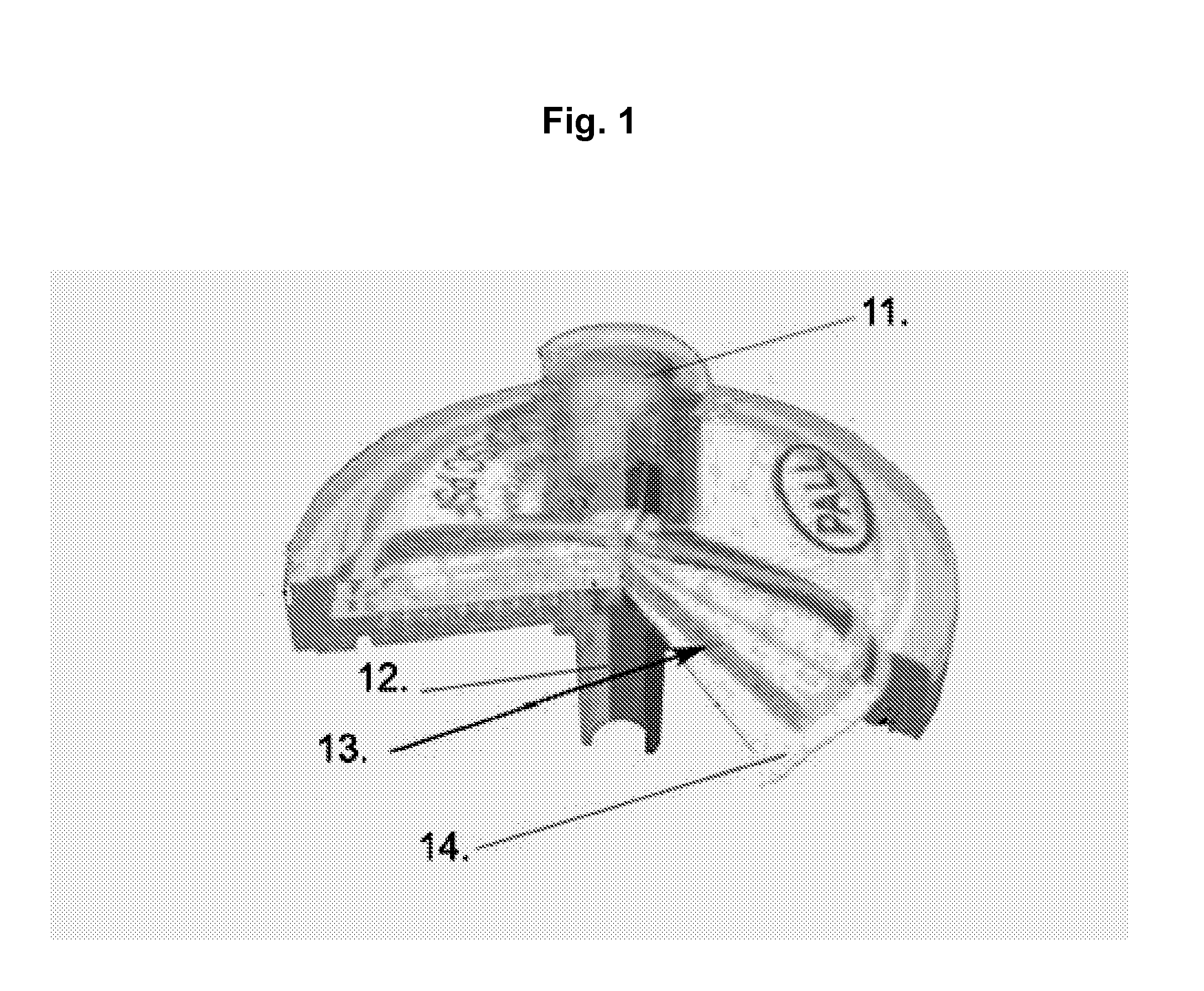
[0138]A number of different form factors are included as examples of integrated filter Assay Cartridge devices (IFAC). Similarly, there are several methods that can be used to prepare samples for testing in the Integrated BioAssay (IBA). Perhaps the simplest IFAC example device is a syringe filter (e.g., Pall Acrodisc™) as shown in FIG. 1; typically a small round disk device with an inlet and outlet port which may have luer-lock fittings on one or both of the ports. A simple preparation method for these syringe filters useful for testing small sample volumes is to simply add the test volume and detection fluids using a pipettor. As a simple example of the syringe filter's utility as an IFAC, a dilution series of microbial samples (E. coli) was prepared using 13 mm Pall Acrodisc™ syringe filters and a pipettor then tested in the Rapid BioAnalyzer (RBA).

[0139]Serial dilutions of E. coli (ATCC 25922) we...

example 2

Detection of Growth by the Integrated Bioassay—Utility of Derivative Calculations

[0145]The following example is presented to illustrate the type of data that the instrument can acquire and that a single “assay endpoint” can easily lead to incorrect conclusions regarding the presence or absence of viable organisms in unknown environmental samples. Further, the example demonstrates the valuable utility of the derivative calculations used in the IBA approach to determine if and when growth of viable organisms has occurred.

[0146]The fluorescent signal in a test sample can change for several reasons unrelated to actual growth of the target organism. Hydrolysis of the glucuronide or galactopyranoside bond will occur at a slow but measurable rate, releasing the fluorescent reporter conjugates. Samples with glucuronidase or galactosidase enzymes present in samples at the initiation of the assay will contribute to changes in fluorescence. These enzymes can come from senescent target organism...

example 3

Method for Sample Preparation in a Syringe Filter Using Negative Pressure

[0150]Water collected from a drinking water source was assessed for the presence of coliforms and E. coli in a simple, integrated manner using the integrated filter device (IFAC) and bioassay instrument. Compliance with regulatory statutes requires testing of 100 mL of water for the presence (or absence) of any coliform bacteria. The following example describes a simple method for preparation of the syringe version of the IFAC using negative pressure (suction) to process 100 mL samples. Additionally, the example provides a simple description of how the Integrated BioAssay can be used to assess toxicity of a water sample by measuring metabolic changes in a known organism. One-hundred milliliter samples are conveniently prepared and tested in the following manner.

[0151]Two 300 mL samples of tap water were collected into sterile glass bottles, one with 10 mg sodium-thiosulfate and one without thiosulfate. The wate...


PUM

PropertyMeasurementUnit
timeaaaaaaaaaa
volumesaaaaaaaaaa
volumeaaaaaaaaaa

Abstract

The present invention relates to an in vitro assay method and device that provides for detection and measurement of entities in a fluid sample that can be captured and concentrated in a unitized self-contained enclosed filter apparatus that is analyzed in an optical detection instrument for indications of the entity. It provides for analysis of biological material including cells, their enzymes, or other constituents thereof, that can be identified based on an indicator-generating means. The analyses provided for include detection of the presence of the entity, and changes in the entity over time, such as associated with growth and increasing metabolic activity with an expanding population of cells, or decreasing metabolic activity, for example, due to presence of inhibitory or toxic agents.

Description

CROSS-REFERENCE TO RELATED APPLICATIONS[0001]This application claims the benefit of U.S. Provisional Application No. 61106624, which was filed on Oct. 20, 2008, hereby incorporated by reference.STATEMENT REGARDING FEDERALLY SPONSORED RESEARCH[0002]The U.S. Government owns rights in the present invention pursuant to contract number W81XWH-04-C-0149 from the United States Department of Defense through the US Army Medical Research Acquisition Activity.BACKGROUND OF THE INVENTION[0003]The present invention pertains to a set of devices and their related methods to rapidly and simply perform detection, quantitation, and identification of microorganisms in samples collected from a variety of sources including but not limited to drinking water, wastewater, industrial and pharmaceutical process waters or surface extraction solutions.Diverse Needs & Requirements[0004]There are diverse needs for microbiological tests and a diverse array of test methods and devices to assess their presence. In ...

Claims


Application Information

Patent Timeline
Patent Type & Authority Applications(United States)
IPC IPC(8): C12Q1/02
CPCC12Q1/24G01N2001/4088G01N33/52
Inventor PUTNAM, DAVID L.PUTNAM, JASON A.HUBBARD, TODD W.VAN ZEE, JAMES A.
Owner PHOTONIC BIOSYST
Why Eureka
Try Eureka