Method for separation, characterization and/or identification of microorganisms using raman spectroscopy

a raman spectroscopy and microorganism technology, applied in the field of methods and systems for detecting, isolating and/or identifying microorganisms in samples, can solve the problems of bloodstream infection, high morbidity and mortality, and take several days to perform, so as to reduce the risk of handling infectious materials and/or contaminating samples, the effect of characterization and/or identification of microorganisms is faster, and the diagnosis is faster

Inactive Publication Date: 2010-06-03
BIOMERIEUX INC
View PDF30 Cites 40 Cited by
  • Summary
  • Abstract
  • Description
  • Claims
  • Application Information

AI Technical Summary

Benefits of technology

[0014]The present invention provides methods for isolating, characterizing and / or identifying microorganisms in a sample. The methods allow for the characterization and / or identification of microorganisms more quickly than prior techniques, resulting in faster diagnoses (e.g., in a subject having or suspected of having septicemia) and identification of contaminated materials (e.g., foodstuffs and pharmaceuticals). The steps involved in the methods of the invention, from obtaining a sample to characterization and / or identification of microorganisms, can be carried out in a very short time frame to produce clinically relevant actionable information, e.g., in less than about 120 minutes. Additionally, the methods of the invention can be fully automated, thereby reducing the risk of handling infectious materials and / or contaminating the samples.
[0034]In one embodiment, the separation is carried out by layering the test sample over a density cushion in a container and centrifuging the container to pellet the microorganisms while the sample medium remains on top of the density cushion. In another embodiment, the container has an optical window at the bottom and / or sides so that the microorganism pellet can be interrogated spectroscopically. The microorganisms can be identified by comparing the spectrum of the pellet to a spectrum or spectra, or spectroscopic properties predicted, of known microorganisms. The ability to identify microorganisms directly in the pellet without further handling enhances the safety of microbial identification.

Problems solved by technology

Bloodstream infections are associated with high morbidity and mortality, yet current diagnostic methods, of culture followed by biochemical identification and antibiotic susceptibility testing, can take several days to perform.
Typically, empiric therapy is initiated based on clinical symptoms, and test results only impact clinical decisions when the initial therapy fails.
Molecular amplification methods have been proposed to fill this need, but serious challenges to this approach remain.
These direct-from-the-bottle tests are not appropriate for all microorganisms (e.g., Gram-positive cocci), are not validated by the test manufacturers, and generally take 3-8 hours to provide results.
Raman spectroscopy has the potential to allow for identification of microorganisms very quickly, but may encounter interference from the many highly fluorescent and absorptive compounds present in liquid microbiological culture media and in clinical samples such as blood or combinations thereof.
However, the '135 application does not discuss identification and quantification of microorganisms from complex biological samples, such as blood.
However, these methods have several drawbacks when attempting to separate and characterize microorganisms from complex samples such as blood-containing culture media.
The resultant microbial preparations often contain contaminating red blood cells, platelets, lipid particles, plasma enzymes and cellular debris, which can cause poor results.
These methods are also very labor-intensive and unsafe due to steps which can result in aerosol exposure of potentially dangerous pathogens to the user.

Method used


Image

  • Method for separation, characterization and/or identification of microorganisms using raman spectroscopy
  • Method for separation, characterization and/or identification of microorganisms using raman spectroscopy
  • Method for separation, characterization and/or identification of microorganisms using raman spectroscopy

Examples


example 1

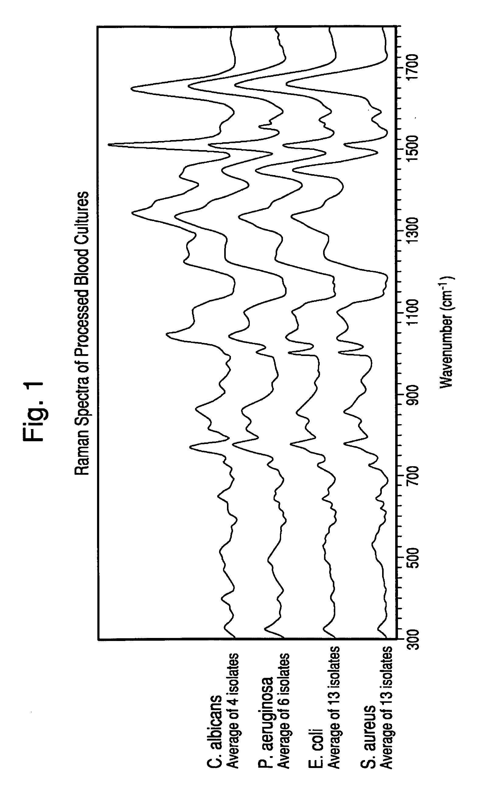
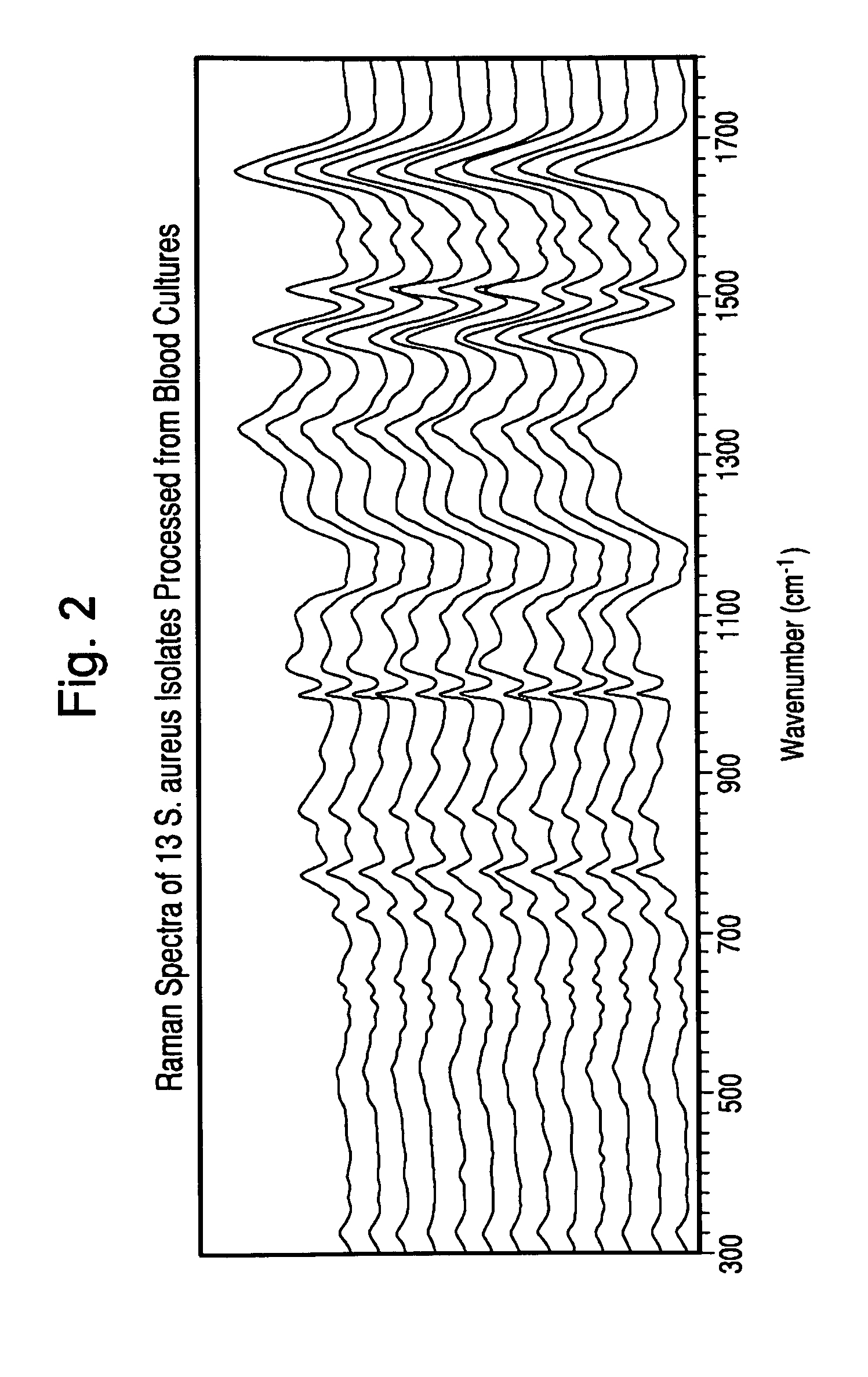
Lysis-Centrifugation Method for Identification of Microorganisms from Blood Cultures by Raman Spectroscopy.

[0115]Microorganisms were “seeded” at a low inoculum into BacT / ALERT® SA bottles containing 10 mLs of human blood. Blood culture broth samples were removed from bottles within a few minutes of being flagged positive by the BacT / ALERT® 3D Microbial Detection System. Broth samples were processed to separate microorganisms from blood and media components that could interfere with subsequent analysis as follows:

[0116]4.0 mL of broth from freshly positive blood cultures was combined with 2.0 mL Lysis Buffer (0.45% Brij® 97 in 0.3M CAPS, pH 11.7), vortex mixed for 5 seconds and then incubated in a 37° C. waterbath for 90 seconds. After incubation, 0.95 mL of lysate was layered on top of 0.5 mL of density cushion (14% w / v Iohexol, 0.005% Pluronic F-108 in 10 mM Hepes, pH 7.4) in each of four 1.5 mL conical centrifuge tubes. All four tubes were then centrifuged for 2 minutes at 10,000 ...

example 2

Analysis of Microorganism Specimens Processed from Positive Blood Cultures with Lysis-Centrifugation by Raman Spectroscopy.

[0118]Specimens processed according to the procedure in Example 1 were rapidly thawed at 37° C. (if previously frozen), mixed gently. A portion of the specimen was kept undiluted and the remainder was diluted to 1:2 in purified water. One microliter of each diluted and undiluted specimen was applied to the first surface of a gold-coated microscope slide and allowed to dry under ambient conditions.

[0119]After drying, 25 spectra in a 5×5 grid pattern were acquired for each of the dried specimens with a Model RxN1 Raman microscope (Kaiser Optical Systems Inc., Michigan) at an illumination wavelength of 785 nm. The microscope was equipped with a 40× objective lens, the laser power was set to 400 mw, and each of the 25 individual spectra was acquired with a 5 second accumulation time.

[0120]After acquisition, the spectra were preprocessed to perform dark subtraction, ...

example 3

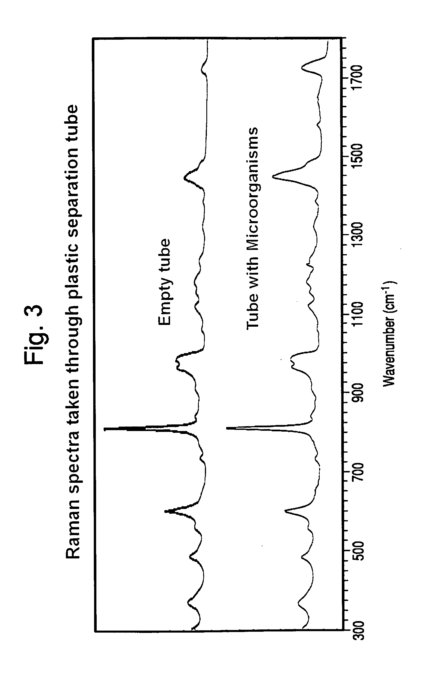
Non-invasive Analysis of Microorganism Specimens Processed from Positive Blood Cultures with Lysis-Centrifugation by Raman Spectroscopy.

[0124]Microorganisms were “seeded” at a low inoculum into BacT / ALERT® SA bottles containing 10 mL of human blood. Blood culture broth samples were removed from bottles within a few minutes of being flagged positive by the BacT / ALERT® 3D Microbial Detection System. The samples were treated as follows:[0125]1. A 2.0 mL sample of positive broth was mixed with 1.0 mL of selective lysis buffer (0.45% w / v Brij® 97+0.3M CAPS, pH 11.7), then placed in a 37° C. water bath for 1 minute.[0126]2. A 1.0 mL sample of lysate was overlayed onto 0.5 mL of density cushion (24% w / v cesium chloride in 10 mM Hepes ph 7.4+0.005% Pluronic F-108) contained in a custom-built optical separation tube. A polypropylene ball was present on the surface of the density cushion to facilitate loading without disturbing the two aqueous phases.[0127]3. The optical separation tube was s...


PUM

PropertyMeasurementUnit
temperatureaaaaaaaaaa

Abstract

The present invention is directed to a method for separating, characterizing and / or identifying microorganisms in a test sample. The method of the invention comprises an optional lysis step for lysing non-microorganism cells that may be present in a test sample, followed by a subsequent separation step. The method may be useful for the separation, characterization and / or identification of microorganisms from complex samples such as blood-containing culture media. The invention further provides for Raman spectroscopic interrogation of the separated microorganism sample to produce measurements of the microorganism and characterizing and / or identifying the microorganism in the sample using said Raman spectroscopic measurements.

Description

CROSS REFERENCE TO RELATED APPLICATION[0001]This application claims the benefit of U.S. Provisional Patent Application No. 61 / 110,187, entitled, “Method and System for Detection and / or Characterization of a Biological Particle in a Sample”, filed Oct. 31, 2008, which is incorporated herein.FIELD OF THE INVENTION[0002]The present invention relates to methods and systems for detecting, isolating and / or identifying microorganisms in a sample. In particular, the present invention is directed method for the rapid characterization and / or identification of a microorganism using Raman spectroscopic techniques.BACKGROUND OF THE INVENTION[0003]The detection of pathogenic microorganisms in biological fluids should be performed in the shortest possible time, in particular in the case of septicemia for which the mortality remains high in spite of the broad range of antibiotics which are available to doctors. The presence of biologically active agents such as a microorganism in a patient's body f...

Claims


Application Information

Patent Timeline
Patent Type & Authority Applications(United States)
IPC IPC(8): C12Q1/04C12Q1/02
CPCC12Q1/04G01N21/35G01N21/3581G01N33/6848G01N21/64G01N21/65G01N21/359G01N21/47C12M1/34
Inventor CLAY, BRADFORDHYMAN, JONESWALSH, JOHNTHORPE, THURMANRONSICK, CHRISTOPHER
Owner BIOMERIEUX INC
Why Eureka
Try Eureka