
Noninvasive Diagnosis of Fetal Aneuploidy by Sequencing

a fetal aneuploidy and sequencing technology, applied in the field of molecular diagnostics, can solve the problems of imposing small but potentially significant risks to both the fetus and the mother, limited reliability of non-invasive screening of fetal aneuploidy using maternal serum markers and ultrasound, and difficulty in measuring aneuploidy, so as to achieve more robust and statistically significant results.

Active Publication Date: 2010-06-03
THE BOARD OF TRUSTEES OF THE LELAND STANFORD JUNIOR UNIV
View PDF43 Cites 186 Cited by
  • Summary
  • Abstract
  • Description
  • Claims
  • Application Information

AI Technical Summary

Benefits of technology

"The present invention is a method for analyzing a maternal sample, such as peripheral blood, to detect abnormal distribution of fetal chromosomes. The method uses a non-invasive method that does not require the sequence difference between fetal and maternal DNA. It involves obtaining fetal DNA from the maternal sample and mapping the DNA fragments to chromosomes using short reads. The method can detect abnormalities in chromosome portions as small as 6% of the fetal DNA concentration. It can also be used to characterize the plasma DNA in unprecedented detail and to obtain information regarding polymorphisms and mutations. The method is accurate and reliable, and it can be used as a universal non-invasive diagnostic test for fetal aneuploidy."

Problems solved by technology

These procedures impose small but potentially significant risks to both the fetus and the mother (2).
Non-invasive screening of fetal aneuploidy using maternal serum markers and ultrasound are available but have limited reliability (3-5).
However, measuring aneuploidy remains challenging due to the high background of maternal DNA; fetal DNA often constitutes <10% of total DNA in maternal cell-free plasma (13).
However, these methods are limited to specific populations because they depend on the presence of genetic polymorphisms at specific loci.

Method used


Image

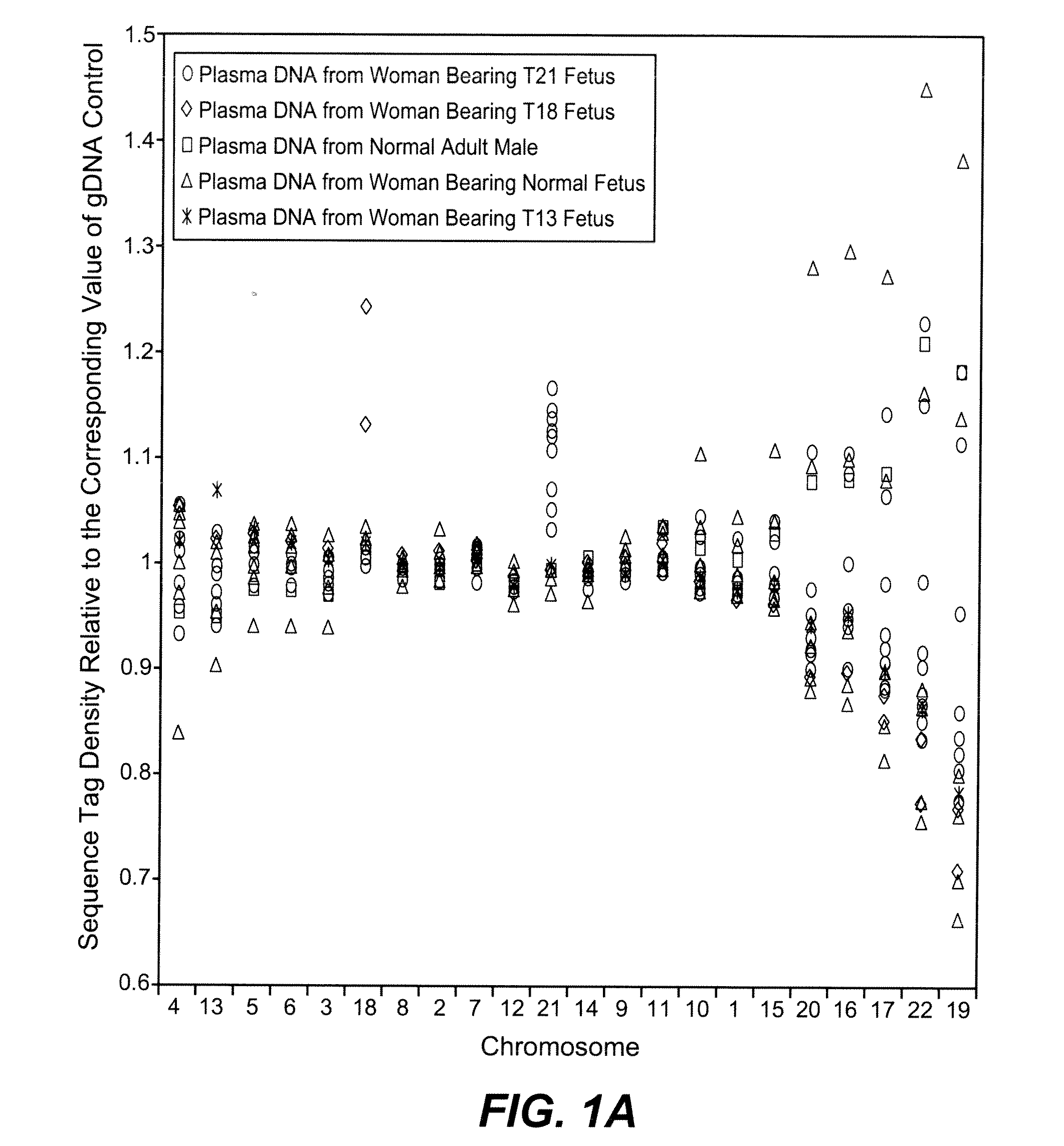
  • Noninvasive Diagnosis of Fetal Aneuploidy by Sequencing
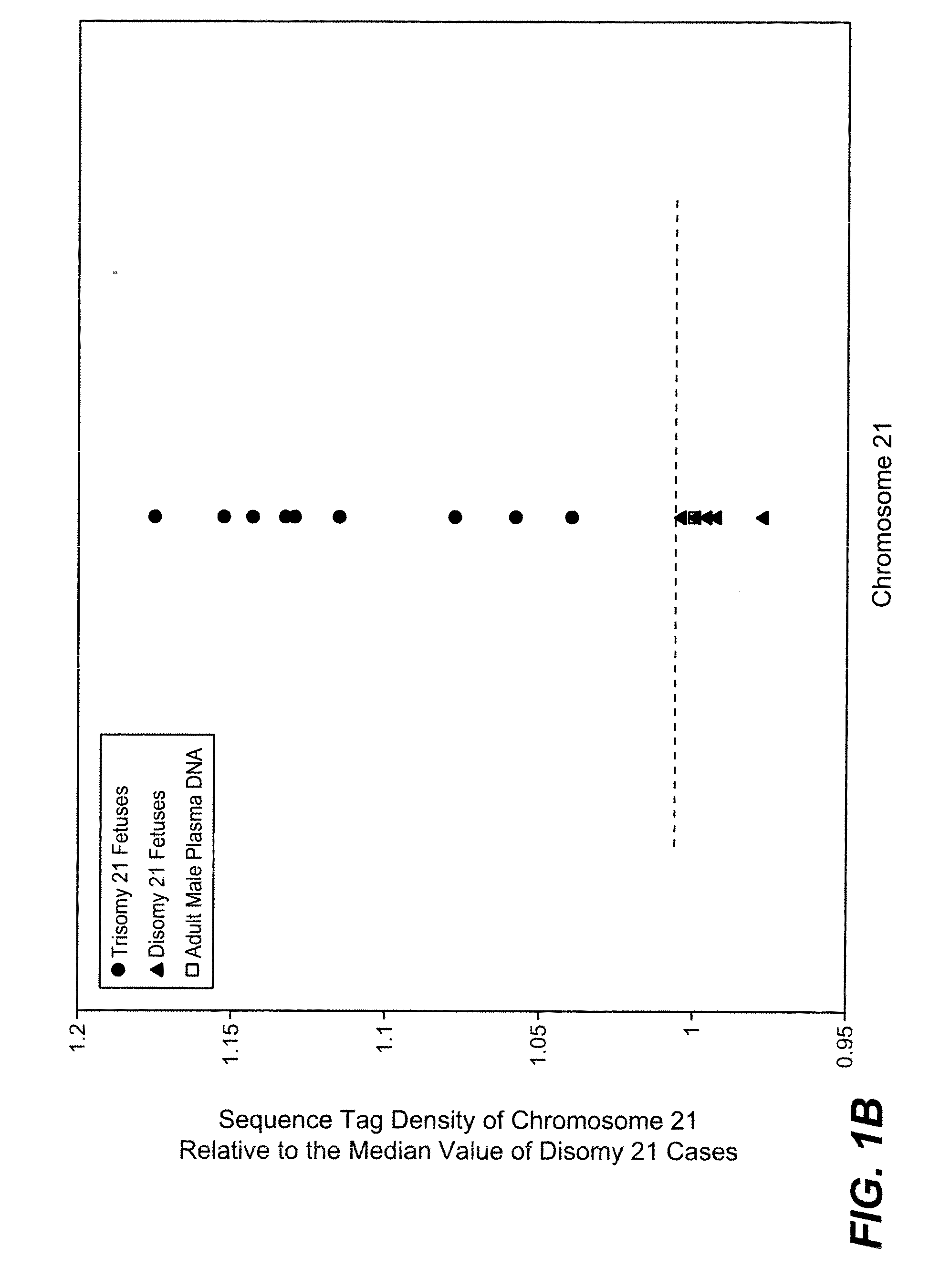
  • Noninvasive Diagnosis of Fetal Aneuploidy by Sequencing
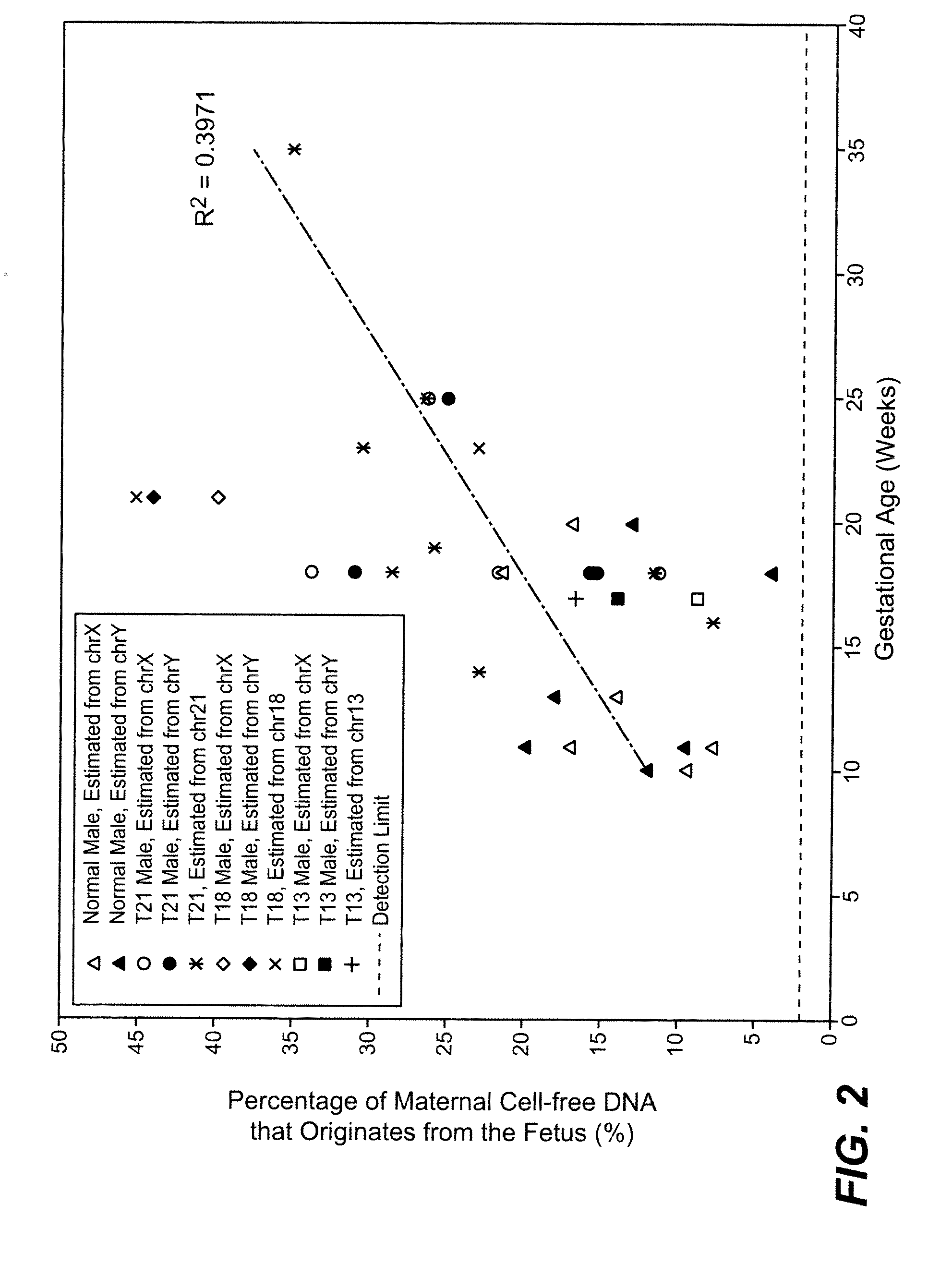
  • Noninvasive Diagnosis of Fetal Aneuploidy by Sequencing

Examples


example 1

Subject Enrollment

[0083]The study was approved by the Institutional Review Board of Stanford University. Pregnant women at risk for fetal aneuploidy were recruited at the Lucile Packard Children Hospital Perinatal Diagnostic Center of Stanford University during the period of April 2007 to May 2008. Informed consent was obtained from each participant prior to the blood draw. Blood was collected 15 to 30 minutes after amniocentesis or chorionic villus sampling except for 1 sample that was collected during the third trimester. Karyotype analysis was performed via amniocentesis or chorionic villus sampling to confirm fetal karyotype. 9 trisomy 21 (T21), 2 trisomy 18 (T18), 1 trisomy 13 (T13) and 6 normal singleton pregnancies were included in this study. The gestational age of the subjects at the time of blood draw ranged from 10 to 35 weeks (Table 1). Blood sample from a male donor was obtained from the Stanford Blood Center.

example 2

Sample Processing and DNA Quantification

[0084]7 to 15 ml of peripheral blood drawn from each subject and donor was collected in EDTA tubes. Blood was centrifuged at 1600 g for 10 minutes. Plasma was transferred to microcentrifuge tubes and centrifuged at 16000 g for 10 minutes to remove residual cells. The two centrifugation steps were performed within 24 hours after blood collection. Cell-free plasma was stored at −80 C until further processing and was frozen and thawed only once before DNA extraction. DNA was extracted from cell-free plasma using QIAamp DNA Micro Kit (Qiagen) or NucleoSpin Plasma Kit (Macherey-Nagel) according to manufacturers' instructions. Genomic DNA was extracted from 200 μl whole blood of the donors using QIAamp DNA Blood Mini Kit (Qiagen). Microfluidic digital PCR (Fluidigm) was used to quantify the amount of total and fetal DNA using Taqman assays targeting at the EIF2C1 locus on chromosome 1 (Forward: 5′ GTTCGGCTTTCACCAGTCT 3′ (SEQ ID NO: 1); Reverse: 5′ C...

example 3

Sequencing

[0085]A total of 19 cell-free plasma DNA samples, including 18 from pregnant women and 1 from a male blood donor, and genomic DNA sample from whole blood of the same male donor, were sequenced on the Solexa / Illumina platform. ˜1 to 8 ng of DNA fragments extracted from 1.3 to 5.6 ml cell-free plasma was used for sequencing library preparation (Table 1). Library preparation was carried out according to manufacturer's protocol with slight modifications. Because cell-free plasma DNA was fragmented in nature, no further fragmentation by nebulization or sonication was done on plasma DNA samples.

[0086]Genomic DNA from male donor's whole blood was sonicated (Misonix XL-2020) (24 cycles of 30 s sonication and 90 s pause), yielding fragments with size between 50 and 400 bp, with a peak at 150 bp. ˜2 ng of the sonicated genomic DNA was used for library preparation. Briefly, DNA samples were blunt ended and ligated to universal adaptors. The amount of adaptors used for ligation was 50...


PUM

PropertyMeasurementUnit
lengthaaaaaaaaaa
concentrationaaaaaaaaaa
nucleic acidaaaaaaaaaa

Abstract

Disclosed is a method to achieve digital quantification of DNA (i.e., counting differences between identical sequences) using direct shotgun sequencing followed by mapping to the chromosome of origin and enumeration of fragments per chromosome. The preferred method uses massively parallel sequencing, which can produce tens of millions of short sequence tags in a single run and enabling a sampling that can be statistically evaluated. By counting the number of sequence tags mapped to a predefined window in each chromosome, the over- or under-representation of any chromosome in maternal plasma DNA contributed by an aneuploid fetus can be detected. This method does not require the differentiation of fetal versus maternal DNA. The median count of autosomal values is used as a normalization constant to account for differences in total number of sequence tags is used for comparison between samples and between chromosomes.

Description

CROSS-REFERENCE TO RELATED APPLICATIONS[0001]This application claims priority from U.S. Provisional Patent Application No. 61 / 098,758, filed on Sep. 20, 2008, and U.S. Utility patent application Ser. No. 12 / 560,708, filed Sep. 16, 2009, both of which are hereby incorporated by reference in their entirety.STATEMENT OF GOVERNMENTAL SUPPORT[0002]This invention was made with U.S. Government support under NIH Director's Pioneer Award DP1 OD000251. The U.S. Government has certain rights in this invention.REFERENCE TO SEQUENCE LISTING, COMPUTER PROGRAM, OR COMPACT DISK[0003]Applicants assert that the text copy of the Sequence Listing is identical to the Sequence Listing in computer readable form found on the accompanying computer file. Applicants incorporate the contents of the sequence listing by reference in its entirety.BACKGROUND OF THE INVENTION[0004]1. Field of the Invention[0005]The present invention relates to the field of molecular diagnostics, and more particularly to the field o...

Claims


Application Information

Patent Timeline
Patent Type & Authority Applications(United States)
IPC IPC(8): C12Q1/68G06F19/00G16B20/10G16B30/00
CPCC12Q1/6869C12Q1/6883G01N2800/387C12Q2600/156G01N33/48C12Q2545/114C12Q2537/143
Inventor FAN, HEI-MUN CHRISTINAQUAKE, STEPHEN R.
Owner THE BOARD OF TRUSTEES OF THE LELAND STANFORD JUNIOR UNIV
Learn More