Complement inhibition for improved nerve regeneration

a technology of complement inhibition and nerve regeneration, applied in the field of methods and medicaments, can solve the problems of loss of neuronal function, degeneration of transected axons, slow regeneration,

Active Publication Date: 2010-06-10
REGENESANCE
View PDF8 Cites 76 Cited by
  • Summary
  • Abstract
  • Description
  • Claims
  • Application Information

AI Technical Summary

Benefits of technology

[0011]In the Examples herein we have observed activation of the complement (C)-system in the rat during WD and in human nerve biopsies of chronic demyelinating neuropathies. The present invention is based on the surprising finding that axonal regeneration is enhanced in rats that are deficient in the complement C6 factor. This surprising finding opens new ways to promote axonal regeneration by manipulation of the complement system and / or macrophage activation.
[0029]The larger C5b fragment binds sequentially to later components of the complement cascade, C6, C7, C8 and C9 to form the C5b-9 membrane attack complex (“MAC”). The lipophylic C5b-9 MAC can directly lyse erythrocytes, and in greater quantities it is lytic for leukocytes and damaging to tissues such as muscle, epithelial and endothelial cells. In sublytic amounts, the C5b-9 MAC can stimulate upregulation of adhesion molecules, intracellular calcium increase and cytokine release. In addition, at sublytic concentrations the C5b-9 MAC can stimulate cells such as endothelial cells and platelets without causing cell lysis. The non-lytic effects of C5a and the C5b-9 MAC are comparable and interchangeable.
[0035]Yet another membrane-bound complement receptor for use in the methods and / or medicaments of the invention is Crry-Ig (Quigg et al., 1998). Crry is a membrane complement inhibitor with decay accelerating activity at the 3 convertase level, inhibiting both the classical and alternative pathway of complement. It also possesses cofactor activity comparable to that of CR1 for the factor I-mediated cleavage of C3b and C4b. Crry-Ig is a recombinant, soluble protein with an increased half-life (40 hours) due to fusion of Crry with the Fc portion of a non-complement-activating mouse IgG1 partner. Overall, Crry-Ig is potent complement inhibitor.
[0041]All of the wholly and partially human antibodies are less immunogenic than wholly murine MAbs, and the fragments and single chain antibodies are also less immunogenic. All these types of antibodies are therefore less likely to evoke an immune or allergic response. Consequently, they are better suited for in vivo administration in humans than wholly animal antibodies, especially when repeated or long-term administration is necessary. In addition, the smaller size of the antibody fragment may help improve tissue bioavailability, which may be critical for better dose accumulation in acute disease indications, such as tumour treatment.
[0049]For oral administration, the inhibitor can be administered in solid dosage forms, such as capsules, tablets, and powders, or in liquid dosage forms, such as elixirs, syrups, and suspensions. Active component(s) can be encapsulated in gelatine capsules together with inactive ingredients and powdered carriers, such as glucose, lactose, sucrose, mannitol, starch, cellulose or cellulose derivatives, magnesium stearate, stearic acid, sodium saccharin, talcum, magnesium carbonate and the like. Both tablets and capsules can be manufactured as sustained release products to provide for continuous release of medication over a period of hours. Compressed tablets can be sugar coated or film coated to mask any unpleasant taste and protect the tablet from the atmosphere, or enteric-coated for selective disintegration in the gastrointestinal tract. Liquid dosage forms for oral administration can contain colouring and flavouring to increase patient acceptance.
[0050]The inhibitors are however preferably administered parentally. Suitable carriers for parental formulations include saline, buffered saline, dextrose, and water. Typically compositions for parenteral administration are solutions in sterile isotonic aqueous buffer. Sterilisation is readily accomplished by filtration through sterile filtration membranes, prior to or following lyophilisation and reconstitution. A typical composition for intravenous infusion could be made up to contain 10 to 50 ml of sterile 0.9% NaCl or 5% glucose optionally supplemented with a 20% albumin solution and an appropriate amount (1 to 1000 μg) of the inhibitor. A typical pharmaceutical composition for intramuscular injection would be made up to contain, for example, 1-10 ml of sterile buffered water and 1 to 1000 μg of the of the inhibitor. Methods for preparing parenterally administrable compositions are well known in the art and described in more detail in various sources, including, for example, Remington's Pharmaceutical Science (15th ed., Mack Publishing, Easton, Pa., 1980) (incorporated by reference in its entirety for all purposes). Where necessary, the composition may also include a solubilising agent and a local anaesthetic such as lignocaine to ease pain at the site of the injection. Generally, the ingredients will be supplied either separately or mixed together in unit dosage form, contained in a hermetically sealed container such as an ampoule or sachette indicating the quantity of active agent in activity units. Where the composition is to be administered by infusion, it can be dispensed with an infusion bottle containing sterile pharmaceutical grade ‘Water for Injection’ or saline. Where the composition is to be administered by injection, an ampoule of sterile water for injection or saline may be provided so that the ingredients may be mixed prior to administration.

Problems solved by technology

It may lead to separation of the neuron from its targets, resulting in loss of neuronal function.
Because most neuronal proteins are synthesised in the soma and carried to the axon by specialised axonal transport systems, degeneration of the transected axons has long been thought to result from starvation of necessary proteins and other materials.
However, regeneration is slow (2-2.5 mm / day) and the environment of a degenerated nerve is full of factors which inhibit the growth of the axon and the necessary growth factors can be limiting or even absent.
Thus the delayed clearance of myelin in complement-depleted animals is expected to result in impaired axonal regeneration.

Method used


Image

  • Complement inhibition for improved nerve regeneration
  • Complement inhibition for improved nerve regeneration
  • Complement inhibition for improved nerve regeneration

Examples


example 1

1. Example 1

Improved Post-Traumatic Nerve Recovery in Complement Component C6 Deficient Rats as Compared to Wild-Type Rats

1.1 Electron Microscopic Analysis

[0068]We have explored the role of the complement system in acute and chronic nerve injury and during regeneration. As a model we used the complement C6 deficient PVG rat strain (Bhole and Stahl, 2004) and compared this with wild type PVG rats. Since the complement system has many functions we chose an animal model in which only the most terminal effectors of the complement cascade was defective.

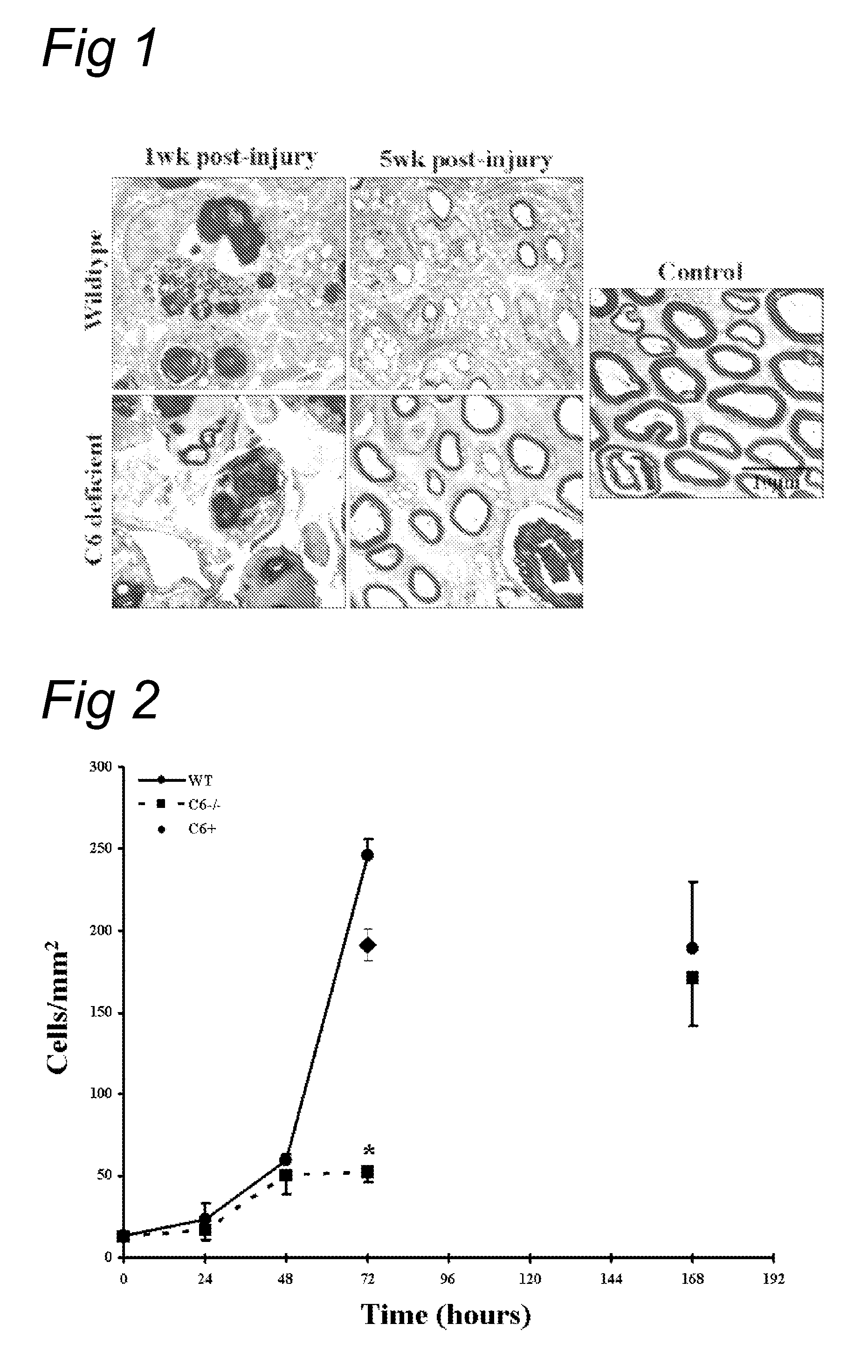
[0069]The effect of complement inhibition on nerve regeneration was studied in the acute model of nerve crush (Glass, 2004). The right sciatic nerve was crushed for 30 seconds in wild type as well as in the C6-deficient PVG rats. Tibial nerve was then analyzed 1 and 5 weeks after injury (see FIG. 1).

[0070]At one week, electron microscopy shows equally severe degeneration in wild type and in C6 deficient rats. At 5 weeks, the C6 deficient r...

example 2

Inhibition of Complement Activation after Nerve Crush by Human C1-Inhibitor

[0082]We tested whether recombinant human C1-inhibitor (rhC1INH; obtained from Pharming, Leiden, The Netherlands) is able to inhibit the rapid (1 hr) complement activation after nerve crush. FIG. 5 shows C1q, C4c and C3c immunostaining of injured wild type rat sciatic nerves treated with rhC1INH or vehicle (PBS) alone at 1 hour after nerve crush. High immunoreactivity for C1q is present in all crushed nerves, confirming C1q up-regulation after the crush injury. C4c and C3c immunoreactivity was detected in the PBS-treated nerves as expected but no C4c and C3c immunoreactivity was detected in the nerves from the rhC1INH treated rats sacrificed at 1 hr post-injury. This demonstrates effective blockade of the complement cascade by rhC1INH after crush and suggests that the alternative pathway of complement activation is not involved in the crush injury model of Wallerian degeneration. Thus, activation of the compl...

example 3

The Effect of Soluble CR1 on Post Traumatic Nerve Regeneration

[0083]Next, we tested the effect of soluble CR1 (sCR1) on post traumatic nerve regeneration. sCR1 inhibits the C3 / C5 convertase, and thereby affects both the classical and alternative pathway of the complement system.

[0084]Wild type PVG rats were treated with soluble CR1 (TP10 from Avant Immunotherapeutics, Inc.) at a dose of 15 mg / kg / day (TP10 soluble CR1 was obtained from Prof. P. Morgan, Cardiff, UK). Control rats were treated with the same volume (600 μl) of vehicle alone (PBS). Soluble CR1 or PBS was delivered i.p. 24 hours before the crush and every following day for a maximum of 8 injections (up to day 6 after crush). The sciatic nerve of the right leg was crushed and the left leg served as control. Both histology and sensory function were analysed.

[0085]FIG. 6 shows that in a functional analysis with the footflick test a faster recovery of the sensory function is seen in the sCR1-treated animals compared to the PB...


PUM

PropertyMeasurementUnit
currentsaaaaaaaaaa
timeaaaaaaaaaa
half lifeaaaaaaaaaa

Abstract

The present invention relates to methods and medicaments used for treating conditions that require axonal regeneration, e.g. in mammals affected by injury or disease of the central or peripheral nervous system. The medicaments used in these methods facilitate axonal regeneration by inhibition of the complement system. Conditions requiring axonal regeneration that may be treated in accordance with the invention include physical injuries as well as neurodegenerative disorders of the peripheral or central nervous system.

Description

FIELD OF THE INVENTION[0001]The present invention relates to methods and medicaments used for treating conditions that require axonal regeneration, e.g. in mammals affected by injury or disease of the central or peripheral nervous system. The medicaments used in these methods promote axonal regeneration by inhibition of the complement system.BACKGROUND OF THE INVENTION[0002]Axon degeneration occurs frequently in many types of chronic neurodegenerative diseases and in injuries to axons caused by toxic, ischemic, or traumatic insults. It may lead to separation of the neuron from its targets, resulting in loss of neuronal function. One model of axon degeneration is the self-destructive process observed at the distal portion of a transected axon upon injury, termed Wallerian degeneration (WD) as first described by Waller (1850). In the process of WD, if a nerve fiber is cut or crushed, the part distal to the injury (i.e. the part of the axon separated from the neuron's cell nucleus) wil...

Claims


Application Information

Patent Timeline
Patent Type & Authority Applications(United States)
IPC IPC(8): A61K39/395A61K31/7105A61K31/24A61K38/02A61K31/19A61P25/00
CPCA61K31/19A61K31/245C12N15/1137A61K38/177C07K16/40A61K31/727A61K2039/505A61P25/00A61P25/02A61P25/28A61P43/00
Inventor BAAS, FRANKRAMAGLIA, VALERIA
Owner REGENESANCE
Why Eureka
Try Eureka