
Glycoprotein Profiling of Bladder Cancer

a bladder cancer and glycoprotein technology, applied in the direction of fluid pressure measurement, liquid/fluent solid measurement, peptide measurement, etc., can solve the problems of inability to rapidly provide, interobserver variation, and relatively high cost, and achieves simple, rapid, reliable, accurate and cost-effective effects

Inactive Publication Date: 2010-07-22
UNIV OF FLORIDA RES FOUNDATION INC
View PDF6 Cites 27 Cited by
  • Summary
  • Abstract
  • Description
  • Claims
  • Application Information

AI Technical Summary

Benefits of technology

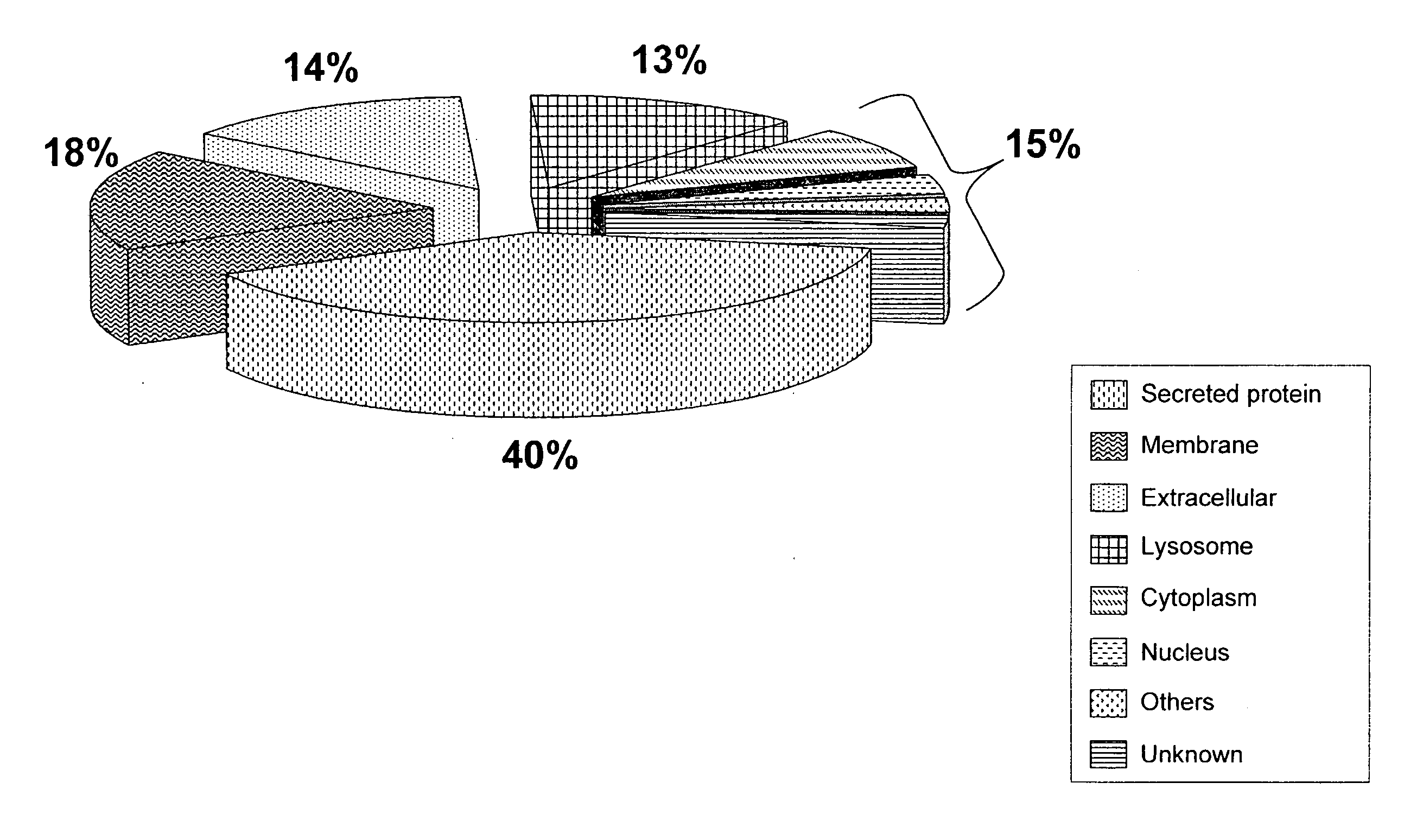
[0012]The present invention concerns biomarkers of bladder cancer in biological samples, such as human urine, and rapid, high-sensitivity methods to profile the N-linked glycoprotein component in naturally micturated human urine specimens. Con A affinity chromatography coupled to nanoflow liquid chromatography was utilized to separate the complex peptide mixture prior to a linear ion trap MS analysis. Of 186 proteins identified with high confidence by multiple analyses, 40% were secreted proteins, 18% membrane proteins and 14% extracellular proteins. In this study, the presence of several proteins appeared to be associated with the presence of bladder cancer, including alpha-1B-glycoprotein (A1BG) that was detected in all tumor-bearing patient samples but in none of the samples obtained from non-tumor bearing individuals. The combination of Con A affinity chromatography and nano-LC / MS / MS provides an initial investigation of N-glycoproteins in complex biological samples and facilitates the identification of potential biomarkers of bladder cancer in non-invasively obtained human urine.
[0018]In another aspect, the invention relates to a simple, rapid, reliable, accurate and cost effective test for bladder cancer biomarkers in voided urine, similar to currently available in-home pregnancy tests that could be used by subjects at home, in a physicians' office, or at a patient's bedside, e.g., at a health care facility. In one embodiment, the test is a method for detecting and, optionally, measuring, one or more biomarkers listed in Table 4 in urine, comprising: (a) obtaining a sample of urine from a subject; (b) contacting the sample with a binding agent that binds to any biomarker in the sample; (c) separating biomarker-bound binding agent; (d) detecting a signal associated with the separated binding agent from (c); and (e) comparing the signal detected in step (d) with a reference signal which corresponds to the signal given by a sample from a subject with a biomarker level equal to a threshold concentration. In one embodiment, the threshold concentration is between 0 ng / ml or pg / ml and an upper limit of ng / ml or pg / ml.

Problems solved by technology

Early detection remains one of the most urgent issues in bladder cancer research.
Furthermore, results are not available rapidly, it is prone to inter-observer variation, and it is relatively expensive.
Unfortunately, these tests also suffer from high false-positive rates and thus there is no protein test available to date that can replace urine cytology (Hautmann, S. et al.
Thus, detecting bladder cancer using diagnostic markers still remains a challenge.
However, only a limited number of proteomic studies utilizing the newer technologies have been conducted in the analysis of urological cancers, largely because of the lack of defined methodologies that can reduce the complexity of the sample and rapidly and accurately identify specific proteins.
Proteomics is regarded as a sister technology to genomics, however, although the pattern of gene activity may be abnormal in a tissue with a pathological lesion, there can be a poor correlation between the level of the transcription of different genes and the relative abundance within the tissue of the corresponding proteins.
Although a number of dysregulated proteins have been identified in a variety of tissue-based studies, it is disappointing that no reliable markers have been identified for transitional cell carcinoma, the most common type of bladder cancer.
Unfortunately, in a larger cohort of patients, the diagnostic accuracy of calreticulin in urine was vulnerable to urinary tract infections (Kageyama, S. et al.
By pooling samples, one is likely to lose the individual information of the intrinsic components present in urine from a single patient at a given time.
Current methods in the non-invasive detection and surveillance of bladder cancer via urine analysis include voided urine cytology (VUC) and some diagnostic urinary protein biomarkers; however, due to the poor sensitivity of VUC and high false-positive rates of currently available protein assays, detection of bladder cancer via urinalysis remains a challenge.

Method used


Image

  • Glycoprotein Profiling of Bladder Cancer
  • Glycoprotein Profiling of Bladder Cancer
  • Glycoprotein Profiling of Bladder Cancer

Examples


example 1

Urinary Protein Content

[0166]In the present study, the glycoprotein profile of urine obtained from patients was investigated and compared with cystoscopically confirmed bladder cancer or no evidence of bladder cancer (Table 1). Protein concentration of normal urine is estimated to be less than 100 mg / L (Adachi, J. et aL Genome Biol, 2006, 7(9):R80). The measurements of urinary protein concentrations from non tumor-bearing patients in the present study were consistent with the reported value (average of 31 mg / L) except for one patient that showed a sign of proteinuria, (defined as >150 mg / day protein) most likely due to a high level of hematuria. The average protein concentration urine from tumor-bearing patients was 127 mg / L. The greater amount of protein recovered from bladder cancer patients' urine meant that 7-15 mL of urine was typically sufficient for this analysis; however, a larger volume of asymptomatic patient urine was required, i.e., 15-30 mL. Agarose-bound Concanavalin A...

example 2

Mass Spectrometric Analysis

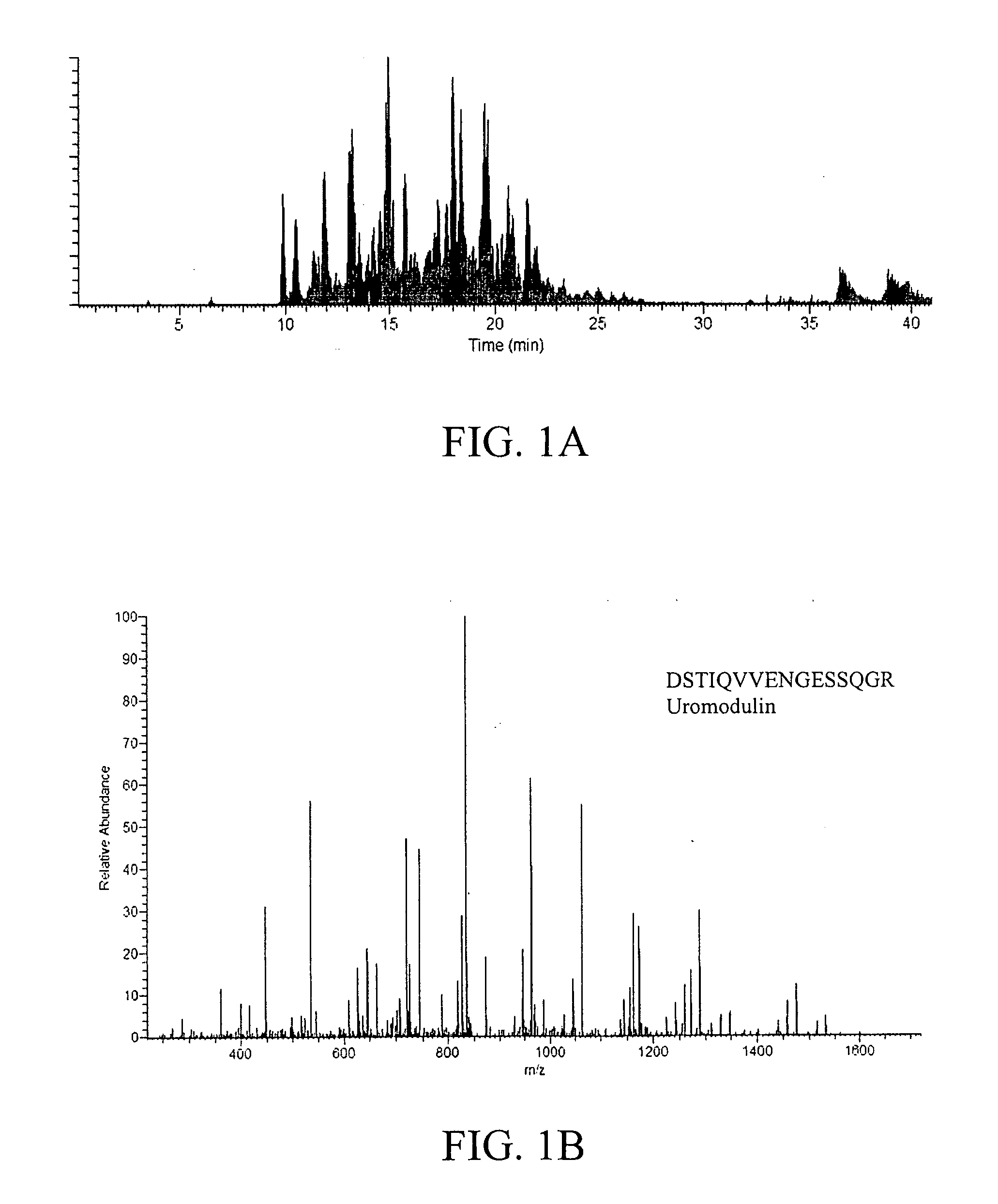
[0167]Con A bound proteins were subjected to mass spectrometric analysis. A semi-shotgun approach was used where the bound fractions were enzymatically digested and analyzed by nano-LC / MS / MS. For each scan, the three most abundant peptides were sequenced. FIG. 1A is a representative nano-LC / MS / MS base peak chromatogram, showing the detection of the more abundant ions across a 40-min gradient separation. The peak capacity of the nano-LC separation was estimated to be ˜60 to 120 based on examining the full-width-at-half-maximum (fwhm). This implies that >60 peptides can be sequenced within a single run; however, it is important to note that multiple peptides were detected within a single resolved peak from all experimentally based peak chromatograms, which is commonly observed from LC / MS / MS analysis of highly complex samples. FIG. 1B shows a representative MS / MS spectrum of a peptide sequence from uromodulin, one of the most abundant proteins in urine, ident...

example 3

Glycoprotein Identification

[0168]Both tryptic digest fractions and tryptic digest / PNGase F fractions were analyzed and their results were combined to increase confidence in protein identification. Removing glycans from digested peptides with PNGase F is reported to provide a stronger signal for the peptide ions compared to the glycan remaining intact (Wang, Y. et al. Glycobiology, 2006, 16(6):514-23). Based on the current results, it was indeed found that glycopeptides were poorly identified when only the tryptic digest fractions were analyzed, but after tryptic digest / PNGase F treatment, a number of glycopeptides were positively identified from the same fraction. In the experiments of this study, all SEQUEST search parameters and data filtering were the same, except that 1 Da was allowed for modification of Asn for deglycosylated digests. Table 3 lists the peptides that were not detected in the fraction with the tryptic digestion alone, where # denotes the glycosylated site of the ...


PUM

PropertyMeasurementUnit
Concentrationaaaaaaaaaa

Abstract

The present invention relates to a method for the diagnosis, prognosis, and monitoring of bladder cancer, such as early or late stage bladder cancer, by detecting in a urine sample from a subject at least one biomarker for bladder cancer identified herein, such as alpha-1B-glycoprotein, haptoglobin, serotransferrin, or alpha-1-antitrypsin. The biomarkers may be detected and, optionally, measured using an agent that detects or binds to the biomarker protein or an agent that detects or binds to encoding nucleic acids, such as antibodies specifically reactive with the biomarker protein or a portion thereof. The invention further relates to kits for carrying out the methods of the invention. The invention further relates to a device for the rapid detection of one or more bladder cancer biomarkers in urine and methods for rapidly measuring bladder cancer biomarkers in urine.

Description

CROSS-REFERENCE TO RELATED APPLICATION[0001]The present application claims the benefit of U.S. Provisional Application Ser. No. 60 / 914,404, filed Apr. 27, 2007, which is hereby incorporated by reference herein in its entirety, including any figures, tables, nucleic acid sequences, amino acid sequences, and drawings.GOVERNMENT SUPPORT[0002]The subject matter of this application has been supported by a research grant from the National Institutes of Health under grant number RO1 CA108597. Accordingly, the government has certain rights in this invention.BACKGROUND OF THE INVENTION[0003]Cancer of the urinary bladder is among the five most common malignancies world-wide (Pisani, P. et al. Int J Cancer, 1999, 83(1):18-29). Transitional cell carcinomas (TCCs) are the most common urothelial tumors in Western countries and constitute approximately 95% of all cases (Aben, K. K. and Kiemeney, L. A. Eur Urol, 1999, 36(6):660-72). Early detection remains one of the most urgent issues in bladder c...

Claims


Application Information

Patent Timeline
IPC IPC(8): G01N33/574C12Q1/68G01N27/26
CPCC12Q1/6886C12Q2600/158G01N2333/8125G01N2333/4728G01N2333/79G01N33/57438
Inventor GOODISON, STEVEROSSER, CHARLES JOEL
Owner UNIV OF FLORIDA RES FOUNDATION INC
Learn More