Motion-attenuated contrast-enhanced cardiac magnetic resonance imaging and related method thereof

a magnetic resonance imaging and contrast-enhanced technology, applied in the field of improvement, to achieve the effect of improving visualization of subendocardial infarcts, retarding enhancement contrast to the myocardium, and improving contrast-enhanced cardiac magnetic resonan

Active Publication Date: 2010-07-29
UNIV OF VIRGINIA ALUMNI PATENTS FOUND
View PDF7 Cites 23 Cited by
  • Summary
  • Abstract
  • Description
  • Claims
  • Application Information

AI Technical Summary

Benefits of technology

[0012]An aspect of an embodiment of the present invention provides a dark-blood technique for contrast-enhanced cardiac magnetic resonance, improving visualization of subendocardial infarcts or perfusion abnormalities that may otherwise be difficult to distinguish from the bright blood pool. In one implementation of an embodiment of the invention, the dark-blood preparation is performed using a driven-equilibrium fourier transform (DEFT) preparation with motion sensitizing gradients which attenuate the signal in the ventricular cavities related to incoherent phase losses resulting from non-steady flow within the heart. This dark-blood preparation preserves the underlying contrast characteristics of the pulse sequence causing a myocardial infarction to be bright while rendering the blood pool dark. When applied to perfusion imaging, this dark-blood preparation will help eliminate artifacts resulting from the juxtaposition of a bright ventricular cavity and relatively dark myocardium.
[0013]An aspect of an embodiment of the present invention involves the combination of an inversion rf pulse to impart post-gadolinium delayed enhancement contrast to the myocardium and a DEFT preparation with motion sensitization gradients to suppress the blood signal in the ventricular cavity. As the motion in the heart cavity has non-steady flow characteristics, an aspect of an embodiment of the invention involves the application of small gradients to suppress the signal from the ventricular cavity. Furthermore the timing of our preparation has been optimized to prevent loss of signal in the myocardial wall resulting from strain. This is a problem that specifically applies to imaging of the heart. Additionally, applying these techniques for contrast enhancement requires careful timing, as applying the DEFT preparation in certain configurations will result in a brighter, rather than darker blood pool signal.
[0014]While other techniques have been used to suppress the blood signal the technique associated with aspects of an embodiment of present invention has advantages over these known techniques. The other techniques which suppress the blood signal based on its T1 relaxation properties require precise knowledge of the T1 relaxation time of the blood pool and myocardium which are increasing continuously after the administration of gadolinium contrast. The motion sensitive preparation associated with aspects of an embodiment of the present invention is only dependent on the incoherent motion of the blood pool in the ventricular cavity to suppress the signal from the blood pool. Other techniques are dependent on the movement of blood out of the imaging plane prior to image acquisition, whereas motion-sensitive preparation of an embodiment of the present invention does not require movement of blood out of the imaging plane for adequate blood suppression. Finally, motion sensitive preparation of an embodiment of the present invention preserves the excellent contrast properties of IR-FLASH, and is robust to changes in T1 of the blood, and myocardium.
[0015]Another aspect of an embodiment of the present invention involves, but not limited thereto, the combination of a DEFT preparation with first-pass contrast-enhanced perfusion imaging of the heart. Conventional techniques do not apply motion-sensitization techniques to imaging of first-pass perfusion with gadolinium-based contrast agents. This aspect of an embodiment of the present invention involves the use of motion-sensitizing techniques in combination with first pass-perfusion techniques to mitigate the dark-rim artifact. As the intensity of the dark-rim artifact is related to the difference in intensity of the bright blood pool and darker myocardium, attenuating the blood pool signal may eliminate this type of artifact. Suppression of the dark-rim artifact during contrast-enhanced first-pass perfusion imaging is not a characteristic of any conventional techniques and should improve the diagnostic accuracy of myocardial perfusion imaging techniques. This is especially important, for example, as false positive stress myocardial perfusion studies frequently result in patients undergoing additional expensive and invasive diagnostic procedures such as coronary angiography.

Problems solved by technology

This is a problem that specifically applies to imaging of the heart.
Additionally, applying these techniques for contrast enhancement requires careful timing, as applying the DEFT preparation in certain configurations will result in a brighter, rather than darker blood pool signal.

Method used


Image

  • Motion-attenuated contrast-enhanced cardiac magnetic resonance imaging and related method thereof
  • Motion-attenuated contrast-enhanced cardiac magnetic resonance imaging and related method thereof
  • Motion-attenuated contrast-enhanced cardiac magnetic resonance imaging and related method thereof

Examples


Embodiment Construction

MRI System

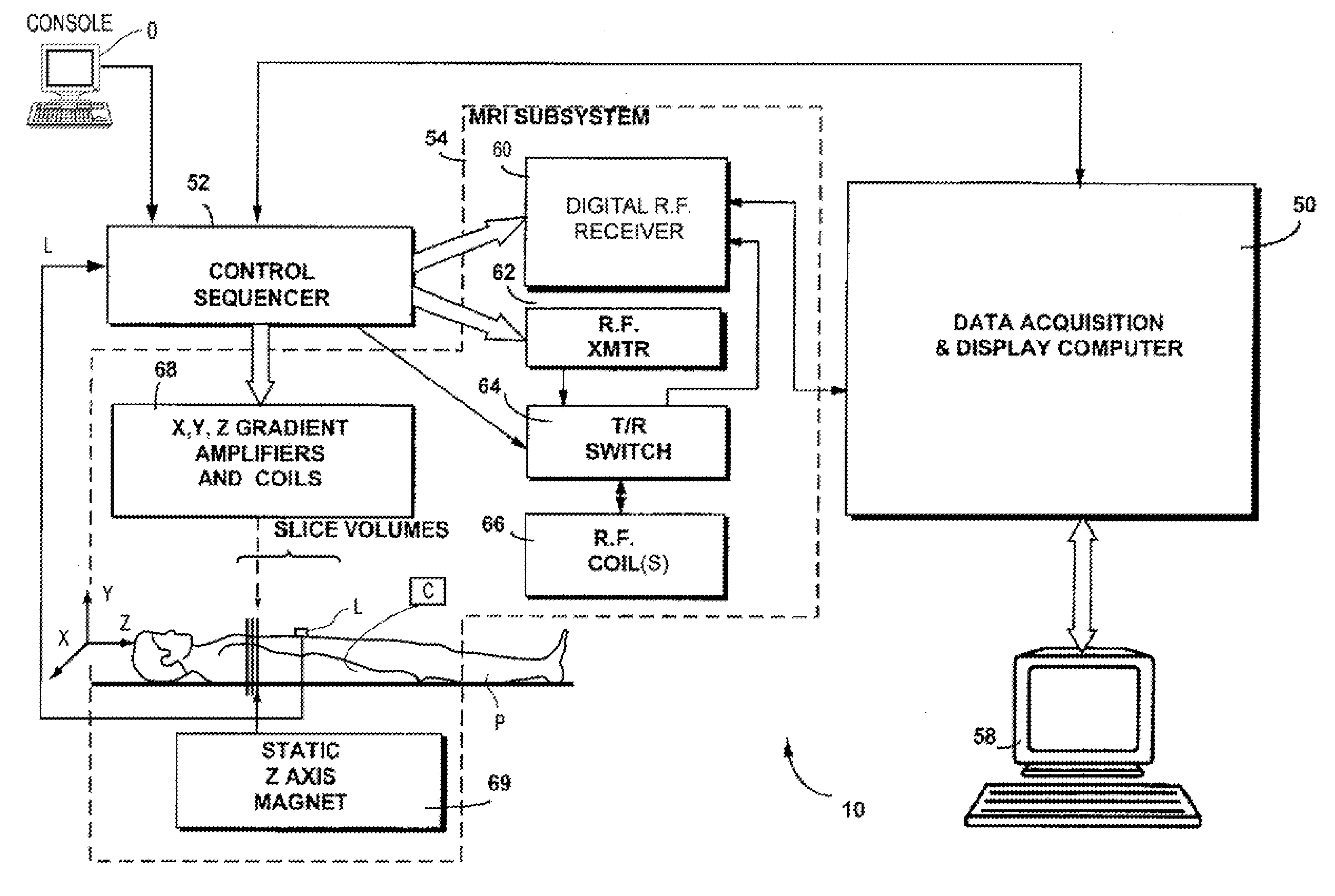
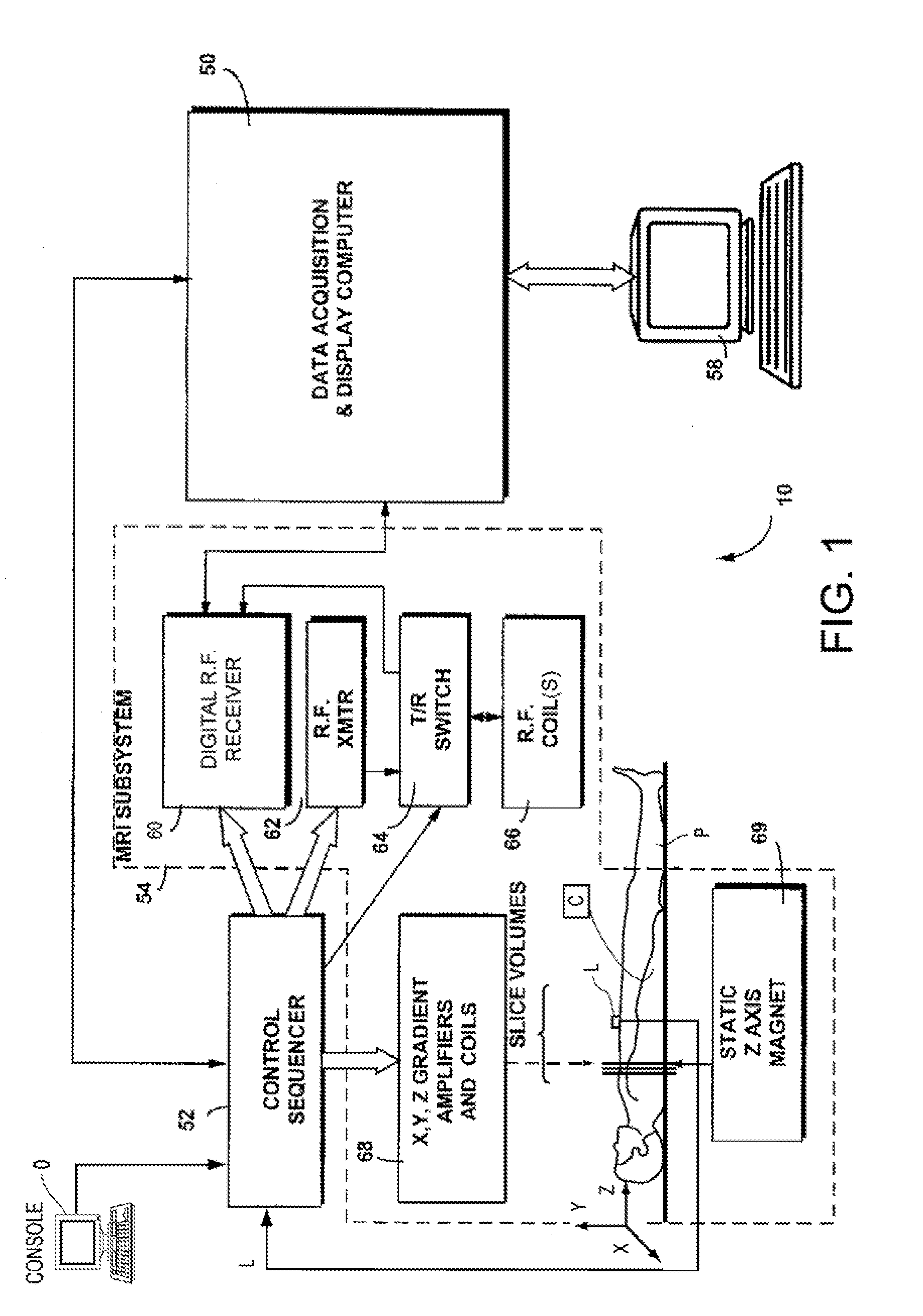
[0032]An embodiment of the present invention may be implemented on any commercial MRI system without necessarily requiring any additional hardware. FIG. 1 illustrates an example such MRI system 10 including a data acquisition and display computer 50 coupled to an operator console 0, a MRI real-time control sequencer 52, and a MRI subsystem 54. The MRI subsystem 54 may include XYZ magnetic gradient coils and associated amplifiers 68, a static Z-axis magnet 69, a digital RF transmitter 62, a digital RF receiver 60, a transmit / receive switch 64, and RF coil(s) 66. A dedicated phased-array coil and Electrocardiogram (ECG) leads L may be used for cardiac applications for synchronization of the control sequencer 52 with the electrical signals of the heart of patient P. The MRI Subsystem 54 may be controlled in real time by control sequencer 52 to generate magnetic and radio frequency fields that stimulate nuclear magnetic resonance (“NMR”) phenomena in a patient P (e.g., a human...


PUM


Abstract

A system and method for providing a dark-blood technique for contrast-enhanced cardiac magnetic resonance, improving visualization of subendocardial infarcts or perfusion abnormalities that may otherwise be difficult to distinguish from the bright blood pool. In one technique the dark-blood preparation is performed using a driven-equilibrium fourier transform (DEFT) preparation with motion sensitizing gradients which attenuate the signal in the ventricular cavities related to incoherent phase losses resulting from non-steady flow within the heart. This dark-blood preparation preserves the underlying contrast characteristics of the pulse sequence causing a myocardial infarction to be bright while rendering the blood pool dark. When applied to perfusion imaging, this dark-blood preparation will help eliminate artifacts resulting from the juxtaposition of a bright ventricular cavity and relatively dark myocardium.

Description

RELATED APPLICATIONS[0001]This application claims the benefit of priority of U.S. Provisional Application Ser. No. 61 / 148,249, filed on Jan. 29, 2009, entitled “Motion-Attenuated Contrast-Enhanced Cardiac Magnetic Resonance Imaging and Related Method thereof; the disclosure of which is hereby incorporated by reference herein in its entirety.FIELD OF THE INVENTION[0002]The present invention is directed to an improved technique for magnetic resonance imaging (MRI). More specifically, aspects of the present invention are directed to techniques that improve contrast during cardiovascular magnetic resonance imaging by suppressing the blood pool signal, rendering the heart cavities dark and improving visualization of heart anatomic structures, scars in the heart, and myocardial perfusion.BACKGROUND OF THE INVENTION[0003]Coronary artery disease (CAD) affects millions of people and heart disease remains the number one cause of death worldwide. Given the importance and prevalence of this typ...

Claims


Application Information

Patent Timeline
Patent Type & Authority Applications(United States)
IPC IPC(8): A61B5/055
CPCA61B5/0402A61B5/055A61B5/7257G01R33/56366G01R33/5601G01R33/5602G01R33/5607A61B5/7285A61B5/318
Inventor SALERNO, MICHAELEPSTEIN, FREDERICK H.KRAMER, CHRISTOPHER M.
Owner UNIV OF VIRGINIA ALUMNI PATENTS FOUND
Why Eureka
Try Eureka