
Ophthalmic drug delivery system and applications
a drug delivery system and ophthalmic technology, applied in the field of ophthalmic drug delivery system and applications, can solve the problems of inability to get medicines through the systemic route into the eye itself, use of eye drops, unacceptable systemic side effects, etc., and achieve the effects of appropriate positioning, stability, movement and comfor
- Summary
- Abstract
- Description
- Claims
- Application Information
AI Technical Summary
Benefits of technology
Problems solved by technology
Method used
Image

Examples
example 1
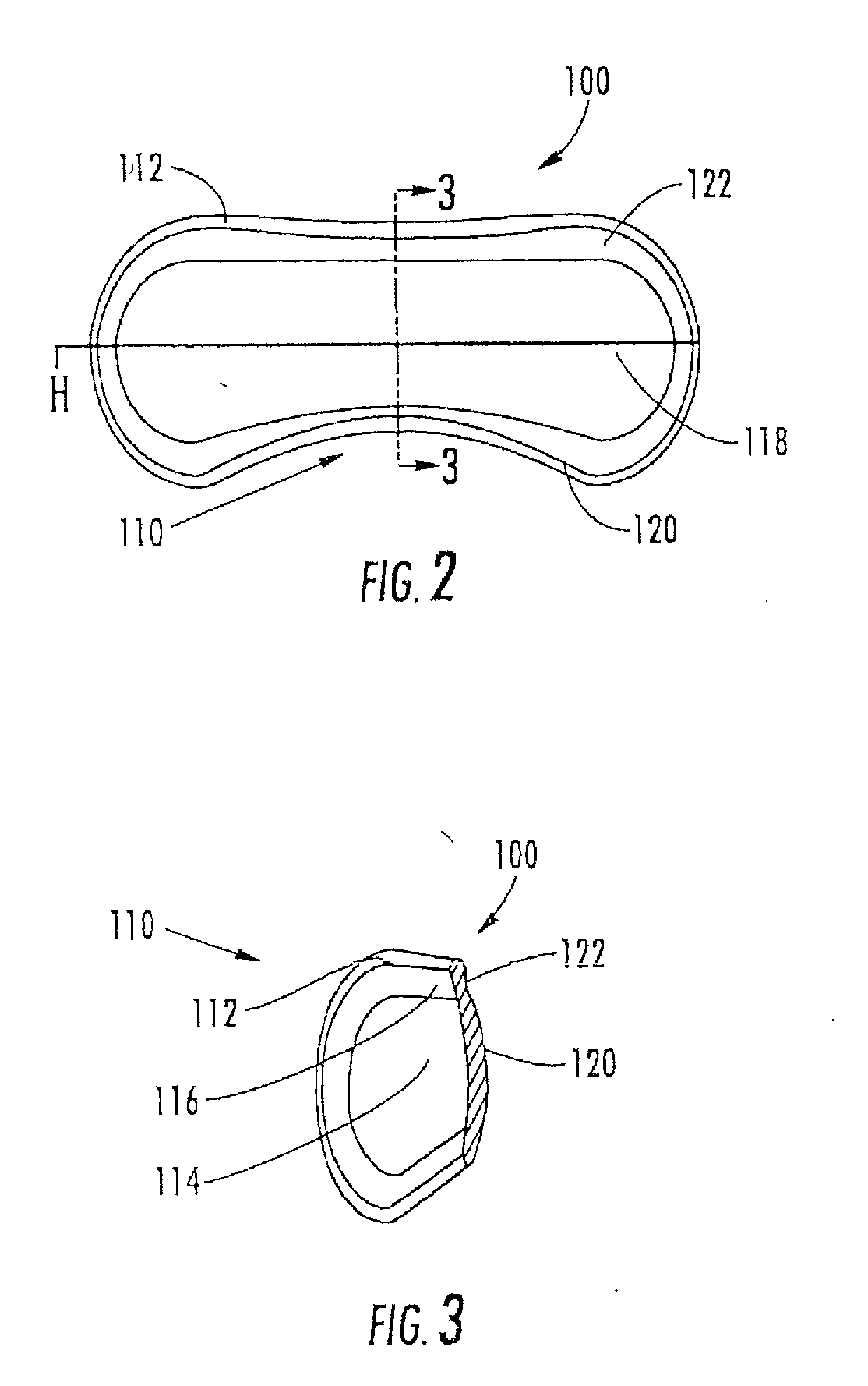
[0162]The aspects of the device of Example One are shown in FIGS. 6-8. The overall shape of this invention is greater horizontally than vertically, and can appear as an oval in as shown in the front elevation view of FIG. 6. It is preferred that the shape be symmetrical about the vertical meridian, such that the lateral halves are mirror images. This aspect allows for the same device design to be used in the right and left eyes (in the same orientation), and on the superior or inferior sclera of an eye. The base curve 114 radius is chosen to fit the sclera 50. The center thickness is greatest in the horizontal centerline, with tapering to a defined minimal, mostly uniform edge thickness around the entire edge perimeter of the ellipse where the anterior surface 207 and posterior surface, 209 meet. This entails a significantly tonic shape on a fairly spherical base curve with a uniform edge radius. Size can range from about 10 mm to about 25 mm in width by about 5 mm to about 12 mm in...
example 2
[0163]The aspects of the device of Example two are shown in FIGS. 6-8. The general geometric parameters were discussed in Example One. A prototype device was constructed from silicone elastomer. The overall width was 21.0 mm, the height was 7.8 mm and the center thickness was 1.5 mm. The toric front surface radii were 5.0 mm vertical meridian and 12.0 mm horizontal meridian. The base curve radius was 12.4 mm. The overall device volume was 150 μm. This device was placed on the superior sclera of a subject's eye. The device was stable in the eye with slight rotation observed. The comfort of the device was reported to be good.
example 3
[0164]The aspects of the device of Example Three are shown in FIGS. 6-8. The general geometric parameters were discussed in Example One. A prototype device was constructed from silicone elastomer. The overall width was 24.5 mm, the height was 10.0 mm, and the center thickness was 2.3 mm. The toric front surface radii were 6.0 mm vertical meridian by 12.5 mm horizontal meridian. The overall device volume was 385
The device was placed on the superior sclera of a subject's eye. The device tended to move slightly to a nasal position. The comfort was rated at “slight awareness”.
PUM
| Property | Measurement | Unit |
|---|---|---|
| thickness | aaaaa | aaaaa |
| thickness | aaaaa | aaaaa |
| radius | aaaaa | aaaaa |
Abstract
Description
Claims
Application Information