Fluorescently labeled fusion protein for assaying adenosine triphosphate

Active Publication Date: 2010-09-16
THE NEW IND RES ORG +2
View PDF2 Cites 10 Cited by
  • Summary
  • Abstract
  • Description
  • Claims
  • Application Information

AI Technical Summary

Benefits of technology

[0017]ATP is the cell's energy currency and to know the amount of ATP in living cells is medically and biologically important. However, until now, since there have been no practical techniques for the measurement, it has been almost unknown whether ATP concentration in living cells is constantly maintained or dynamically fluctuated, or whether ATP concentration differs from one cell to the next.
[0018]ATP concentration can be measured with the ratio of donor to acceptor fluorescence by FRET technique through the use of the fluorescence labelled fusion protein of the present invention as a fluorescent protein probe. The method has major advantage that quantitative capability is not affected by the amount of protein, unlike in t

Problems solved by technology

However, in the case where luciferase is expressed in the cells to measure ATP concentration in a living organism, quantitative measurement is difficult and thus following the change in ATP concentration with a high temporal resolution is difficult, because of the drawbacks that luminescence of luciferase is dark and also the amount of luminescence is dependent on the concentration of protein.
The method enables quantitative measurement of ATP in solution, but the operation is so complicated that

Method used


Image

  • Fluorescently labeled fusion protein for assaying adenosine triphosphate
  • Fluorescently labeled fusion protein for assaying adenosine triphosphate
  • Fluorescently labeled fusion protein for assaying adenosine triphosphate

Examples


example 1

Preparation of Fluorescence Labelled Fusion Protein

1) ε Protein

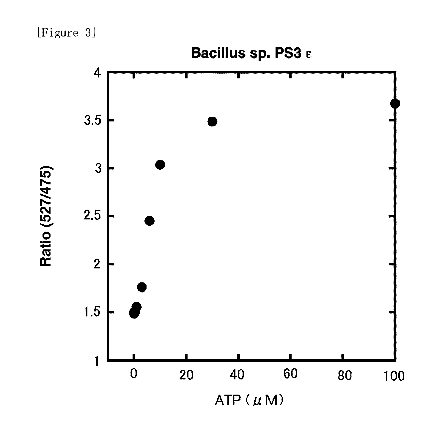
[0058]a. ε protein derived from Bacillus sp. PS3 (SEQ. ID. NO: 1)

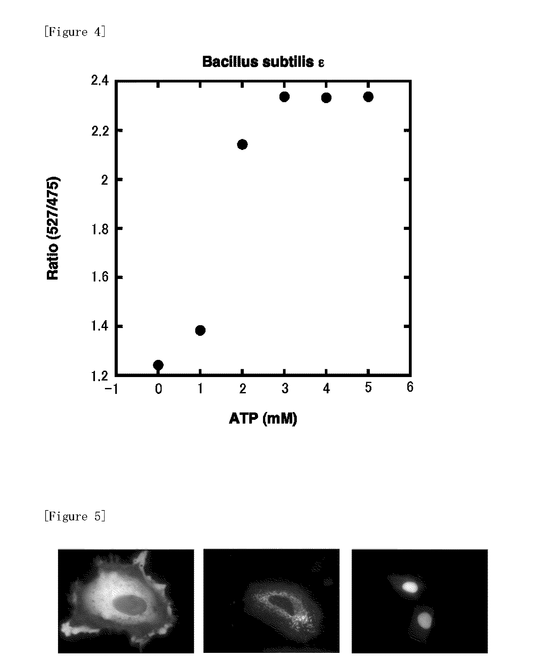
[0059]b. ε protein derived from Bacillus subtilis (SEQ. ID. NO: 2)

[0060]Each ε protein described above was obtained in the same way as that in described in J. Biological Chemistry 2003; 278, 36013-36016. Alanine and asparagine were attached to the C-terminal of the amino acid sequence derived from Bacillus subtilis represented by SEQ. ID. NO: 2 described above. Further, in a protein consisting of the amino acid sequence represented by SEQ. ID. NO: 1 derived from Bacillus sp. PS3 described above, hydrophobic amino acid residue parts (Va19 / Va142 / Phe69 / Leu78: SEQ. ID. NO: 1) necessary for interaction with γ protein, which are the other subunits, were substituted by hydrophilic amino acid residues. The altered proteins obtained from a and b described above were termed as A and B, respectively.

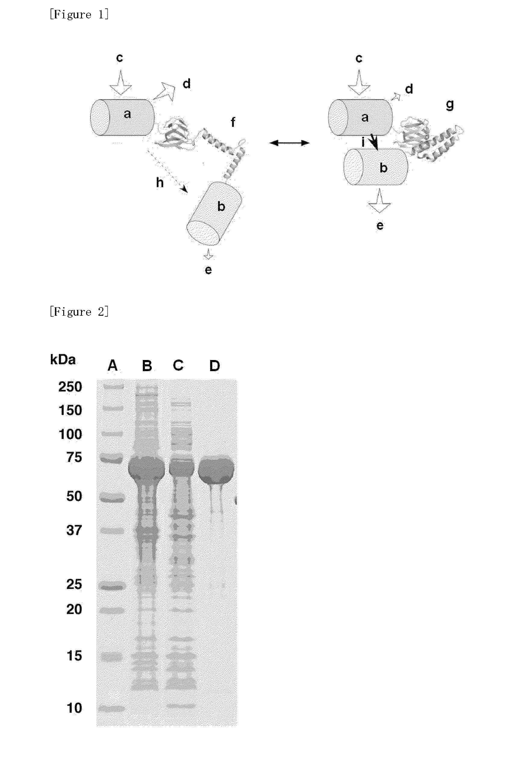
2) Preparation of Fluorescence labelled Fusion Protein

[0061]cDNA enco...

example 2

Preparation of In Vivo Expression Plasmid of Fluorescence Labelled Fusion Protein

1) ε Protein

[0070]Alanine and asparagine were attached to the C-terminal side of the amino acid sequence of ε protein b (ε protein derived from Bacillus subtilis: SEQ. ID. NO: 2) described in Example 1. Further, hydrophobic amino acid residue parts necessary for interaction with y protein, which are the other subunits, were substituted by hydrophilic amino acid residues (V9T / V42K / F67T / L78N: SEQ. ID. NO: 2). The altered protein obtained like this was termed as B1.

2) Preparation of In Vivo Expression Plasmid of Fluorescence Labelled Fusion Protein

[0071]cDNA encoding CFP was attached to the 5′ terminal of cDNA encoding altered protein B1 and cDNA encoding YFP was attached to the 3′ terminal.

[0072]DNA encoding ε protein was amplified by PCR method and then the terminal portion was cleaved with restriction enzyme ClaI and EcoRI. DNA encoding CFP was amplified by PCR method likewise and then the terminal port...


PUM

PropertyMeasurementUnit
Concentrationaaaaaaaaaa
Resonance energyaaaaaaaaaa
Fluorescenceaaaaaaaaaa

Abstract

The object of the present invention is to provide a substance, which is easy to handle and enables the measurement of ATP with a high sensitivity regardless of the concentration of protein, and further a measuring method of ATP using the substance. Such object is solved with a fluorescence labelled fusion protein obtained by attaching two types of fluorescent substances of potential donor and acceptor for fluorescence resonance energy transfer (FRET) respectively to a protein which can cause structural changes depending on ATP binding, namely ε protein, which is the subunit of ATP synthetase, and further solved by contacting the fluorescence labelled fusion protein with a subject substance and then measuring the fluorescence spectra.

Description

TECHNICAL FIELD[0001]The present invention relates to a fluorescence labelled fusion protein for measuring adenosine triphosphate and a measuring method of adenosine triphosphate using the fluorescence labelled fusion protein as a fluorescent protein probe.BACKGROUND ART[0002]Adenosine triphosphate (hereinafter simply referred to as “ATP”) is the energy currency in living organisms.[0003]Until now, the most commonly used measuring method of ATP has been luciferin-luciferase system and a small amount of ATP in a solution can be measured selectively with this system (Nonpatent Document Nos. 1 to 3). Reagents and kits regarding ATP measurement by luciferin-luciferase system already have been commercially available and commonly used. For example, LL100-1 / ATP Luminescence Kit (TOYO INK GROUP), ATPlite ATP detection system (Perkin Elmer) and the like is included. However, in the case where luciferase is expressed in the cells to measure ATP concentration in a living organism, quantitative...

Claims


Application Information

Patent Timeline
IPC IPC(8): C12Q1/68C12Q1/02C12N9/00C12N15/63C07H21/04
CPCC07K14/00C07K14/32C07K2319/60C12N9/14G01N2333/914G01N33/542G01N33/573G01N33/582G01N33/533
Inventor NOJI, HIROYUKIIMAMURA, HIROMIIINO, RYOTAYAMADA, YASUYUKI
Owner THE NEW IND RES ORG
Why Eureka
Try Eureka