
Device for local intraluminal transport of a biologically and physiologically active agent

a technology of biologically and physiologically active agents, applied in the direction of catheters, prostheses, therapy, etc., can solve the problems of drug loss in the blood stream, late thrombosis, and inability to obtain desired efficacy

Inactive Publication Date: 2011-01-06
TERUMO KK
View PDF16 Cites 59 Cited by
  • Summary
  • Abstract
  • Description
  • Claims
  • Application Information

AI Technical Summary

Benefits of technology

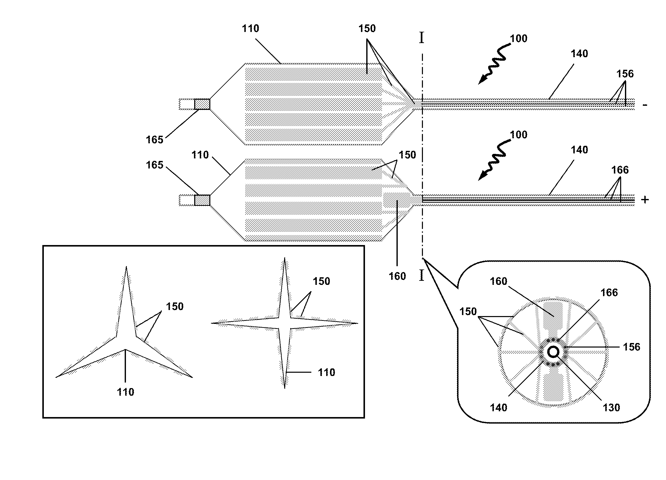
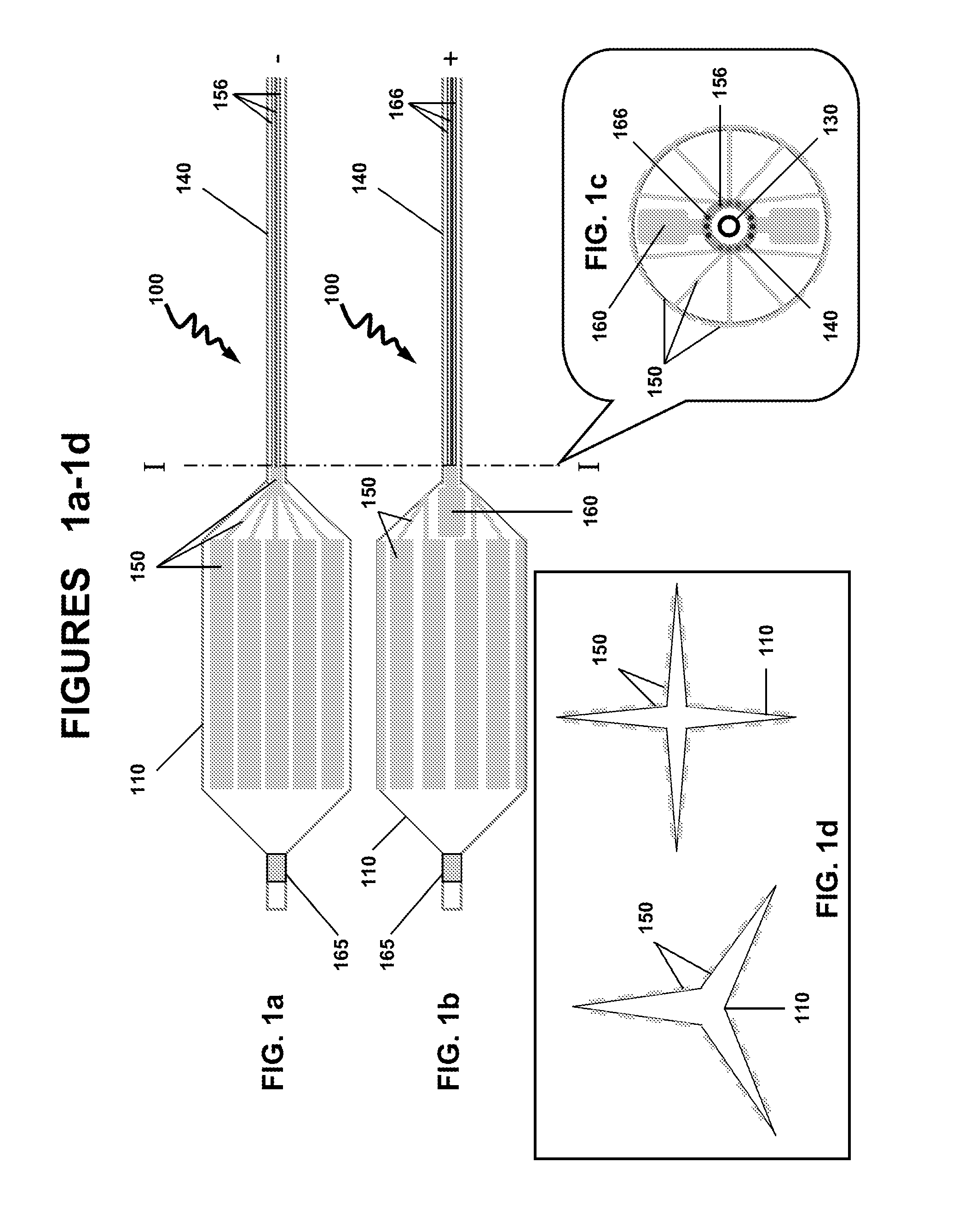
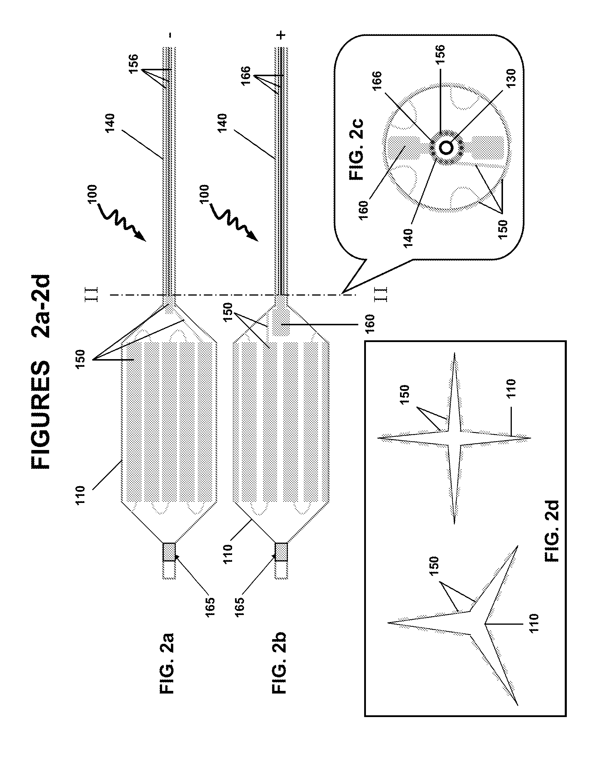
[0010]In a particular variation of the above, the hydrophobic fragment and the hydrophilic fragment is —[—(C5-18alkylenyl)m-L-(CH2CH2O)n—]p—, wherein L is a linker selected from the group consisting of —C(O)O—, —C(O)NH—, —OC(O)O—, —OC(S)O—, —OC(O)NH—, —NR1C(O)O—, —SC(O)O—, —SC(O)S—, —NR1C(NR1)O— and —NR1C(O)NR1—, wherein each R1 is independently H or C1-3alkyl, and where m is 1, 2 or 3, n is 1 to 90, and p is 1 to 10. In a particular variation of the above device, the biologically active agent is selected from the group consisting of a carcinostatic, an immunosuppressive, an antihyperlipidemic, an ACE inhibitor, a calcium antagonist, an integrin inhibitor, an antiallergic, an antioxidant, a GPIIb/IIIa antagonist, retinoid, flavonoid, carotenoid, a lipid improvement agent, a DNA synthesis inhibitor, a tyrosine kinase inhibitor, an antiplatelet, a vascular smooth muscle antiproliferative agent, an anti-inflammatory agent, a biological material, an interferon and a NO production accelerator. In one aspect, the biologically active agents are substantially water soluble agents or water soluble drugs. The biologically active agents may include antithrombotics, antiproliferatives, anti-inflammatory agents, smooth muscle cell migration inhibitors and restenosis-reducing agents. Particular biologically active agents include paclitaxel, sirolimus, simvastatin and rapamycin. In certain aspects, the total load of the biologically active agents may be about 1-1,000 μg, 1-250 μg, 1-100 μg, 1-50 μg, 1-25 μg, 1-10 μg or about 5 μg, the dose of which depends on the nature and biological activity of the agents. The calculation of the dosages are previously known to one skilled in the art. In another variation of the above device, the dilating member is a self-expandable scaffold or a shape memory scaffold. In one variation, the dilating member is circumferentially loaded with a continuous gold layer. In another variation, the dilating member is partially loaded with a continuous gold layer. In a particular variation of the above device, the dilating member is a balloon and the gold surface layer comprises discontinuous rectangle-shaped gold layers. In yet another variation, the dilating member is a balloon and the gold surface layer comprises discontinuous wave-shaped gold layers.
[0011]In another embodiment, there is provided a method for the controlled delivery of a biologically active agent to an intraluminal surface using a drug delivery device, wherein the device comprises: an elongated insertion member having a proximal end and a distal end; a dilating member comprising a proximal end and a distal end, and an inner surface and an outer surface, wherein the proximal end of the dilating member is attached to the distal end of the elongated insertion member, and wherein a part of the surface of the dilating member is coated with a gold surface layer; a biodegradable substrate comprising the biologically active agent, wherein the substrate is covalently bonded to the gold surface layer by a gold-sulfur (Au—S—) bond; an electrical lead having a first end and a second end, the first end connected to the gold surface layer, wherein the electrical lead is configured to pass an electrical current to the gold surface layer; and wherein the controlled delivery and release of the substrate comprising the biologically active agent is initiated by a electrical current reduction and cleavage of the Au—S bond; the method comprises inserting the device into the lumen and advancing the device until the dilating member is in a desired region of the intraluminal surface; expanding the dilating member to contact the outer surface of the dilating member with the vessel wall; and passing an electrical current to the electrical lead sufficient to reduce and cleave the Au—S bond and releasing the biodegradable substrate comprising the biologically active agent over a controlled time period. In one aspect, the controlled time period is between 0.1 and 120 seconds, or between 5 and 30 seconds, between 10 and 20 seconds, or between 1 and 10 seconds, between 1 and 20 seconds, between 1 and 30 seconds, or between 30 and 60 seconds, between 40 and 60 seconds or between 50 and 60 seconds. In one aspect, the release of the substrate comprising the biologically active agent from the device may be performed at low electrical currents. The electrical current are generated at biologically safe levels. The release of the substrate may be performed using electrochemically programmed methods to release the agent at the desired levels, rate. The release of the substrate may be programmed to provide the biological agent at the desired concentrations. The programmed release of the substrate from the gold surface may be biased at about −1.5 V (vs. Ag/AgCl) f...

Problems solved by technology

However, drug-eluting stents are known to cause late thrombosis, and patients in whom such a stent has been implanted must take dual anti-platelet regime for prolonged periods.
Since vulnerable plaque is not stenosis, treatment with a stent is not appropriate, and local therapeutic drug administration by catheter has been studied; but there is a problem in that the desired efficacy cannot be obtained, due to shortage of drug release period, the loss of the drug into the bloodstream and the trauma while the catheter is being introduced into the patient.
This process results in the lost of the drug in the blood stream when the drug is released inside the patient.

Method used


Image

  • Device for local intraluminal transport of a biologically and physiologically active agent
  • Device for local intraluminal transport of a biologically and physiologically active agent
  • Device for local intraluminal transport of a biologically and physiologically active agent

Examples


example

NP-3000-Bio Preparation Conjugated with Rapamycin

[0065]Nanoparticles are produced using a single emulsion technique in which 10 mL of a 25-mg / mL solution of the polymer and various amounts of drug in dichloromethane is homogenized for 2 min in 250 mL of a 0.1% aqueous PVA solution (PVA 88% hydrolyzed, PolyScience Inc., Warrington, Pa.). The resulting emulsion is stirred for 4 h to allow the dichloromethane to evaporate. The nanoparticles are collected by centrifugation at 5,000 rpm for 10 min and washed three times in distilled water and then lyophilized.

Immobilization on the Gold (Au) Surface Layer:

[0066]The device upon which an ultrathin gold film has been formed or deposited upon, is submerged for 18 hours in a 1-mM ethanol solution of HOOC-PEG-C5-18alkylenyl-SH (or also 11-carboxyl-1-undecanethiol), that induces the formation of a self-assembled monolayer (SAM) on the gold surface. A gold-sulfur bond (Au—S) is formed between the thiol group (—SH) and the gold surface, wherein th...


PUM

PropertyMeasurementUnit
Volumeaaaaaaaaaa
Timeaaaaaaaaaa
Hydrophilicityaaaaaaaaaa

Abstract

Provided herein is a drug delivery device and the method of us for the intraluminal controlled delivery of a biologically active agent comprising a dilating member comprising a proximal end and a distal end, and an inner surface and an outer surface, wherein a part of the outer surface of the dilating member is coated with a gold surface layer; a biodegradable substrate comprising the biologically active agent, wherein the substrate is covalently bonded to the gold surface layer by a gold-sulfur (Au—S—) bond; an electrical lead having a first end and a second end, the first end connected to the gold surface layer, wherein the electrical lead is configured to pass an electrical current to the gold surface layer; and wherein the controlled delivery and release of the sub strate comprising the biologically active agent is initiated by an electrical current reduction and cleavage of the Au—S bond.

Description

TECHNICAL FIELD[0001]The present invention generally relates to a device for local intraluminal transport of a biologically and physiologically active agent, and more specifically, relates to a device to be inserted intralumenally into the body, e.g., via a blood vessel, for local therapeutic release of a biologically and physiologically active agent.BACKGROUND DISCUSSION[0002]Drug-eluting stents are in wide use for treatment of blood vessel stenosis and the like. A drug-eluting stent dilates the blood vessel stenosis and also releases a small amount of a drug that prevents restenosis, as the stent has a surface coated with the drug for this purpose. However, drug-eluting stents are known to cause late thrombosis, and patients in whom such a stent has been implanted must take dual anti-platelet regime for prolonged periods. Attention has also focused recently on vulnerable plaque forming in less nonstenosed blood vessels as a cause of sudden death and blood vessel total occlusion. S...

Claims


Application Information

Patent Timeline
IPC IPC(8): A61N1/30A61F2/82A61F2/966A61M25/10
CPCA61F2/958A61F2250/0001A61F2250/0067A61L29/10A61L29/16A61L31/088A61N1/306A61L2300/604A61L2300/62A61M25/10A61M25/1027A61M2025/105A61M2025/1088A61L31/16
Inventor ISHII, NAOKI
Owner TERUMO KK
Learn More