Method for decellularization

a tissue engineering and decellularization technology, applied in the field of tissue engineering constructs and tissue engineering methods, can solve the problems of less etoh concentrations that can be used, and achieve the effects of less calcification, reduced inflammation, and improved tensile strength and elastic modulus

Inactive Publication Date: 2011-07-07
CHILDRENS MERCY HOSPITAL
View PDF8 Cites 40 Cited by
  • Summary
  • Abstract
  • Description
  • Claims
  • Application Information

AI Technical Summary

Benefits of technology

[0052]In another embodiment of the present invention, a decellularized tissue is provided. This decellularized tissue can be prepared using any method described herein. Preferably, after the decellularization process, the tissue will contain little or no dsDNA. Advantageously, tissues decellularized using methods of the present invention exhibit less calcification after implant when compared to tissues prepared not according to the methods of the present invention. Another characteristic of a tissue decellularized in accordance with the present invention is that the tissue has a reduced inflammatory response when compared to tissues prepared not according to method of the present invention. Advantageously, tissues decellularized in accordance with methods of the present invention have a higher ultimate tensile strength and elastic modulus when compared to tissues not prepared according to the methods of the present invention.

Problems solved by technology
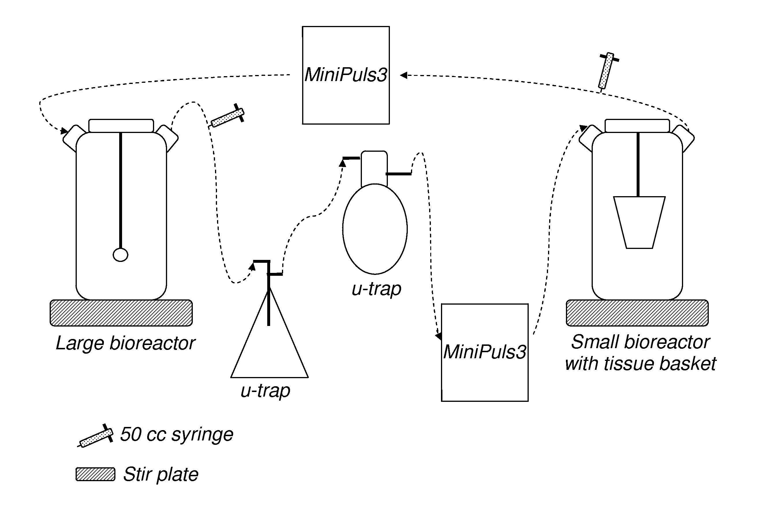
Likewise, the greater amount of time the tissue is exposed to EtOH, the lesser the concentration of EtOH can be used.

Method used


Image

  • Method for decellularization
  • Method for decellularization
  • Method for decellularization

Examples


example 1

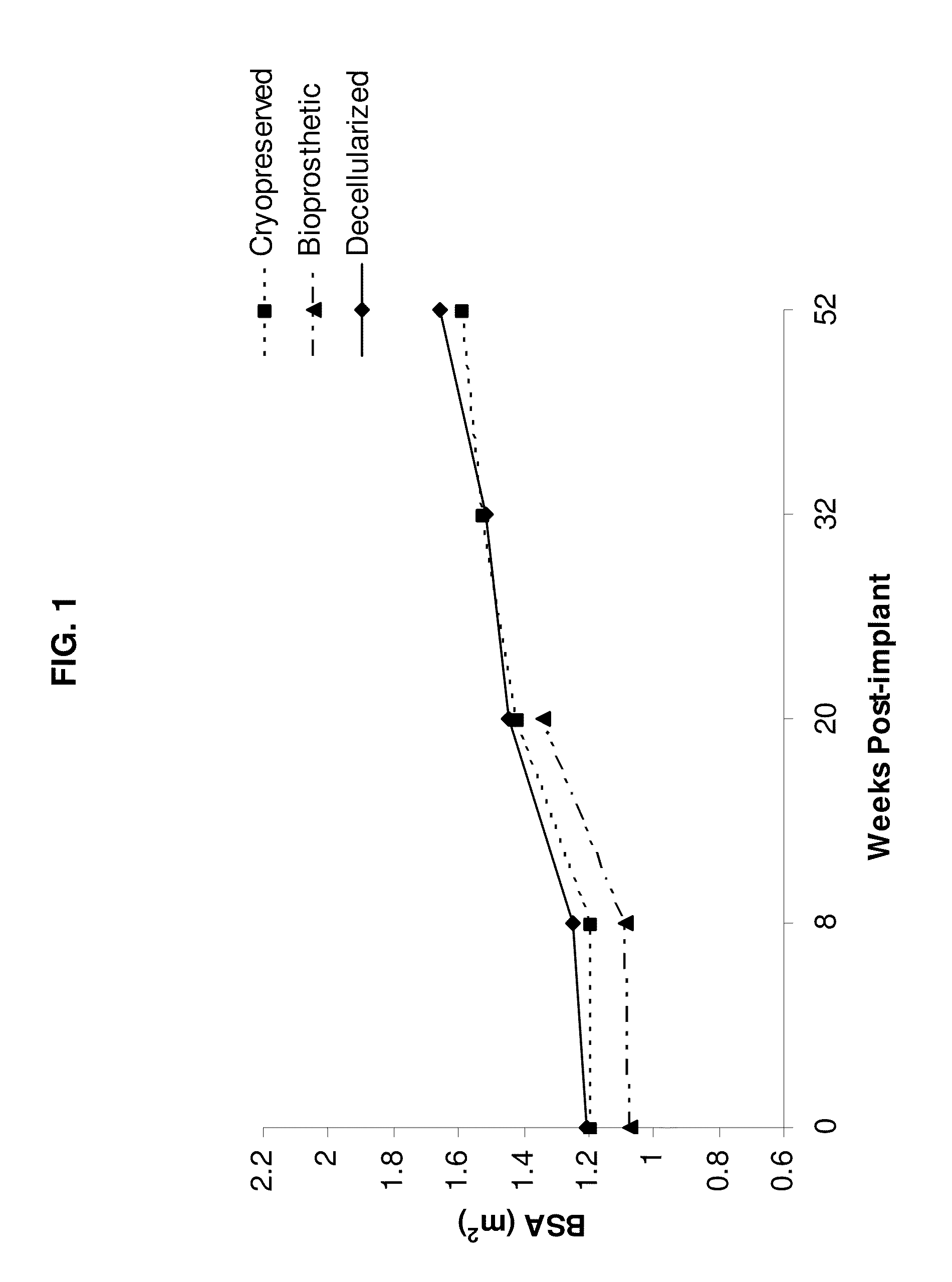
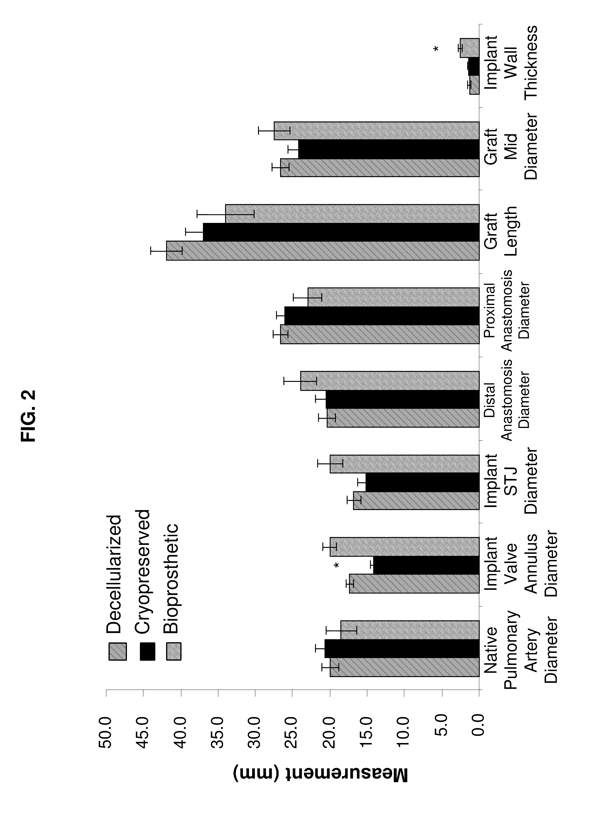
[0070]This example illustrates analysis of the differences between decellularized, cryopreserved, and bioprosthetic heart valves.

Materials and Methods

Animals

[0071]All animal procedures were carried out under protocols approved by the Institutional Animal Care and Use Committee and animals received humane care in compliance with the Guide for Care and Use of Laboratory Animals (NIH Publication #85-23). Nineteen female domestic sheep (ovis ares; Suffolk / North Country Cheviot; 160±9d, 46.5±9 kg) were divided into three treatment groups. Group 1 sheep (n=8) were implanted with cryopreserved homografts further treated with a series of steps resulting in the decellularization of the tissue. Group 2 sheep (n=6) were implanted with cryopreserved homografts and Group 3 sheep (n=4) were implanted with a commercially available glutaraldehyde-preserved porcine aortic root bioprostheses (Freestyle, Medtronic, Minneapolis, Minn.). Sheep were survived for either 20 wk (Group 1, n=4; Group 2, n=3; ...

example 2

[0106]This examples illustrates one embodiment of the decell process of the present invention.

Materials and Methods

Solutions Used:

[0107]a. Triton X®-100 (Triton): 0.05% Triton X®-100 solution a 1:2000 dilution derived from 100% Triton X®-100 detergent (Sigma T8787) in ddH2O. Each valve will need 200 mL of this solution, which can be made ahead of time.[0108]For 2 L use 1 mL 100% Triton-X®, 1999 mL ddH2O.[0109]b. N-lauroylsarcosine Sodium Salt Solution (NLS): 1% NLS Solution a 1:20 dilution derived from 20% Sodium Laureth Sulfate (Sigma—L7414) in ddH2O. Each valve will need 200 mL of this solution, which can be made ahead of time.[0110]For 2 L use 100 mL 20% NLS, 1900 mL ddH2O[0111]c. Hypertonic Salt Solution (HSS): 1% NaCl (Fisher—BP358-1), 12.5% D-Mannitol (Sigma—M9647), 5 mM MgCl2 (Sigma—M2643), 500 mM KCl (Sigma P4504) in NS (Normal Saline). Each valve will need 200 mL of this solution which can be made ahead of time.[0112]For 2 L use 2 L NS, 18 gm NaCl, 2.03 gm MgCl2, 74.3 gm KC...

example 3

[0124]This example illustrates the multi-anionic detergent / enzyme pH controlled reciprocating osmolality mammalian heart valve decellularization method for the creation of ECM Scaffold to be used for heart valve tissue engineering.

Materials and Methods

[0125]Hearts were aseptically harvested during multi organ donor harvest. The transport solution were sterile lactated ringers or PBS solution with 4× amphotericin B*=4 ug / ml and 4× penicillin / streptomycin*=400 IU / mL) were provided in advance. *standard tissue culture medium concentrations were Ampho (250 ug / mL) at 8 ug / mL=2 mL / 500 mL and Pen / Strep (10 K IU / mL) at 100 IU / mL=5 mL / 500 mL.

[0126]The valves were dissected in a laminar flow safety cabinet using sterile technique and stored, individually, in sterile 250 mL bottles with fresh transport solution at 4° C. for a maximum of 72 hours. All solutions and solvents used were sterile. Next a muscle shelf debridement protocol was performed.

[0127]On the first processing day, the valves we...


PUM

PropertyMeasurementUnit
timeaaaaaaaaaa
timeaaaaaaaaaa
timeaaaaaaaaaa

Abstract

The present invention provides for decellularized tissue and method for decellularizing tissue. The method generally comprises the steps of obtaining a harvested tissue, performing a muscle shelf debridement, treating the tissue with an enzyme, washing the tissue with a detergent, and performing an organic solvent extraction on the tissue. The tissues decellularized according to the present invention have several advantages including removing more of the residual cell debris, dsDNA, and chemicals, as well as exhibiting less calcification and better ultimate tensile strength than tissues prepared not according to the method of the present invention.

Description

RELATED APPLICATIONS[0001]This application relates to and claims priority to U.S. Provisional Patent Application No. 61 / 258,666, which was filed on Nov. 6, 2009, the contents and teachings of which are incorporated herein by reference.FIELD OF INVENTION[0002]The field of invention relates to a treatment and method of treatment for tissues and tissue engineering as well as the product produced by such methods and treatments. Specifically, the invention relates to the field of tissue engineered constructs wherein the cells of the tissue are removed in preparation for engineering or cell seeding.BACKGROUND OF INVENTION[0003]Numerous types of tissue engineered constructs and vascular grafts have been produced over the last few decades. Previous tissue constructs have included man-made polymers as substitutes for various portions of the organ to which the tissue belongs. Materials such as Teflon and Dacron have been used in various configurations such as scaffoldings, tissue engineered b...

Claims


Application Information

Patent Timeline
Patent Type & Authority Applications(United States)
IPC IPC(8): C12N5/071C12N5/077A61K35/12
CPCA61F2/2415A61K35/12C12N1/08
Inventor HOPKINS, RICHARD A.
Owner CHILDRENS MERCY HOSPITAL
Why Eureka
Try Eureka