Nanoparticle sensor for measuring protease activity and method for manufacturing the same

a technology of protease activity and nanoparticles, applied in the direction of instruments, peptides/protein ingredients, peptides, etc., can solve the problems of insufficient introduction of quantitatively imaging and analyzing the activity and expression of specific proteases or non-invasively imaging the expression level of proteases in vivo, and the development of related techniques is urgently needed. , to achieve the effect of inhibiting fluorescence emission and high extinctive activity

Active Publication Date: 2011-09-01
KOREA INST OF SCI & TECH +2
View PDF1 Cites 17 Cited by
  • Summary
  • Abstract
  • Description
  • Claims
  • Application Information

AI Technical Summary

Problems solved by technology

Most of them are generated in an inactive state and specifically activated because numerous proteases may badly affect cells.
For example, serious results may be caused by shortage or overexpression of a specific protease, which may result in cancer, arthritis, neurodegenerative disorder, cardiovascular disease, autoimmune disease and the like.
If the cell apoptosis does not normally is work or is damaged by pathogenic organ, the result may be fatal, and developed into incurable diseases such as cancer, Alzheimer's disease, AIDS and the like.
However, any technology for quantitatively imaging and analyzing activities and expressions of specific proteases or non-invasively imaging expression level of proteases in vivo has not been introduced.
Thus, development of related techniques is urgently needed.
However, such methods need multi-stage measurement protocols, and thus are inefficient in economical and temporal aspects in use for screening a lot of drugs, such as developing novel drugs.
Furthermore, those methods are configured to detect expression of a specific protease in the body or quantitatively analyzing the expression level of the specific protease, thereby being impossible to be used for early diagnosis of diseases.
If high fluorescence is contained in a background, an image resolution in a target biomolecule or biologic tissue cannot help being lowered.
However, the self-quenching based on the FRET is performed when the fluorophores are within a very short distance therebetween and does not exhibit superior fluorescence quenching rate.
In addition, the single molecule of fluorophore-peptide substrate-quencher is difficult to emit high fluorescence, capable of being imaged only in vivo, which may result in difficulty of a real-time measurement of protein expression in vivo.

Method used


Image

  • Nanoparticle sensor for measuring protease activity and method for manufacturing the same
  • Nanoparticle sensor for measuring protease activity and method for manufacturing the same
  • Nanoparticle sensor for measuring protease activity and method for manufacturing the same

Examples


example 1

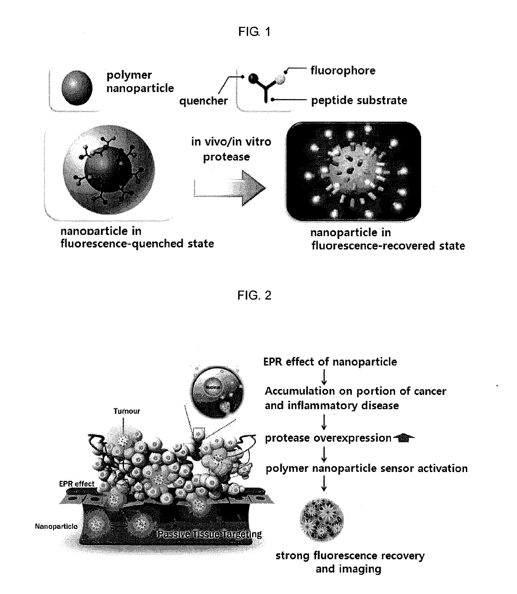
Preparation of Fluorophore-Peptide-Quencher-Nanoparticle Derivative

[0078]1-1. Preparation of Fluorophore-Peptide-Quenching Derivative

[0079]First, to prepare a fluorophore- and quencher-conjugated peptide, the peptide was prepared by Fmoc strategy according to a solid phase synthesis, and the fluorophore and the quencher were chemically conjugated to the prepared peptide sequence.

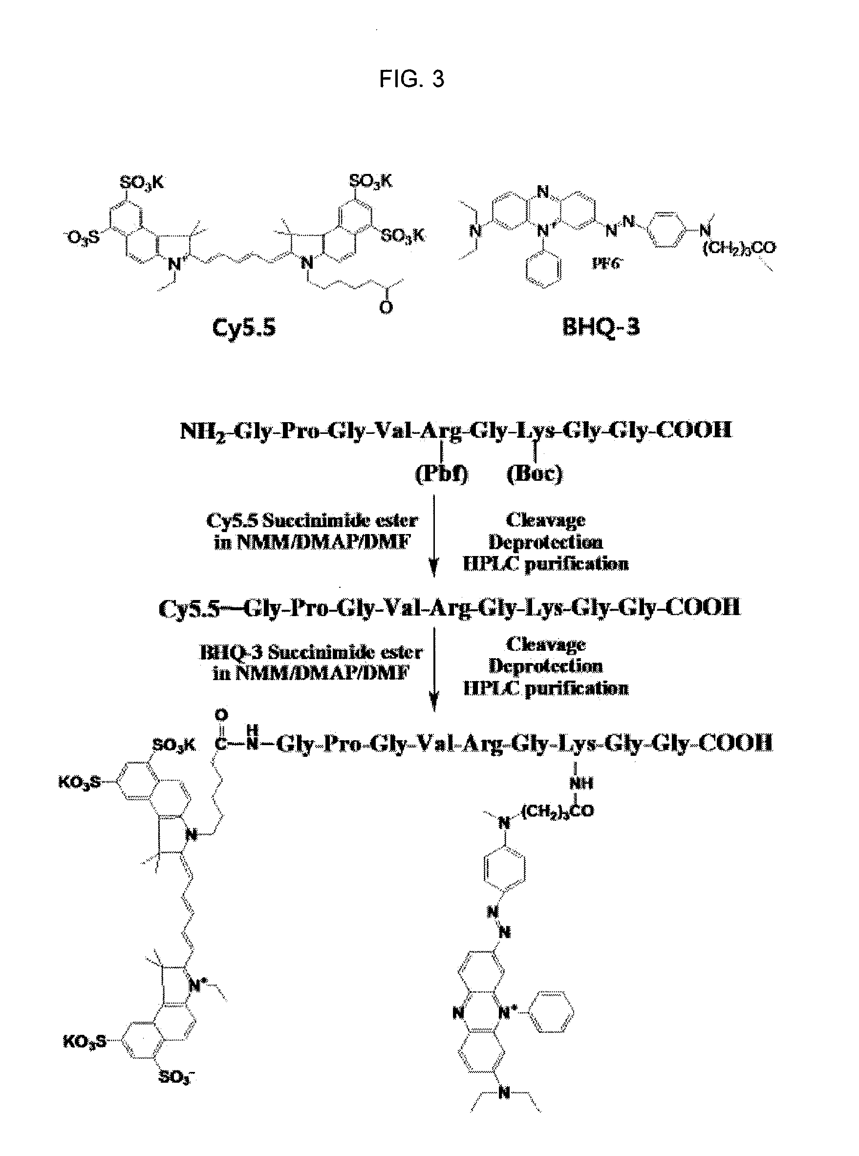
[0080]As the fluorophores, Cy5.5 (ex / em, 670 / 690), TRITC (ex / em, 547 / 572) and FITC (ex / em, 490 / 520) were used. As the quenchers for quenching fluorescence emitted from each of Cy5.5, TRITC and FITC fluorophores, BHQ-3 (abs. 620 nm-730 nm), BHQ-2 (abs. 550 nm-650 nm) and BHQ-1 (abs. 480 nm-580 nm) (Biosearch Technologies Inc.) were used, respectively.

[0081]As one example, a process of synthesizing a fluorophore-peptide-quencher, which emitted fluorescence by being selectively lysed by MMP-2, was as follows (see FIG. 3).

[0082]NH2-Gly-Pro-Leu-Gly-Val-Arg(Pbf)-Gly-Lys(Boc)-Gly-Gly-COON as a peptide substrate, se...

experimental example 1

Variation of Optical Properties of Protease Sensor by MMP-Specific Lysing

[0094]Specific selectivities for proteases of the polymer based sensors, which was prepared in Example 1 and specific to MMPs, were observed. Each of the sensors (PN1, PN2 and PN3) was added in a mixture containing proteases, thereby measuring fluorescent intensities (FIs) recovered according to time.

[0095]In detail, PN1, PN2 and PN3 (10 μg / Ml each), which specifically reacted with MMPs to recover fluorescence, were added in MMP-2, MMP-3, MMP-7, MMP-9 or MMP-13 (1 μg / Ml) all in an active state, and emission of fluorescence by zymolysis was observed. For activating each MMP, the MMPs were added in TCNB solution (0.1 M of Tris, 5 mM of calcium chloride, 200 mM of NaCl and 0.1% of brij), in which p-aminophenyl mercuric acid were mixed, followed by reaction at 37° C. for 1 hour. The activated MMPs respectively reacted with a polymer nanoparticle based sensor in 200 μl of solution in a 96-well at 37° C., thereby obs...

example 2

Fluorescence Recovery and Imaging of Protease Activity Measuring Sensor According to Concentration of Protease

[0099]The sensors prepared in Example 1 were used to quantitatively analyze concentrations of proteases. In detail, according to the same method as in Example 1, activated MMP-2s respectively in concentrations of 0, 0.55, 1, 3, 7 and 14 nmol / L were added in the PN1 sensor and reacted with each other at 37° C. for 60 minutes, thereby observing emission levels of fluorescence (fluorescent intensities) using a fluorescence spectrometer and a digital imaging system (Kodak Image Station 4000 mM).

[0100]As shown in FIG. 5A, fluorescence emitted from the PN1 sensor was increased in proportion to the concentration of the added MMP-2 protease. It was thusly proved that a specific protease concentration within a specimen could be quantitatively detected by measuring the fluorescence intensity of an experimental group, and imaging thereof could be carried out using an optical imaging sy...


PUM

PropertyMeasurementUnit
wavelengthsaaaaaaaaaa

Abstract

Disclosed are a nanoparticle sensor for measuring protease activity, for protease imaging, and a method for preparing the same. More specifically, the present invention relates to a nanoparticle sensor for measuring protease activity in which a fluorophore- and a quencher-conjugated peptide substrate is conjugated to a biocompatible polymer nanoparticle. The peptide substrate is specifically lysed by a protease. The sensor according to the present invention is capable of inhibiting emission of fluorescence with high extinctive activity of the quencher on a fluorescent material. But strong fluorescence is specifically emitted only if the peptide substrate is lysed by a specific protease. Therefore, the sensor is especially useful as a method for screening a novel drug such as a protease overexpression inhibitor, and early diagnosis of incurable diseases and various diseases such as autoimmune diseases including cancer, osteoarthritis, rheumatoid arthritis and dementia.

Description

FIELD OF THE INVENTION[0001]This specification relates to a nanoparticle sensor for measuring protease activity for protease imaging and a method for manufacturing the same.BACKGROUND OF THE INVENTION[0002]Most of cells in human bodies consist of proteins. Most of them are generated in an inactive state and specifically activated because numerous proteases may badly affect cells. That is, the proteases control activities or lives of proteins through hydrolysis of irreversible peptide bond. Examples of such control may include assigning positions of proteins inside or outside cells or separating them from cell surfaces, activating or deactivating various growth factors, hormones, cytokines, enzymes and proteases, converting agonists into antagonists and the like.[0003]Proteases serve to control various cell functions in a broad sense. Since such control is derived by lysing bioactive substances, the functions and roles of the proteases are very important for living of every life. For...

Claims


Application Information

Patent Timeline
Patent Type & Authority Applications(United States)
IPC IPC(8): C07K9/00B82Y5/00
CPCC12Q1/37G01N33/54346Y10S977/773Y10S977/953Y10S977/807Y10S977/811Y10S977/774B82Y5/00B82Y15/00
Inventor KWON, ICK-CHANCHOI, KUI-WONKIM, KWANG-MEYUNGYOUN, IN-CHANLEE, SEUL-KIPARK, KYEONG-SOONMOON, DAE-HYUKCHI, DAE-YOONLEE, SEUNG-JINMYUNG, SEUNG-JAE
Owner KOREA INST OF SCI & TECH
Why Eureka
Try Eureka