Method for healing bone fracture using transfected chondrocytes

a bone fracture and chondrocyte technology, applied in the field of bone fracture healing using transfected chondrocytes, can solve the problems of fracture healing complex process, limited wide clinical application, and short-term effects of high cos

Inactive Publication Date: 2011-09-22
TISSUEGENE INC
View PDF2 Cites 4 Cited by
  • Summary
  • Abstract
  • Description
  • Claims
  • Application Information

AI Technical Summary

Benefits of technology

[0031]c) introducing the connective tissue cell into the fracture site, and allowing the fracture to heal.

Problems solved by technology

Osteoporosis, which is characterized by low bone mass and microarchitectural deterioration of bone structure resulting in bone fractures, is a common health problem among increasing number of the elderly.
Fracture healing is a complex process and remains poorly understood.
Recently, the therapeutic value of TGF-β has been reported (Critchlow et al., Bone, 521-527, 1995; and Lind et al., A Orthop Scand, 64 (5): 553-556, 1993), but its short-term effects and high cost have limited wide clinical application.
A high rate of failure has been associated with these conventional therapies.
One major disadvantage of these methods is the requirement of a large amount of recombinant proteins to achieve therapeutic effects due to the short duration of action of the therapeutic proteins in vivo.
Fractures in elderly individuals often require surgery and can lead to increased morbidity and mortality.
The failure rate of conventional internal fixation is high in osteoporotic bone fractures, due to the decreased holding power of plate-and-screw fixation.1 Although autogenous cancellous bone from the iliac crest provides the best healing power for bone fractures, surgeons were forced to use bone allografts because of complications.2 The shortage of allograft donors spurred the development of three categories of synthetic graft substitutes.3,4 Osteoconductive matrix materials provide a microenvironment that supports growth of osteoprogenitor tissue, but they do not actively stimulate the bone formation process.
This is due, at least in part, to the short half-life of BMPs, and, therefore, efficacy is limited without repeated administration.

Method used


Image

  • Method for healing bone fracture using transfected chondrocytes
  • Method for healing bone fracture using transfected chondrocytes
  • Method for healing bone fracture using transfected chondrocytes

Examples


example 1

Experimental Procedure for Bone Regeneration

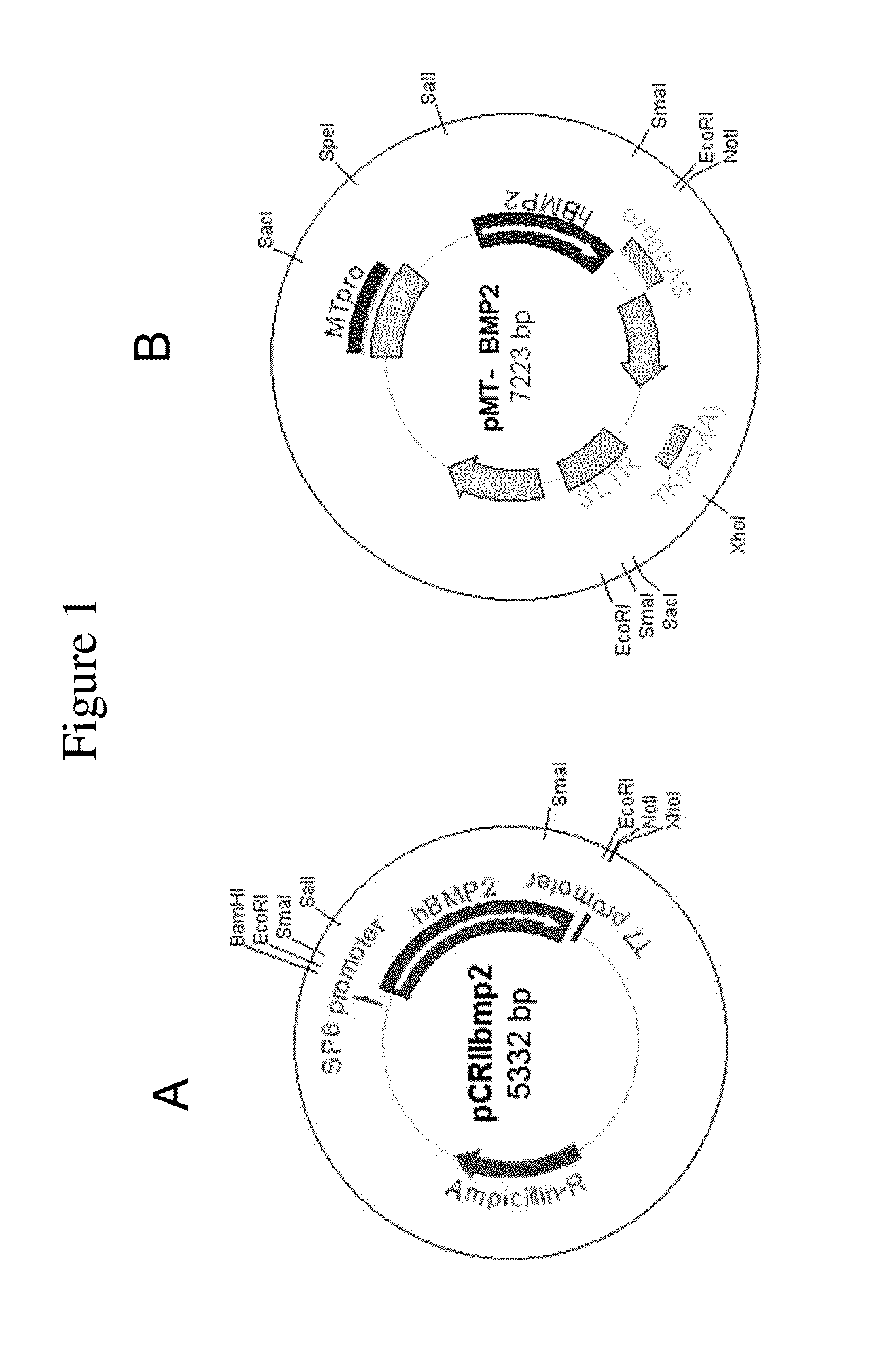
[0158]Human BMP2 gene was cloned by PCR (polymerase chain reaction) with human fetal brain cDNA and two primers. 5′ primer was 5′-TCCCAGCGTGAAAAGAGAGACTGC-3′ (SEQ ID NO:1) and 3′ primer was 5′-TTTTGCTGTACTAGCGACACCCACAACC-3′ (SEQ ID NO:2). After the PCR with GC-rich PCR system (Roche), cloning into pCRII-TOPO vector was done using TOPO TA cloning kit (Invitrogen) (FIG. 1A). For cloning into retroviral vector, pCRIIbmp2 DNA was cut with Sal I and Not I. Human BMP2 cDNA insert (˜1.2 kb) was ligated into pMTMLV with Sal I and Not I overhangs (FIG. 1B).

[0159]Packaging cell line GP-293 cell (5×10⁵ cells / p60 culture dish) was cultured one day before transfection. pMTMLV or pMT-BMP2 was transfected to GP-293 cell using Fugene (Roche). 48 hours after the transfection, neomycin was added to the culture media for the selection of neomycin resistant cells. Selection was continued for 10 days. Selected 293MT and 293MTBMP2 cells were cultured (5×10⁵ ce...

example 2

Injection of NIH3T3-BMP-2 Cells into Rabbit

[0160]New Zealand white rabbits weighing 2.0-2.5 kg were selected for animal study. The tibia bone was exposed and a defect (2 cm long and 0.5 cm deep) was made with orthopedic surgical instruments. Either control NIH3T3-neo, or NIH3T3-BMP-2 cells (2 ml of 2×10⁶ cells / ml) were injected into the defect area after suturing. At 8 weeks after injection of the cells, radiographic analysis and histological examination were performed.

example 3

Weekly Radiographic Examination

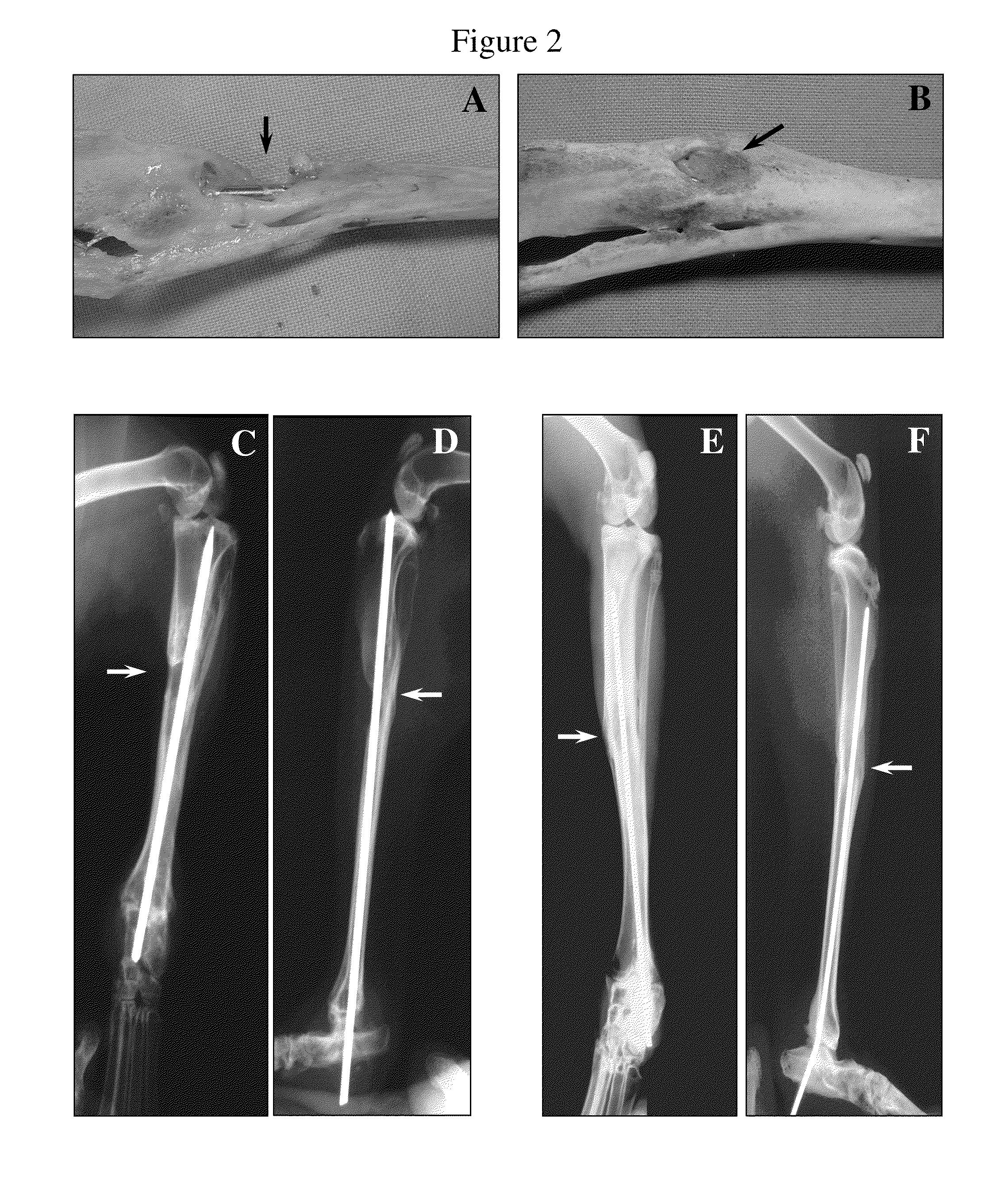
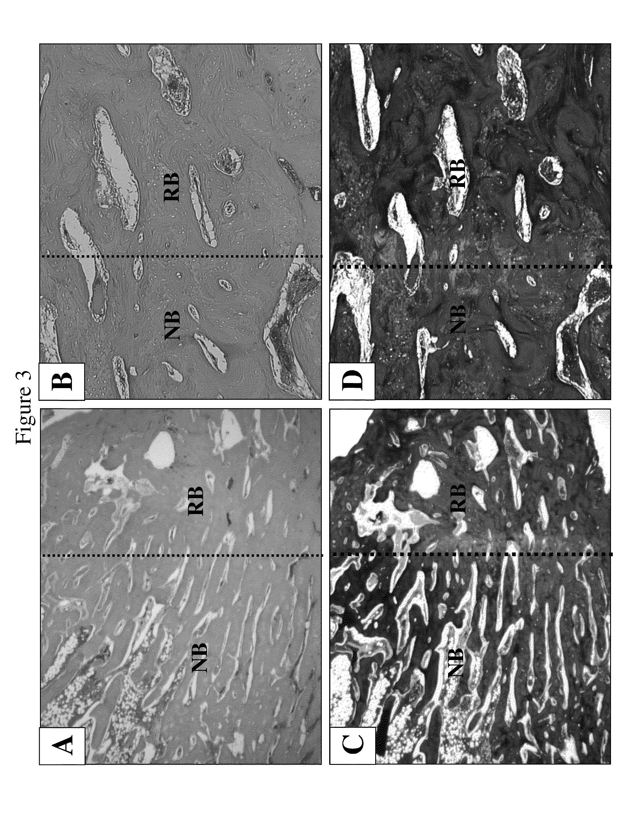
[0161]New Zealand white rabbits weighing 2.0-2.5 kg were selected for animal study. The tibia bone was exposed and a defect (2 cm long and 0.5 cm deep) was made with orthopedic surgical instruments. NIH3T3-BMP-2 cells (2 ml of 2×10⁶ cells / ml) were injected into the defect area in the tibia bone after suturing. Then radiographic analysis was performed at 1, 2, 3, 4, 5, 6, and 7 weeks after injection of the cells. The specimen was harvested at 7 weeks post injection and a picture was taken. Histological examination was carried out after harvest.

[0162]FIGS. 2A-2F show regeneration of bone with NIH3T3-BMP-2 fibroblast cells. FIGS. 1A and 1B show pictures of leg bones after 8 weeks of injection of control NIH3T3 fibroblast cells (A) and NIH3T3-BMP-2 cells (B). FIGS. 2C-2F show radiographic examinations of the control (C & D) and experimental (E & F) leg bones before sacrificing the animals. The bone defect treated with cells expressing BMP-2 proteins was he...


PUM

PropertyMeasurementUnit
timeaaaaaaaaaa
concentrationaaaaaaaaaa
sizeaaaaaaaaaa

Abstract

The application discloses a method for making bone at a bone defect site for a person suffering from low bone mass which includes inserting a gene encoding a protein having bone regenerating function into a connective tissue cell operably linked to a promoter, and transplanting the mammalian cell into the bone defect site, and allowing the bone defect site to make the bone.

Description

BACKGROUND OF THE INVENTION[0001]1. Field of the Invention[0002]The present invention relates to a method of introducing at least one gene encoding a member of the transforming growth factor β superfamily into at least one mammalian connective tissue cell for use in generating or regenerating bone, in particular, to repair fracture in osteoporotic bone or to fuse spine in the mammalian host.[0003]2. Brief Description of the Related Art[0004]Homeostasis of living bone tissue is a dynamic process modulated by regulatory signals such as hormones, and growth and differentiation factors. The growth factors known to stimulate proliferation of bone cells are bone morphogenic proteins (BMPs), transforming growth factor-β proteins (TGF-β), insulin-like growth factors (IGFs), and basic fibroblast growth factors (bFGFs).[0005]Osteoporosis, which is characterized by low bone mass and microarchitectural deterioration of bone structure resulting in bone fractures, is a common health problem among...

Claims


Application Information

Patent Timeline
Patent Type & Authority Applications(United States)
IPC IPC(8): A61K35/32A61P19/08A61K35/33A61K35/35
CPCA61K35/32A61K35/33A61K35/35A61K38/1841C12N5/0652C12N2501/155C12N2510/00A61K38/1875C12N5/0655A61P19/08
Inventor LEE, KWAN HEENOH, MOON JONGYI, YOUNGSUK
Owner TISSUEGENE INC
Why Eureka
Try Eureka