Method for Amplification of Signal in Immunochromatographic Assay and Immunochromatographic Kit Using the Method
Inactive Publication Date: 2011-09-22
INFOPIA CO LTD
View PDF5 Cites 4 Cited by
Summary
Abstract
Description
Claims
Application Information
AI Technical Summary
This helps you quickly interpret patents by identifying the three key elements:
Problems solved by technology
Method used
Benefits of technology
[0013]The signal amplification method of the present invention immobilizes a primary conjugate body to a capture antibody together with an antigen and then binds a secondary conjugate body to the primary co
Problems solved by technology
However, because the detection sensitivity of the related art immunochromatographic
Method used
Image
Click on the blue label to locate the original text in one second.
Reading with bidirectional positioning of images and text.
Examples
Example
Embodiment 1
Composition of Nano Particle-Antibody Conjugate Body
[0042]1. Composition of Primary Conjugate Body
[0043]A 0.1 mL 0.1 M borate buffer (pH 8.5) was added into a 1 mL gold nano-particle colloid solution (BBInternational, 10 nm), a 1 mg / mL first antibody 10 uL was added thereinto, and they were reacted for 30 minutes. After the reaction, a 0.1 ml solution obtained by dissolving a 1% (w / v) bovine serum albumin (BSA) (Sigma) as a connector in a phosphate buffered saline (PBS) (Gibco) was added thereinto, and they were reacted at the normal temperatures for 15 minutes. After the reaction, it was centrifuged at 10,000 rpm at 4° C. for 20 minutes to disperse in a 1 mL BSA (Sigma) solution dissolved in a 10 mM PBS at a 1 mg / mL concentration. The centrifuging / dispersing process was repeated once again, and it was again centrifuged to disperse in a 1 mL PBS, thereby fabricating a primary conjugate body.
[0044]The first antibody may be 4T21, 560 (HyTest) for immunoassay of a troponin ...
Example
Embodiment 2
Fabrication of Immunochromatograph
[0047]1. Method of Fabricating Immunochromatograph
[0048]A nitrocellulose membrane (Millipore, 180 sec) and an absorbing pad (Millipore) were adhered to a plastic pad (Millipore). Thereafter, using a dispenser system (Zeta Co.), a capture antibody (second antibody) 1 mg / mL solution dissolved in a PBS and a goat anti-mouse IgG antibody (Sigma, M8642) 1 mg / mL solution dissolved in a PBS as a contrast group were scribed on the membrane at a speed of 6 cm / sec, thereby forming a detection site and a control line. After the membrane was dried, it was cut by a cutter at intervals of 3 mm.
[0049]The second antibody, i.e., the capture antibody, may be a troponin capture antibody (Hytest) for immunoassay of a troponin I, and may be a myoglobin capture antibody M09983110 (Fitzgerald) for immunoassay of a myoglobin.
[0050]After a sample pad (Millipore, C068) was immersed in a 0.5% Tween 20, a 5% Sucrose, a 0.05% Dextran, a 5% sodium azide aqueous solut...
Example
Embodiment 3
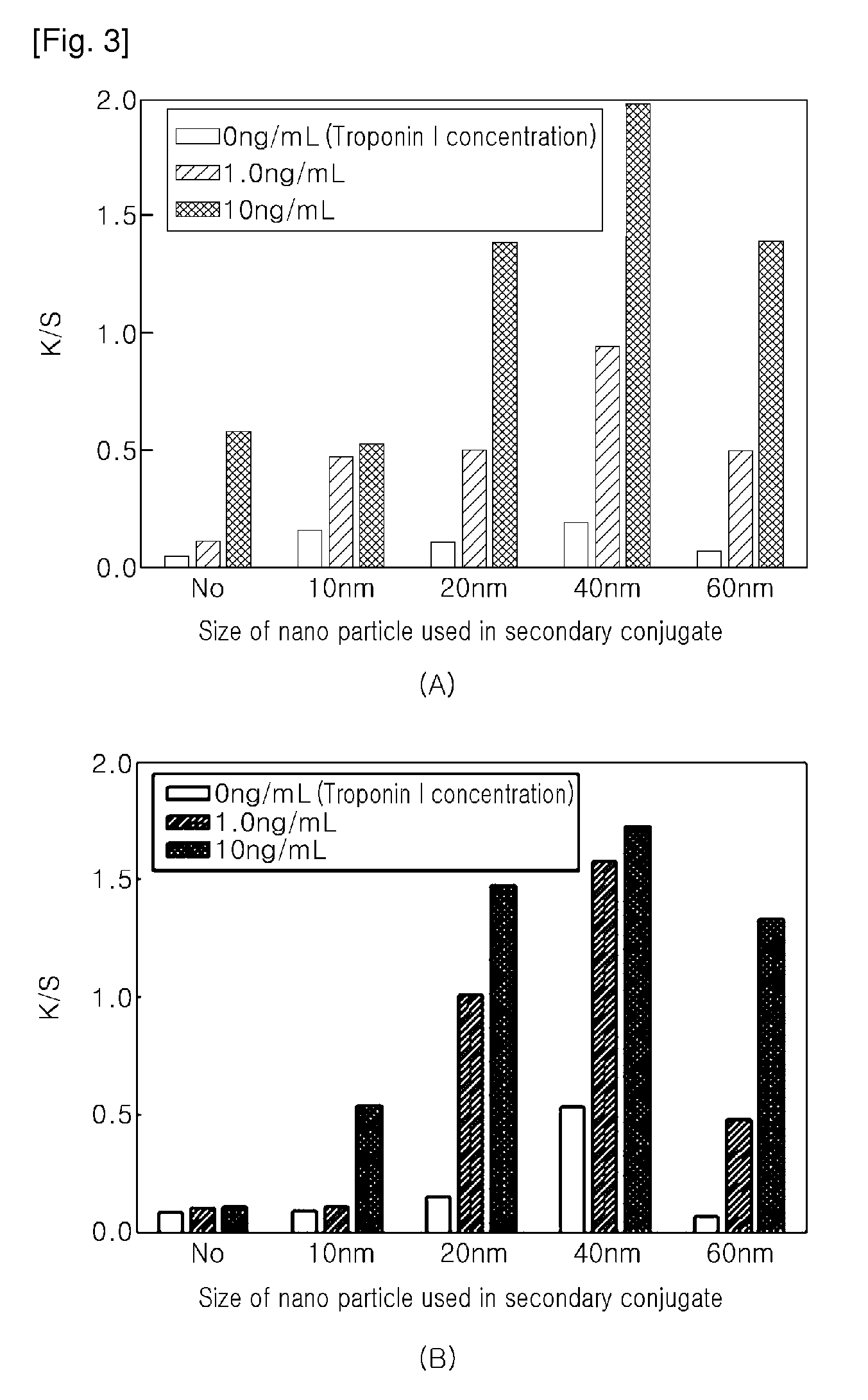
Signal Amplification Effect According to the Size of Gold Nano-Particle
[0054]A primary conjugate body using gold nano-particles with a diameter of 10 nm or 20 nm, and gold nano-particles forming a secondary conjugate body were fabricated in various sizes (10, 20, 40, 60 nm), and the signal amplification effects depending on the gold nano-particle sizes were observed.
[0055]The immunochromatographic kit was immersed in a 96 well plate where a serum (Linear chemicals, Cromatest) 70 μL dissolving a troponin I at a predetermined concentration was immersed, and a measurement was performed. The result of the case of the primary conjugate body having a nano-particle diameter of 10 nm is illustrated in Table 1 and FIG. 3A that illustrates the K / S values depending on the nano-particle sizes (the primary conjugate body has a nano-particle diameter of 10 nm). The result of the case of the primary conjugate body having a nano-particle diameter of 20 nm is illustrated in Table 2 and F...
PUM
Abstract
The present invention relates to a method for amplifying a signal in an immunochro-matographic assay for high-sensitivity detection of an analyte and an immunochromatographic kit using the method, which amplifies a signal by controlling a flow rate by discrimination between the size of a first indicator and the size of a second indicator. According to an aspect of the present invention, a method for amplifying a signal in an immunochromatographic assay includes: binding a primary conjugate body, which has a first antibody binding specifically to a first epitope of an analyte, a connector, and a first indicator to which the first antibody and the connector are bound, to the analyte; binding the analyte bound to the primary conjugate body to an immobilized second antibody binding specifically to a second epitope of the analyte; and binding a secondary conjugate body, which has a third antibody binding specifically to the connector of the primary conjugate body and a second indicator to which the third antibody is bound, to the connector of the primary conjugate body, wherein the primary conjugate body is disposed nearer to the immobilized second antibody than the secondary conjugate body, and the particle of the second indicator is larger than the particle of the first indicator, so that the secondary conjugate body reaches the immobilized second antibody later than the primary conjugate body. According to another aspect of the present invention, an immunochromatographic kit includes: a sample pad to which a liquid sample containing an analyte is applied; a conjugate pad including a primary conjugate body having a first antibody binding specifically to a first epitope of an analyte, a connector, and a first indicator to which the first antibody and the connector are bound, and a secondary conjugate body having a third antibody binding specifically to the connector of the primary conjugate body and a second indicator to which the third antibody is bound, wherein the primary conjugate body is disposed nearer to an immobilized second antibody than the secondary conjugate body and the second indicator is larger than the first indicator so that the secondary conjugate body reaches the immobilized second antibody later than the primary conjugate body; a membrane including a detection site immobilizing thereto the second antibody binding specifically to a second epitope of the analyte to which the primary conjugate body is bound, and a control site for error detection; and an absorbing pad absorbing the liquid sample by a capillary phenomenon. Thus, the present invention can perform signal amplification without separate mechanical control or artificial step-by-step reaction.
Description
TECHNICAL FIELD[0001]The present invention relates to a method for amplifying a signal in an immunochromatographic assay for high-sensitive detection of an analyte and an immunochromatographic kit using the method, and more particularly, to a method for amplifying a signal in an immunochromatographic assay for high-sensitive detection of an analyte and an immunochromatographic kit using the method, which amplifies a signal by controlling a flow rate of indicators by discrimination between the size of a first indicator and the size of a second indicator binding to the first indicator in a sandwich assay method.BACKGROUND ART[0002]An immunochromatographic assay is a method that can test an analyte qualitatively and quantitatively within a short time by using the property that biological materials or chemical materials bind specifically to each other. Particularly, a sandwich-type immunoassay method is well known as immobilizing a first antibody, which is specific to a first epitope of...
Claims
Application Information
Patent Timeline
Application Date:The date an application was filed.
Publication Date:The date a patent or application was officially published.
First Publication Date:The earliest publication date of a patent with the same application number.
Issue Date:Publication date of the patent grant document.
PCT Entry Date:The Entry date of PCT National Phase.
Estimated Expiry Date:The statutory expiry date of a patent right according to the Patent Law, and it is the longest term of protection that the patent right can achieve without the termination of the patent right due to other reasons(Term extension factor has been taken into account ).
Invalid Date:Actual expiry date is based on effective date or publication date of legal transaction data of invalid patent.