
Assay for positioning a feeding tube and method thereof

a feeding tube and positioning technology, applied in the field of medical diagnostics, can solve the problems of inconvenient feeding tube positioning, prolonged feeding period, and harm to patients, and achieve the effect of accurate positioning of feeding tubes

Active Publication Date: 2011-11-03
INGENZA
View PDF7 Cites 9 Cited by
  • Summary
  • Abstract
  • Description
  • Claims
  • Application Information

AI Technical Summary

Benefits of technology

[0016]The present invention describes an improved medical diagnostic test which significantly lowers the current frequency of error for positioning of a feeding tube for administrating food or drugs in patients. The reduction in the frequency of error of tube placement significantly reduces the likelihood of severe harm or death to patients from feeding or drug delivery through an incorrectly positioned feeding tube in patients.

Problems solved by technology

Incorrect placement of a feeding tube and subsequent administration of food or drugs can have serious or life threatening consequences for the patient.
Failure to locate the feeding tube in the stomach can result in harm and even death to the patient, particularly if feeding occurs into the lungs.
Although X-ray examination has an excellent success rate, in practice this verification test is not routinely used for every placement check, as a result of the prohibitive cost, impracticality and harm to the patient due to the quantity and frequency by which confirmation tests are recommended to occur.
Use of X-ray examination to determine feeding tube placement results in longer periods between feedings, overexposure to X-rays and general discomfort associated with X-ray examinations.
Occasionally even the X-ray test can be misinterpreted if the image is not interpreted correctly by a properly trained professional or if the image is complicated by the status of the patient thus making the identification of the tube or its location less certain.
All these factors are detrimental to patient health.
In many instances the use of an X-ray examination to carry out this test is simply unavailable outside of hospitals or larger institutions which can offer this service.
Laboratory testing of bilirubin, pepsin and trypsin concentration levels can be a useful indicator to predict feeding tube location, but such methods are not practical for clinical use, particularly for a bedside or point of care setting (Metheny, N. et al., J Parenter Enteral Nutr, 21: 279 (1997)).
The implementation of tests involving measuring concentration levels of bilirubin, pepsin and trypsin have not been pursued because of the high number of samples requiring analysis, the requirement for rapid delivery and turnaround of results, the provision of a suitable laboratory facility within close proximity to the patient, the overwhelming volume of testing required and the cost burden that would be incurred to deliver this service.
In the absence of a suitably rapid, low cost, convenient, unambiguous and accurate test within a bedside or point of care clinical setting, less accurate methods are sometimes used auscultating for a gurgling sound over the abdomen or visual inspection of the aspirate.
But even with this method, there are many instances where this test is unsuitable.
Since the range for gastric aspirate is typically between pH 0 and pH 7 it can make accurate diagnosis difficult and unreliable, owing to the differences in color of the indicator paper across this range.
In the past litmus paper has been used, but use of the litmus test has recently been criticized for being inaccurate and therefore pH indicator paper should be used in its place.
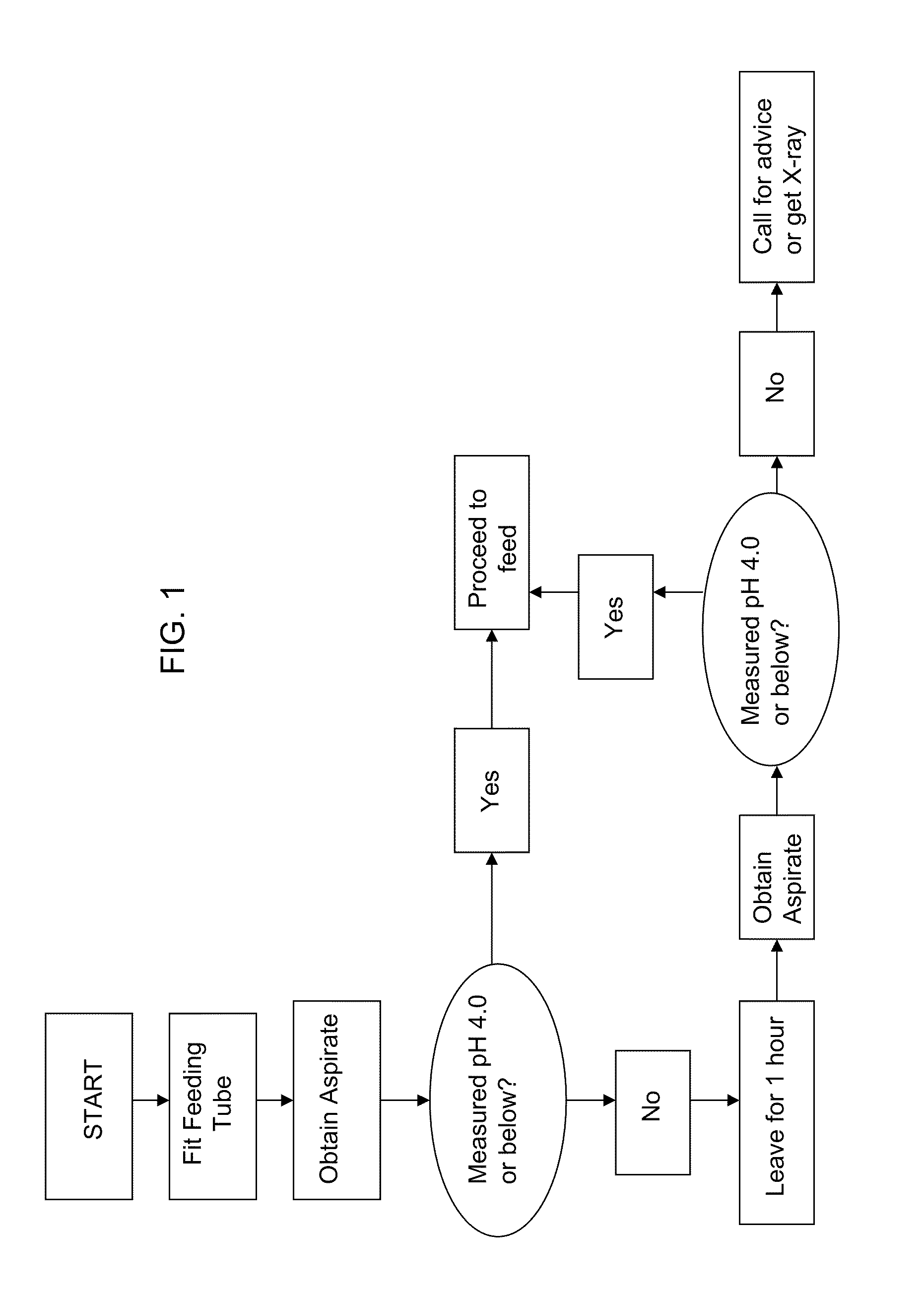
As depicted in Table 1 and FIG. 7, a pH measurement of 4.0 or less results in correct tube placement, but the overall failure rate of the placement tests increases, necessitating additional X-ray tests.
Elevating the pH measurementcut off” to 5.5 enables more tests to indicate stomach placement, thus improving the “pass rate” but with an increased risk that correct location may not be made.
Currently, misplaced naso or orogastric tubes, not detected prior to use is a significant patient safety issue and was the second most reported preventable patient safety incident in a 2009-2010 United Kingdom National Health Service annual report.
Such preventable safety incidents also referred to as “never events”, can lead to hospitals losing important funding.
However, although the chance of misplacement is small, owing to the tremendous number of verification tests carried out each year (estimated to be many millions) the absolute number of misplacements is high.
Accurate determination of the error rate of existing feeding tube misplacements is difficult to obtain since they are poorly reported and depend on how they are defined.
In summary, the results of these practices expose patients fitted with a feeding tube to a high level of risk.
The negative outcomes resulting from the misdiagnosis or lack of diagnosis of the feeding tube placement are dire, causing numerous instances of severe harm and death.
Therefore, the identification of a species or entity which is uniquely present in the stomach and whose use incorporates the desirable features of the feeding tube verification test, as outlined above, is highly desirable. pH is one such indicator and is used in existing tests, but it is associated with a high degree of error and cannot be used in every circumstance.

Method used


Image

  • Assay for positioning a feeding tube and method thereof
  • Assay for positioning a feeding tube and method thereof
  • Assay for positioning a feeding tube and method thereof

Examples


example one

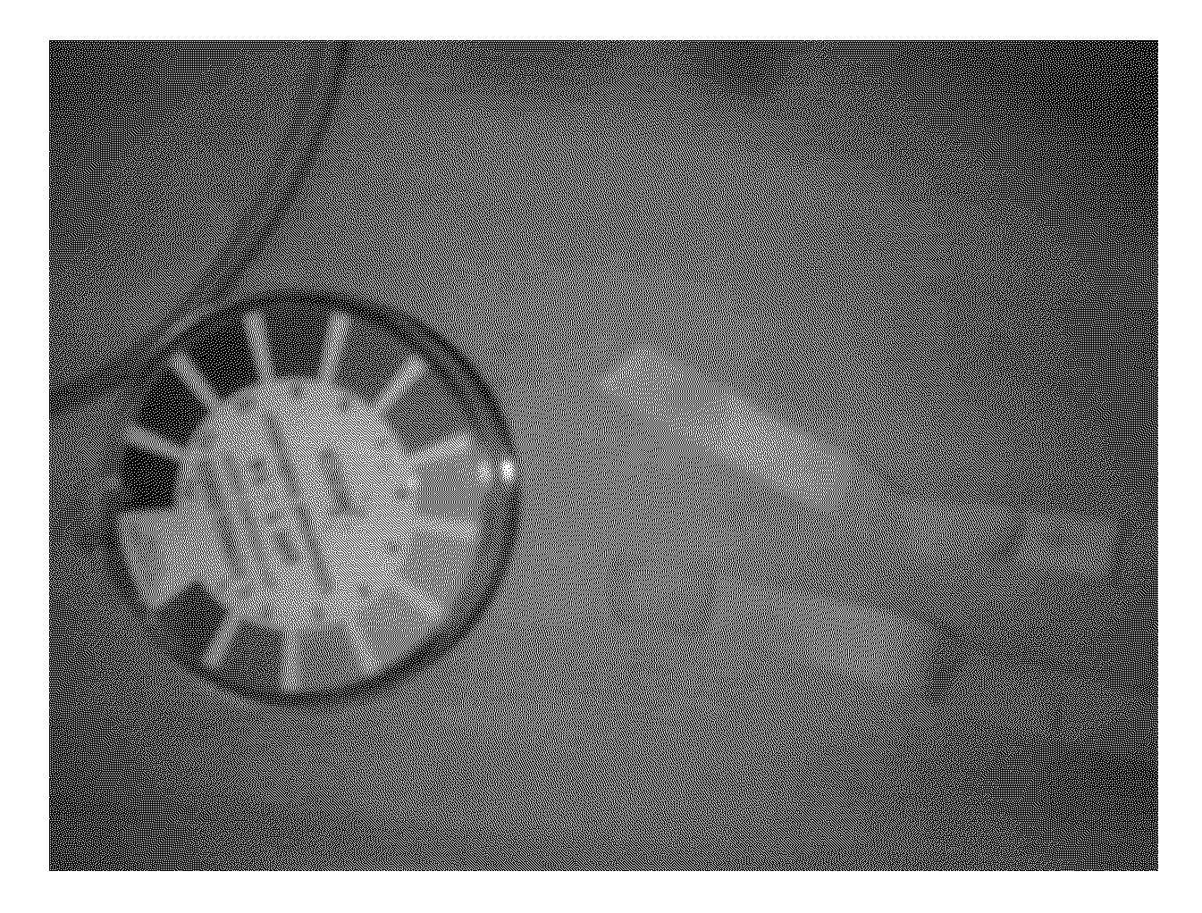
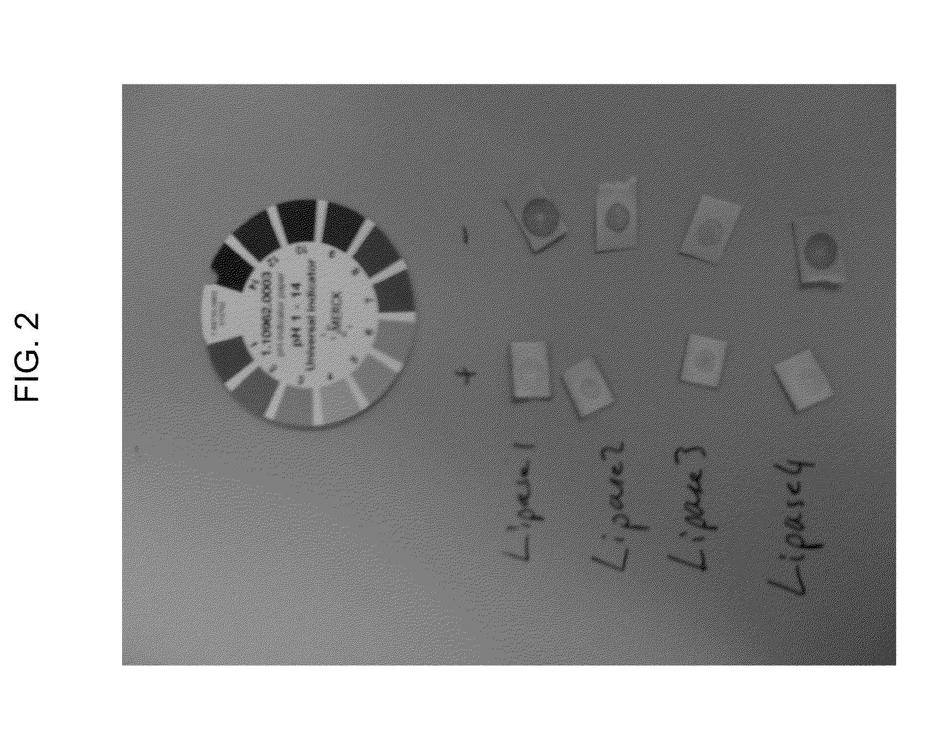
[0040]The assay of the present invention has an ester chemical impregnated into Ph indicator paper. The impregnation was carried out by adding 1 ml of tributyrin from Sigma to 9 ml of water, after which the mixture was vigorously shaken for 30 seconds to disperse tributyrin throughout the water in micro-droplets. The pH indicator paper was immersed into the resulting mixture, removed and then dried to evaporate the residual water. Once dried, the prepared pH indicator paper containing tributyrin had a 20 microlitre drop applied which contained a dissolved lipase from Amano. Alongside this, a second pH indicator paper from the same source, but lacking the impregnated tributyrin had another 20 microlitre drop of the same dissolved lipase applied. After a short period the pH was examined using the reference pH indicator scale provided by the pH indicator paper manufacturer. The pH of the indicator paper containing the tributyrin was pH 4, while the control pH indicator measurement with...

example two

[0041]It is well known in the art that the biochemistry and physiology between rabbit gastric lipase (RGL) and human gastric lipase (HGL) are similar. (See, Gargouri et al. (Biochimica et Biophysica Acta, 1006 (1989) 255-27, “Gastric lipases: biochemical and physiological studies.”) Therefore, a sample containing lyphophilized rabbit stomach extracts (JO4001) were obtained after soaking stomachs in an appropriate buffer. The procedure corresponds to the first step of gastric lipase purification as reported in Carriere et al. (Eur. J. Biochem. (1991) 202:75-83). “Purification and biochemical characterization of dog gastric lipase.”)

[0042]Lipase activity was measured at 5.5 using the pH-stat technique and tributyrin as substrate (See, Enzymology at Interfaces and Physiology of Lipolysis, “Standard assay of gastric lipase using pHstat and tributyrin as substrate”) according to Moreau et al., Biochem Biophys Ascta. 960: 286-93 (1988). “Purification, characterization and kinetic properti...

example three

[0045]A significant aspect of the present invention is providing a point of care test for determining proper placement of a feeding tube which can be readily used in a clinical bedside environment. To make the test easier to read, an alternate pH strip design was tested using yes / no technology similar to home pregnancy test kits. The pH strip was prepared with 0.2% w / v phenol red, 1% w / v tributyrin in methanol. The pH was increased to 9.5 by the addition of sodium hydroxide. The phenol red indictor test shows a color change for a pH below 6.8 (yellow) and no change for a above 8.2 (red). The initial pH before applying the sample is high and thus the phenol red is red in color due to the addition of sodium hydroxide in formulating the strip. Only when the enzyme and substrate are both present is sufficient acid generated in the strip to lower the pH enough to allow the phenol red to convert from a red color to a yellow color. In the absence of enzyme or substrate the acid cannot form...


PUM

PropertyMeasurementUnit
pHaaaaaaaaaa
pHaaaaaaaaaa
pHaaaaaaaaaa

Abstract

The present invention discloses a bedside or point of care assay for properly positioning a feeding tube in the stomach of a patient based on detecting the presence of a hydrolytic enzyme found in the stomach by an ester substrate which can be impregnated on a pH strip. The application of this test reduces the risk of tube misplacement and the resulting harm to the patient that occurs in the event of tube misplacement.

Description

CROSS-REFERENCE TO RELATED APPLICATION[0001]The present application claims priority to and the benefit of U.S. Provisional Application No. 61 / 283,401 filed on Dec. 3, 2009, and is incorporated by reference and made a part hereof.BACKGROUND OF THE INVENTION[0002]1. Field of the Invention[0003]The present invention is generally related to the field of medical diagnostics for verifying the location of a medical device in a patient. More particularly, the present invention relates to the use of an indicator substance which is acted upon, either directly or indirectly, by an enzyme specifically formed or located at the place where the medical device should be positioned. Specifically, the present invention discloses a point of care method and assay for accurately positioning a feeding tube in the stomach of a patient based on detecting the presence of a hydrolase enzyme found in the stomach where the feeding tube should be positioned.[0004]2. Description of the Prior Art[0005]Feeding tub...

Claims


Application Information

Patent Timeline
Patent Type & Authority Applications(United States)
IPC IPC(8): A61B5/157C12Q1/44
CPCA61B17/3415C12Q1/34G01N33/84A61B5/68G01N33/48A61B2010/0061C12Q1/25C12Q1/44A61B5/6846A61M2205/3324A61J15/0069A61J2200/70A61J15/0088A61M1/73
Inventor CARR, REUBENFARLEY, LUCY
Owner INGENZA
Learn More