In Vitro Generation of Hepatocytes from Human Embryonic Stem Cells

a technology of hepatocytes, which is applied in the field of in vitro generation of hepatocytes from human embryonic stem cells, can solve the problems of limiting the use of mouse es cells as a model of human development, human es cells may rapidly differentiate or fail to survive, and cannot prevent the differentiation of human es cells

Inactive Publication Date: 2011-12-22
RELIANCE LIFE SCI PVT
View PDF3 Cites 13 Cited by
  • Summary
  • Abstract
  • Description
  • Claims
  • Application Information

AI Technical Summary

Benefits of technology

[0024]The present disclosure is directed to differentiated cell populations obtained from pluripotent human stem cells, such as human embryonic stem cells (ES). In one embodiment, a differentiated cell population in an in vitro culture is obtained by differentiating pluripotent human stem cells in HepG2 conditioned medium. A further embodiment of the present disclosure depicts a method of generating a differentiated cell population from human pluripotent stem cells by maintaining a culture of undifferentiated pluripotent human stem cells and culturing the culture of undifferentiated pluripotent human stem cells in the presence of HepG2 conditioned medium. Another embodiment of the present disclosure depicts a method of g

Problems solved by technology

Although research with mouse ES cells facilitates the understanding of developmental processes and genetic diseases, significant differences in human and mouse development limit the use of mouse ES cells as a model of human development.
When cultured in a standard culture environment in the absence of a feeder layer, human ES cells may rapidly differentiate or fail to survive.
Unlike murine ES cells, the presence of exogenously added LIF does not prevent differentiation of human ES cells.
In spite of the progress in effectively culturing ES cells, several significant disadvantages with these methods still exist.
The major obstacle of the use of human ES cells in human therapy is that the originally described methods to propagate human ES cells involve culturing the human ES cells on a layer of feeder cells of non-human origin, and in the presence of nutrient serum of non-

Method used


Image

  • In Vitro Generation of Hepatocytes from Human Embryonic Stem Cells
  • In Vitro Generation of Hepatocytes from Human Embryonic Stem Cells
  • In Vitro Generation of Hepatocytes from Human Embryonic Stem Cells

Examples


example 1

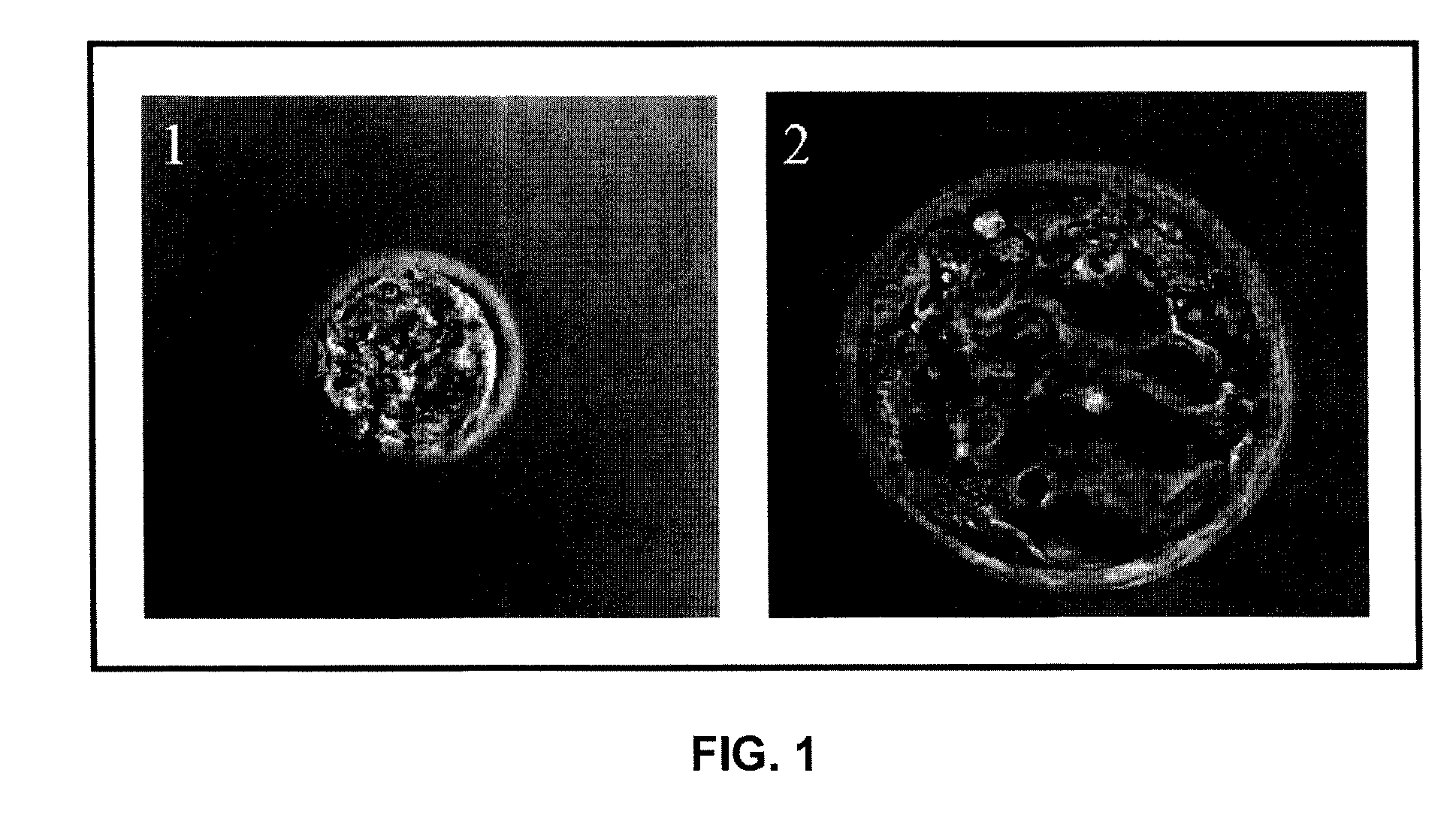
[0099]The present example discloses the preparation of blastocysts by in vitro fertilization.

[0100]1) Isolating Blastocysts

[0101]Blastocyst stage embryos (blastocysts) may be isolated from a variety of sources. These blastocysts may be isolated from recovered in vivo fertilized preimplantation embryos, or from in vitro fertilization (IVF) (for example, embryos fertilized by conventional insemination, intracytoplasmic sperm injection, or ooplasm transfer). Human blastocysts are obtained from couples or donors who voluntarily donate their surplus embryos. These embryos are used for research purposes after acquiring written and voluntary consent from these couples or donors. Alternatively, blastocysts may be derived by transfer of a somatic cell or cell nucleus into an enucleated oocyte of human or non-human origin, which is then stimulated to develop to the blastocyst stage. The blastocysts used may also have been cryopreserved, or result from embryos which were cryopreserved at an ea...

example 2

[0107]The present example discloses the derivation and storage of mouse embryonic fibroblast (feeder) cells.

[0108]1) Procurement of Pregnant Mice and Dissection

[0109]Mouse embryonic fibroblasts (MEFs) may be obtained from inbred C57 Black mice or other suitable strains. In an illustrative method, a mouse at 13.5 days of pregnancy / days post coitum (dpc) is sacrificed by cervical dislocation. The abdomen of the mouse is swabbed with 70% Isopropanol followed by a small incision. The viscera is exposed by pulling apart the abdominal skin in opposite directions. The uterus filled with embryos is seen in the posterior abdominal cavity. The uterus is dissected out with sterile forceps and scissors and placed into 50 ml screw capped conical centrifuge tube containing 20 ml of sterile Dulbecco's phosphate buffered saline, Ca- and Mg-free (GIBCO-BRL, Cat No. 14190-144). Uteri containing embryos are dissected out from all the pregnant animals sacrificed. The uteri are then washed 5-6 times in ...

example 3

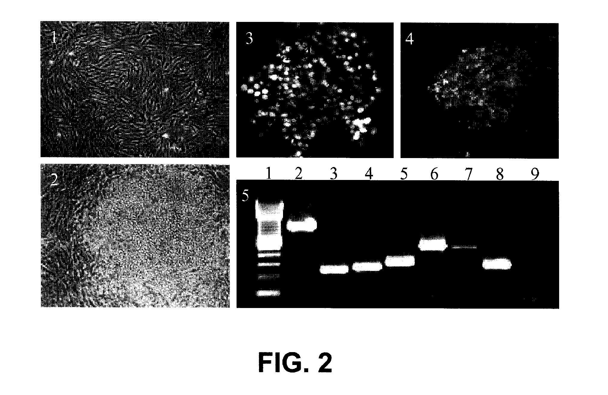
[0118]The present example describes the derivation and maintenance of human ES cells.

[0119]1) Inactivation and Plating of Mouse Embryonic Fibroblast (Feeder) Cells

[0120]The feeder cells stored in liquid nitrogen were thawed and cultured as needed. The vials were thawed by placing the frozen vials in a 37° C. water bath until the contents were semi-thawed. The contents were then collected in a tube and mixed with warm media to dilute the cryoprotectant. The cells were pelleted, resuspended, and plated in fresh MEF media (90% Dulbecco's modified Eagle's medium-High Glucose (GIBCO), 10% Fetal bovine serum (Hyclone), 1 mM L-Glutamine (GIBCO), 1% Non-Essential amino acids (GIBCO) and 0.1 mM β-Mercaptoethanol (Sigma)) in tissue culture flasks. Once the cells reached confluence, they were ready for inactivation. The cells were inactivated by Mitomycin C treatment or by gamma irradiation. Here, the cells were inactivated by Mitomycin C treatment for two and half hours. 10 ng / ml of Mitomycin...


PUM

PropertyMeasurementUnit
Toxicityaaaaaaaaaa
Morphologyaaaaaaaaaa
Enzyme activityaaaaaaaaaa

Abstract

Differentiation of human pluripotent stem cells, such as human embryonic stem cells (hESC), into hepatocytes by in vitro methods is disclosed. The pluripotent stem cells are cultured in conditioned medium from the hepatocarcinoma cell line, HepG2. Specific growth factors and defined media may also be added to the medium for stage specific differentiation of the derived hepatocytes. Hepatocytes differentiated from human pluripotent stem cells may be characterized by fluorescence activated cell sorting (FACS), immunofluorescence analysis (IF), real time polymerase reaction (RT-PCR), and functional assays. The methods disclosed herein are able to differentiate high percentages of hepatocytes from human pluripotent stem cells using the disclosed methods. These differentiated cells may exhibit polygonal shape morphology, typical of hepatocytes, and may express hepatocyte specific genes. The differentiated cells may also be positive for definitive endoderm markers and hepatic markers.

Description

CROSS-REFERENCES TO RELATED APPLICATIONS[0001]This application is a Continuation-In-Part of U.S. patent application Ser. No. 11 / 436,193 filed on May 17, 2006, which claims the benefit of and priority to the provisional Indian Application No. 595 / MUM / 2005, filed May 17, 2005. All of the above-mentioned applications are hereby incorporated by reference.STATEMENT REGARDING FEDERALLY SPONSORED RESEARCH OR DEVELOPMENT[0002]Not applicable.REFERENCE TO A “MICROFICHE APPENDIX”[0003]Not applicable.BACKGROUND OF THE INVENTION[0004]1. Field of the Invention[0005]The present disclosure relates to the isolation, maintenance and propagation of human embryonic stem cells (hESC) from the inner cell mass of surplus embryos. This disclosure also relates differentiating hepatocytes from hESC.[0006]2. Description of Related Art[0007]Pluripotent stem cells that are derived from the inner cell mass of a blastocyst are referred to as embryonic stem cells, while stem cells derived from primordial germ cell...

Claims


Application Information

Patent Timeline
IPC IPC(8): C12Q1/68G01N33/53G01N33/50C12N5/071C12N5/00
CPCC12N5/0606C12N5/067C12N2500/25C12N2501/11C12N2506/02C12N2501/12C12N2501/237C12N2501/39C12N2502/14C12N2501/113
Inventor MANDAL, ARUNDHATISAHA, DEBAPRIYARAVINDRAN, GEETAVISWANATHAN, CHANDRA
Owner RELIANCE LIFE SCI PVT
Why Eureka
Try Eureka