
Lung tissue model

a pulmonary model and tissue culture technology, applied in the field of three-dimensional (3d) pulmonary model tissue culture, can solve the problems of short-lived models in vitro, inability to consider pulmonary tissue models, and difficulty in gas and nutrient exchang

Active Publication Date: 2012-02-23
UNIV OF PECS
View PDF5 Cites 37 Cited by
  • Summary
  • Abstract
  • Description
  • Claims
  • Application Information

AI Technical Summary

Benefits of technology

[0231]In a version the artificial tissue scaffold itself is a support on the surface of which the cells are attached, preferably a porous membrane support. In this version of the scaffold it has a structure specially designed for and useful for cell attachment, e.g. a porous or curved or engrailed or grooved surface to which the cells are attached so that this facilitates the formation of a 3 dimensional structure.
[0242]Thus, a three dimensional tissue culture free of any artificial tissue scaffold does not disintegrate or become disorganized in lack of an artificial tissue scaffold but maintains its three dimensional structure. Even if said three dimensional tissue culture free of any artificial tissue scaffold is cultured and formed on a solid support material, the formation of the three dimensional structure is not assisted by and is not due to attachment of cells to this solid support and it can be separated without destruction of the three dimensional structure.

Problems solved by technology

Though the vascular endothelial cells were obtained from human lung, the artificial tissue prepared by this method was similar to human skin, therefore, it can not be actually considered as a pulmonary tissue model.
The authors observed that in the prior art primary cell lines “of human broncho-epithelial cells and human lung cancers provide a more differentiated model similar to the structure and function of epithelial cells in vivo; however, these models are short-lived in vitro.”
. . create additional problems, including difficulties of gas and nutrient exchange in the third dimension.
. . are purposefully engineered in a way to make tissue build-up and allow cellular interactions, nutrients and gas exchange to occur.” They conclude, however, that “Biotechnology in reconstructive surgery is still a test-tube based laboratory issue .
However, how ATII type differentiation is regulated is still far from understood.
The shortcomings of non-human models in mimicking human tissue characteristics often compromise the success of understanding molecular interactions in human tissues.
Despite the extensive literature of lung models, it appears that the prior art discloses only tissue scaffold based three dimensional models and no simple, tissue scaffold free lung tissue model, comprising at least epithelial cells and fibroblasts, is disclosed in the prior art.

Method used


Image

  • Lung tissue model
  • Lung tissue model
  • Lung tissue model

Examples


embodiments

[0398]Preparation of a 3D Model Tissue Culture

[0399]In the method of the present invention at least pulmonary epithelial cells and mesenchymal cells, preferably fibroblasts are used. The cells are cultured separately in order to obtain viable cultures, then mixed in an appropriate ratio and cocultured in the presence of CO2 under appropriate conditions as will be understood based on the present disclosure and art methods. By setting ratio of the cells and selecting conditions overgrowth of one cell type by another can be avoided.

[0400]In a preferred embodiment said cells are obtained from human subject as primary cells and either de-differentiated or used immediately. De-differentiation can be carried out e.g. by known methods (passages, removing other type of cells, addition of growth factors). If the cells are capable of confluence, they are considered as dedifferentiated.

[0401]Pelleting the cocultured cell mixture is an important step to establish cell-cell contacts and to result...

example 1

Materials and Methods

[0461]Primary SAEC, NHLF and pulmonary HMVEC cells were purchased from Lonza. All cell types were isolated from the lungs of multiple random donors of different sexes and ages. We used SAGM, FGM or EGM-2 medium for the initial expansion of SAEC, NHLF or pulmonary HMVEC, respectively, as recommended by the manufacturer. All types of cell cultures were incubated in an atmosphere containing 5% CO2, at 37° C. For 2D and 3D culturing, pure or mixed cell populations were cultured in a 50-50% mixture of SAGM (Small Airway Growth Medium, Lonza) and complete DMEM. For two and three-cell cultures containing HMVEC cells, the appropriate growth factor supplements for HMVEC cells were added to the 50-50% mixture of SAGM and DMEM. The compositions of cell culture media were prepared in accordance with instructions of the manufacturer. For 2D and 3D culturing, cells were mixed at the indicated ratios and dispensed onto flat-bottom 6 well plates or 96-well V-bottom plates (Sars...

example 2

Experiments for Development of a 3D Lung Tissue Model

[0493]Hanging Drop Model

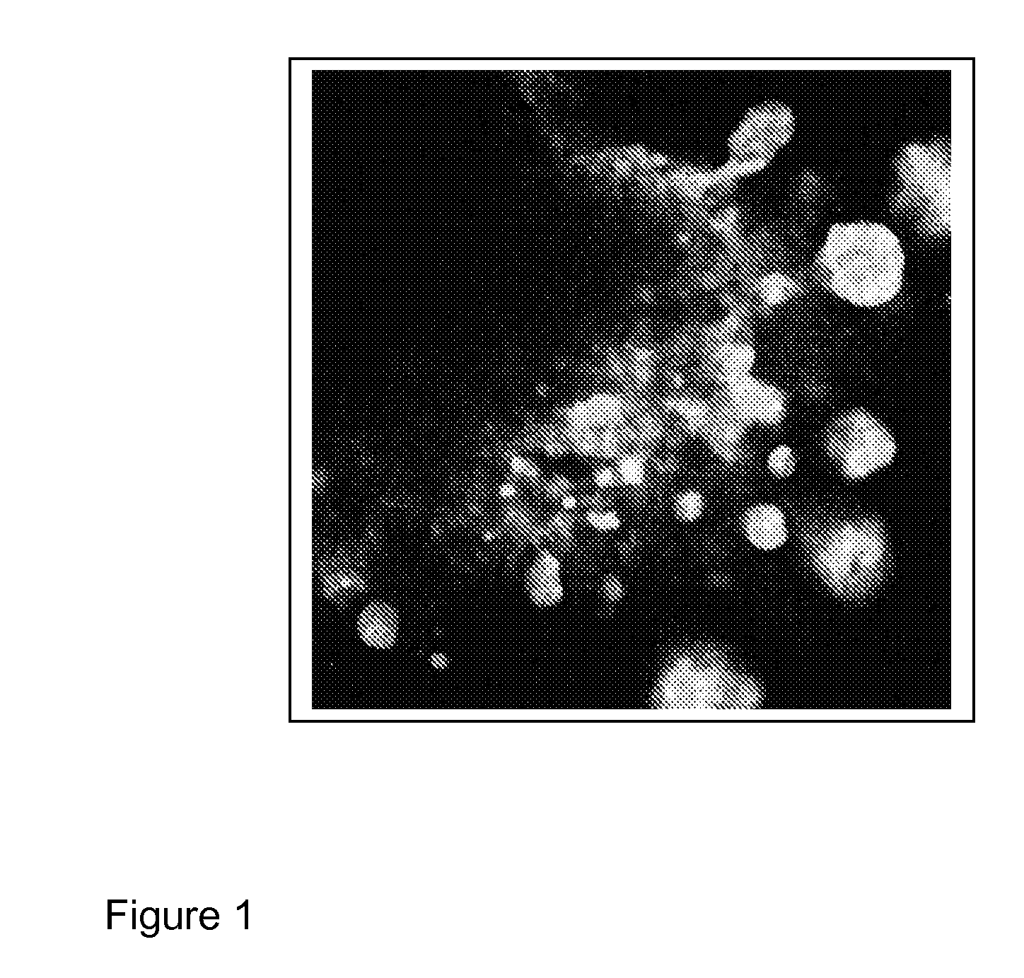
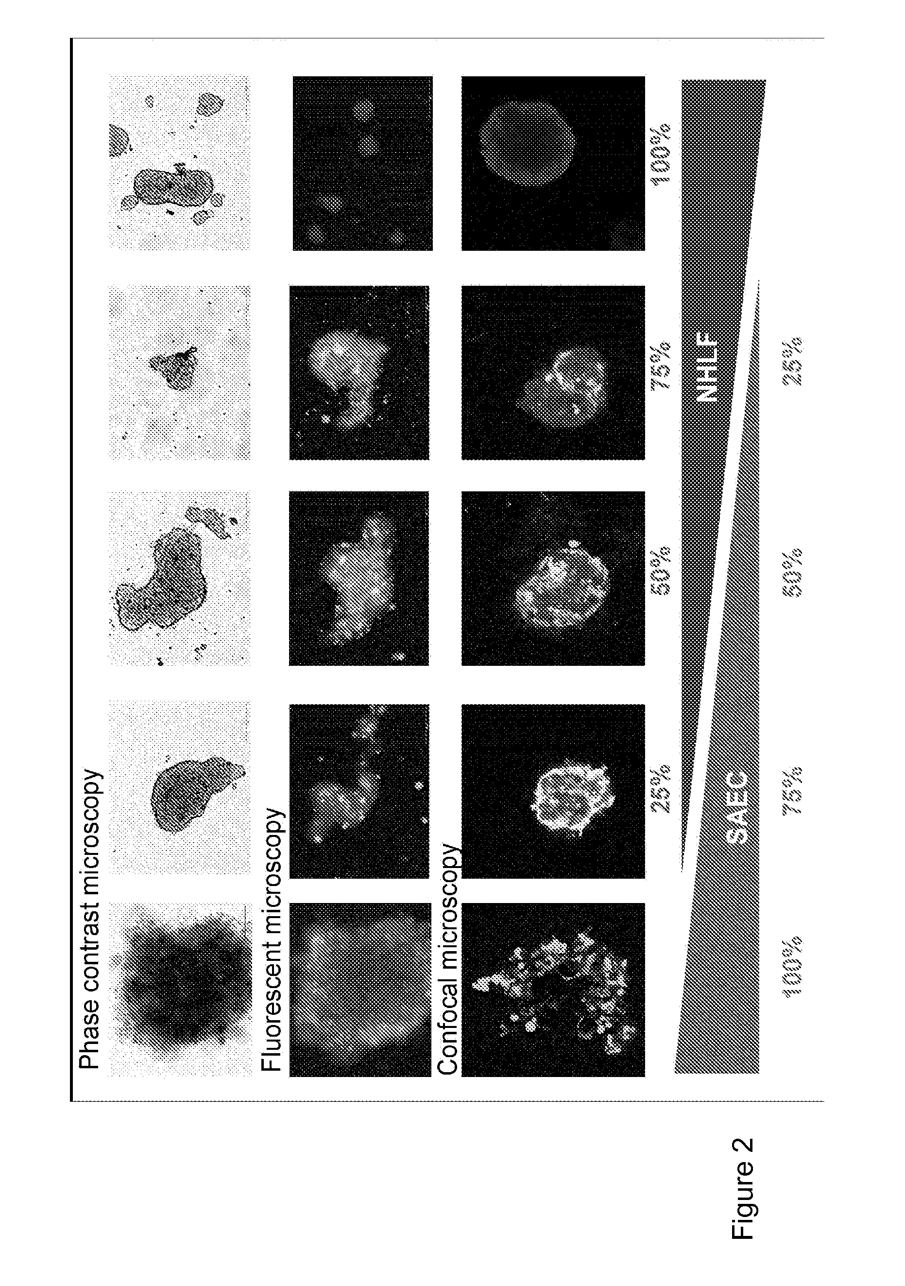
[0494]To simulate human lung structure, we started with a 3D cell aggregate of 100 000 cells, in roughly equal amounts of distinct fibroblast (NHLF) and small airway epithelial cell populations (SAEC), randomly intermixed. Within a day of incubation in a hanging drop assay, the cells generated loose tissue structures. The formation, however, was not stable and was not possible to transfer the generated micro-tissues from the initial culture conditions to another test plate without irrecoverable damage to the tissue structure.

[0495]Pelleted, Matrigel Containing Model

[0496]To improve the stability of mixed lung micro-tissues, 1:1 ratio of SAEC and NHLF were pelleted and grown in the presence of matrigel. Many 3D lung and other tissue models use matrigel to create a 3D structure where various cell types can grow and interact with one another. SAECs and NHLFs were stained with a fluorescent physiological dyes D...


PUM

PropertyMeasurementUnit
Volumeaaaaaaaaaa
Adhesion strengthaaaaaaaaaa

Abstract

The present invention provides for an engineered three dimensional (3D) pulmonary model tissue culture which is free of any artificial scaffold.

Description

FIELD OF THE INVENTION[0001]The present invention provides for an engineered three dimensional (3D) pulmonary model tissue culture which is free of any artificial scaffold. Three dimensional models of healthy lung tissue as well as disease tissues are available. The product according to the invention can be marketed e.g. in the form of tissue cultures, plates or arrays comprising such cultures or kits. The invention is applicable in medical and scientific research, for testing compounds for their effect on lung tissue, for screening, testing and / or evaluating drugs, and in certain cases in diagnostics of lung diseases.BACKGROUND ART[0002]Tissue engineering is a rapidly developing field of biomedical research that aims at repair, replace or regenerate damaged tissues. Due to the latest events of disastrous phase II clinical drug trials (e.g. TGN1412), further goals of tissue engineering include generation of human tissue models for safety and efficacy testing of pharmaceutical compou...

Claims


Application Information

Patent Timeline
IPC IPC(8): C12N5/077G01N33/53C12Q1/68C12Q1/02G01N21/64
CPCC12N5/0688C12N5/0697C12N2501/415C12N2502/1323C12N2502/28G01N2500/00C12N2533/90G01N33/5044G01N33/5088G01N33/57423C12N2503/04
Inventor PONGRACZ, JUDIT ERZSEBETBARTIS, DOMOKOS
Owner UNIV OF PECS
Learn More