Technology and method to study microbial growth and adhesion to host-related surfaces and the host-microbiota interactions

a technology of host-related surfaces and microbial growth, applied in the field of microbial growth and adhesion to host-related surfaces and the interaction of host microorganisms, can solve the problems of limiting the experimental time to 2 hours maximum, time-consuming cell culture growth, and inability to perform high-throughput screening

Inactive Publication Date: 2012-03-08
UNIV GENT
View PDF2 Cites 4 Cited by
  • Summary
  • Abstract
  • Description
  • Claims
  • Application Information

AI Technical Summary

Problems solved by technology

Yet, for reasons of cytotoxicity, cell cultures are very sensitive to co-incubation with mixed microbial slurries, thus limiting the experimental time to 2 hours maximally.
Moreover, the growth of cell cultures is time-consuming and it is not possible to perform high-throughput screenings to evaluate many bacteria or components at the same time.
Although it is possible to trigger mucus production in for example HT29 (Novellvaux et al., 2006) as well as MKN1, MKN7 and MNK45 cell-lines (Linden et al., 2007), it is not possible to monitor colonization and persistence of mixed microbial communities on these cells for reasons of cytotoxicity.
However, only a few investigators have reported expression of membrane-associated mucins in the human oral cavity.
The structural complexity of mucin polymers entails that the complete degradation of them by a single microbial species is unlikely.
The removal of carbohydrates and other components from the glycoprotein compromises the protective function in the gut, especially when the rate of mucus breakdown exceeds the rate of mucus production.
However, in this simplified environment, the control of changing conditions is not possible and only short term experiments can be conducted.
Nevertheless, stability of the microbial community under long term studies is not always possible.
Moreover, in the EnteroMix colon simulator, the volumes are small when compared with the in vivo situation, there is no stabilization of the microbial community and only short experiments can be performed.
Microbiota are allowed to adapt to the conditions for 16 hours, however there is no long-term stabilization of the microbial community and the volumes in the different chambers are small when compared with in vivo situations.
At the same time, however, it lacks the key point that specifically characterizes the mucosal biofilm namely, the anaerobic conditions prevailing at the top of the biofilm and microaerophilic conditions prevailing at the base of the biofilm.
These systems allow the microorganisms to adhere but do not offer the opportunity of studying the gut biofilm formation (Lebeer et al., 2007) and the host-microbial interaction under continuous simulated conditions.
None of the aforementioned models simulating the GI tract has an adequate device to study the mechanisms of bacterial adhesion in response to the host signals and the reciprocal cross-talk.
Animal studies demonstrated that vertebrates possess a broad scala of preserved interactions with the microbes with which they co-evolve (Cheesman and Guillemin, 2007) in particular when maintaining gut epithelial homeostasis, however, mechanistic studies are not always possible.
The in vitro use of cell lines can be limited by the fact that these cells do not produce a mucus layer (Caco-2 cells) or by the fact that pure microbial cultures or only a mix of few strains can be tested, for reasons of cytotoxicity.
Cell cultures are very sensitive to co-incubation with mixed microbial slurries, thus limiting the incubation time and the adaptation of the host and the microbial metabolism.
Although the above described systems are very useful for short-term experiments, they are generally not suited to study the complex properties of the intestinal microflora over long-term studies, due to the cytotoxicity of the microbial cells towards the human cell layer.

Method used


Image

  • Technology and method to study microbial growth and adhesion to host-related surfaces and the host-microbiota interactions
  • Technology and method to study microbial growth and adhesion to host-related surfaces and the host-microbiota interactions
  • Technology and method to study microbial growth and adhesion to host-related surfaces and the host-microbiota interactions

Examples


Embodiment Construction

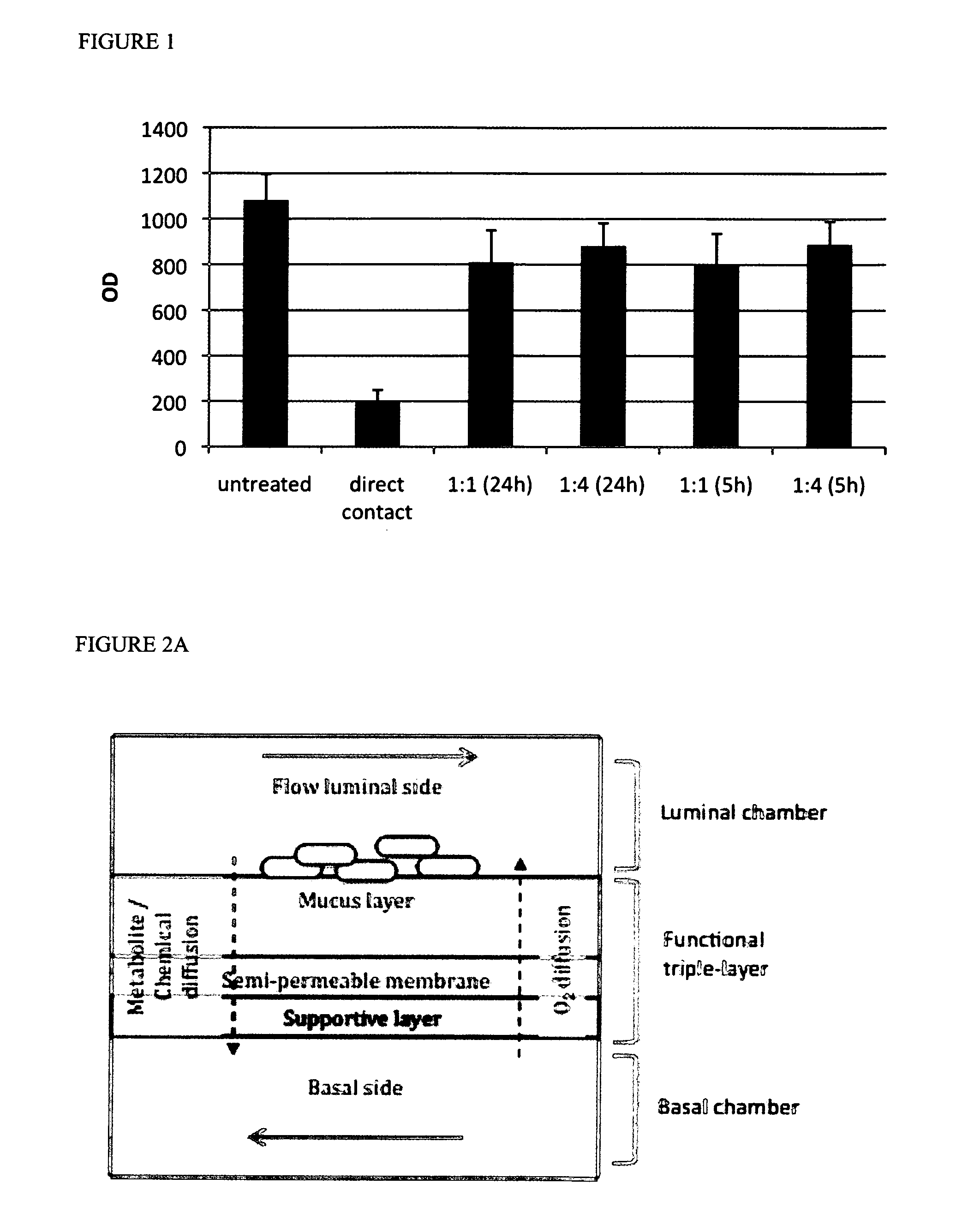
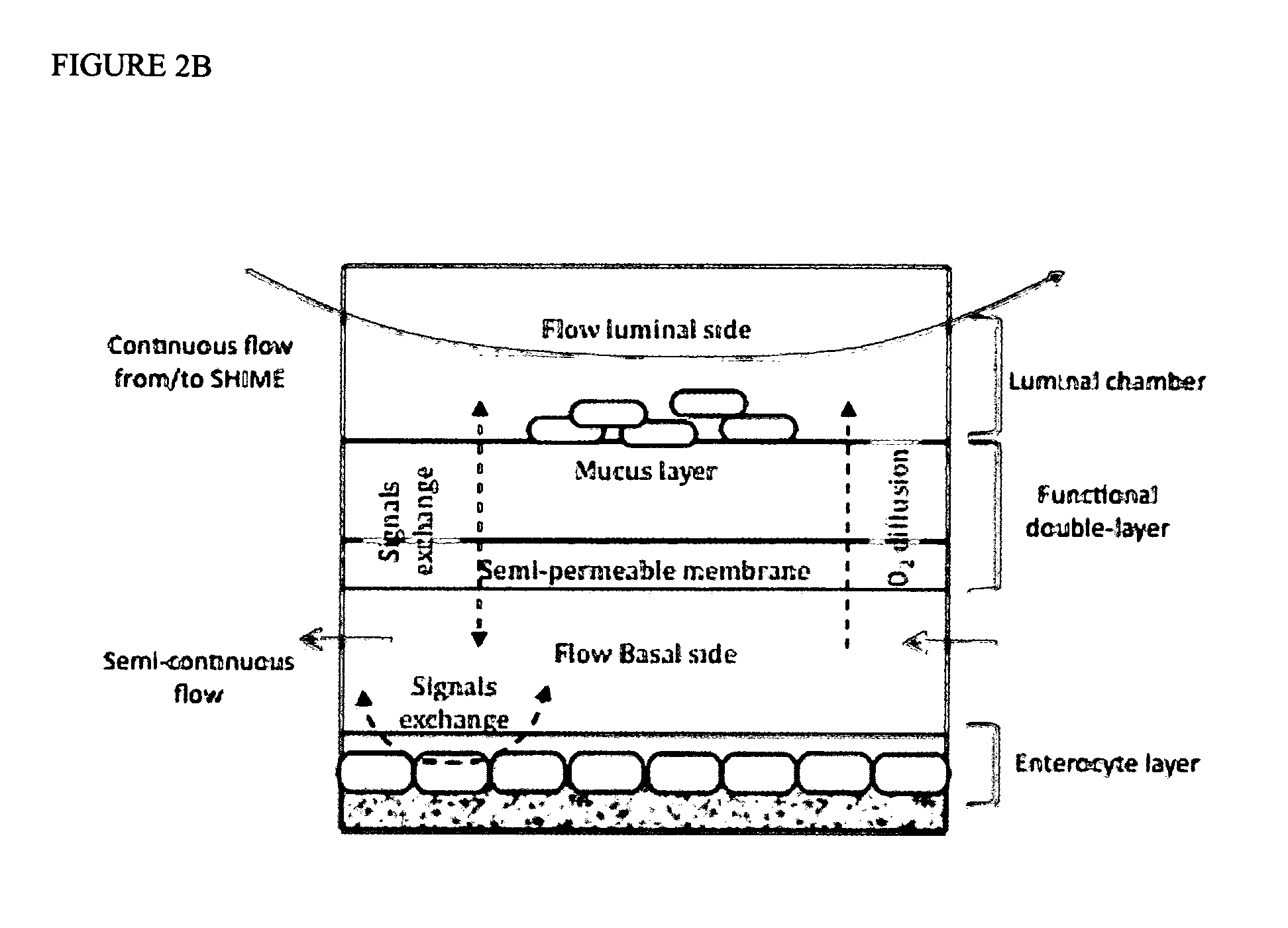
[0062]The adhesion module of the present invention comprises 2 compartments separated by a semi-permeable membrane; said membrane having an artificial mucus layer applied on its luminal side; and said module being characterized by having anaerobic conditions at its luminal side and aerobic conditions at its basal side.

[0063]With the term “module” or “adhesion module” is meant an excipient (e.g. a vessel or a container) wherein the environmental conditions of a mucosal layer are mimicked.

[0064]The combination of the mucus layer and the semi-permeable membrane, for dividing the adhesion module in 2 compartments, is hereinafter also referred to as the functional layer. Said functional layer can be a double, triple or multi-layer, but consists of at least two layers wherein the first layer, i.e. the top layer or the layer in direct contact with the luminal compartment, is the artificial mucus layer and the second layer is the semi-permeable membrane. With the term “luminal compartment” ...


PUM

PropertyMeasurementUnit
thicknessaaaaaaaaaa
partial oxygen pressureaaaaaaaaaa

Abstract

The present invention relates to in vitro adhesion modules that allow growth, stabilization and study of microbial communities that adhere to and colonize host-related surfaces, that mimic transport of chemical compounds across epithelial surfaces and simulate host-microorganism interactions and adaptation. It includes the provision of micromolar amounts of oxygen via the basal side of a mucus layer towards the adhered microorganisms thus establishing the microaerophilic conditions prevailing at the base of a biofilm. It can also include cells, simulating the host, in a chamber on the basal side of a functional layer comprising said mucus layer. The adhesion module of the present invention can be placed between the different compartments of the SHIME—the Simulator of the Human Intestinal Microbial Ecosystem. An extension of the SHIME is made where the duodenum, jejunum and ileum are separately mimicked.

Description

[0001]The present invention relates to in vitro models that allow growth, stabilization and study of microbial communities that adhere to and colonize host-related surfaces and that mimic transport of chemical compounds across epithelial surfaces and that allows the host-microbiota adaption and signal exchange.BACKGROUND OF THE INVENTION[0002]The human body hosts a tremendously diverse microbial community on external and internal host surfaces such as the skin, respiratory tract, mouth, gastrointestinal tract, reproductive tract, urinary tract and eyes. The total number of microbial cells (1014) exceeds the number of host cells (1013) with an order of magnitude and comprises many thousands of species. Their enormous enzymatic diversity, their capacity to trigger host immunological responses and the possibility of modulating physiological processes within the host that are involved in the etiology of cancer, obesity, or cardiovascular diseases, makes these host-associated microorgani...

Claims


Application Information

Patent Timeline
Patent Type & Authority Applications(United States)
IPC IPC(8): C12M3/00
CPCC12M23/34C12M29/04C12N5/0679C12N11/02C12M41/34
Inventor MARZORATI, MASSIMOPOSSEMIERS, SAMVAN DEN ABBEELE, PIETERVAN DE WIELE, TOMVANHOECKE, BARBARAVERSTRAETE, WILLY
Owner UNIV GENT
Why Eureka
Try Eureka